Introduction
Injury to a cerebral venous sinus is a well-recognised complication of neurosurgical and skull base procedures.Reference Gazzeri, Galarza, Fiore, Callovini and Alfieri1–Reference Apra, Kotbi, Turc, Corns, Pages and Souillard-Scemama4 In contrast, it is extremely rare during otological surgery,Reference Zaher Addeen and Al-Mohammad5–Reference Ulug, Basaran, Minareci and Aydin7 as is cerebral venous sinus thrombosis.Reference Sim and Lannigan8
We present a unique case, whereby management of an initial venous sinus injury during routine myringoplasty led to thrombosis of what turned out to be the dominant venous sinus, resulting in fulminant intracranial hypertension. Whilst there have been similar cases reported for operations on the posterior fossa and cerebellopontine angle,Reference Ohata, Haque, Morino, Nagai, Nishio and Nishijima9,Reference Keiper, Sherman, Tomsick and Tew10 to our knowledge, this is the first instance following routine otological surgery. We highlight the need to immediately consider highly asymmetric cerebral venous outflow if severe venous bleeding is encountered during even routine otological surgery, and present a rare example of successful emergency management of this clinical scenario.
Case report
A 40-year-old female underwent right-sided primary myringoplasty via a post-auricular incision to close a tympanic membrane perforation. During elevation of the periosteum from the mastoid portion of the temporal bone, brisk bleeding was encountered. Normal surgical techniques to arrest the bleeding failed, and what was presumed to be a large mastoid emissary vein was compressed using a bismuth iodine paraffin pack, with the intention of later removal.
Upon reintervention a week later, further brisk bleeding was encountered that proved increasingly difficult to control, so once again bismuth iodine paraffin pack compression and overclosure were performed. The patient, upon waking, reported severe headache, dizziness and pulsatile tinnitus. Fundoscopy demonstrated grade IV papilloedema, and a diagnosis of fulminant intracranial hypertension was made. The patient was transferred to our centre for specialist ENT and neurosurgical input.
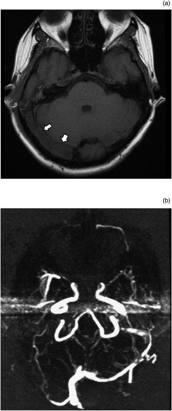
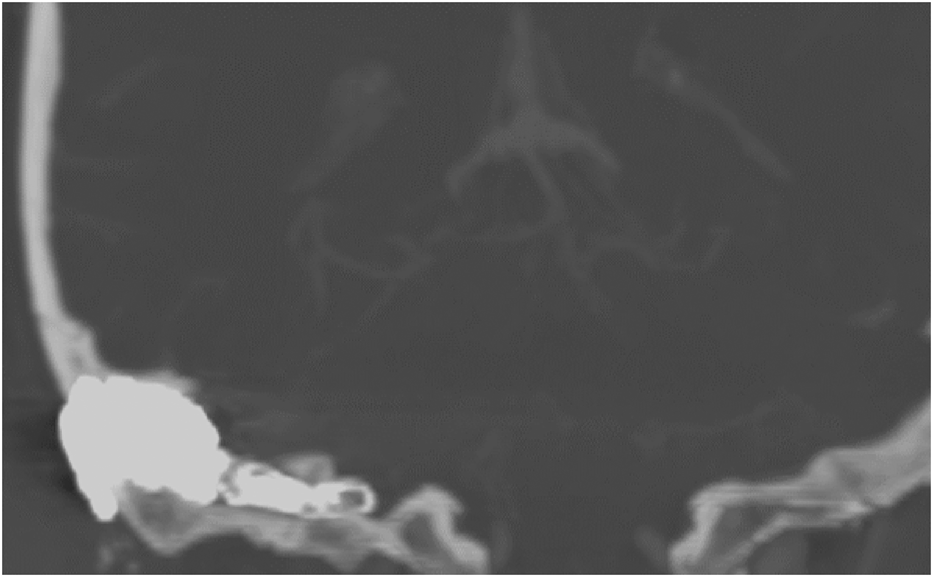
Upon admission to our centre, magnetic resonance imaging and magnetic resonance venography demonstrated acute thrombus throughout the right transverse and sigmoid sinuses, extending into the right jugular vein (Figure 1a, b). Marked right-sided dominance of cranial venous outflow was also noted, with the left transverse and sigmoid sinuses being narrow throughout. Computed tomography (CT) confirmed a bismuth iodine paraffin pack extending from the point of injury to the jugular bulb (Figure 2). It was presumed that there was a pre-existing bony dehiscence overlying the dominant sigmoid venous sinus, and on elevating periosteum, the elevator breached the venous sinus wall.
(a) Axial magnetic resonance imaging scan taken upon admission, showing thrombus in the right transverse sinus (arrows). (b) Magnetic resonance venogram taken upon admission, showing absent flow (due to thrombosis) in the right-sided venous sinus system, and a narrow transverse and sigmoid sinus on the left side.

Coronal computed tomography reformatted image taken upon admission, showing a bismuth iodine paraffin pack extending medially into the right inferior sigmoid sinus.

Given the presence of severe symptoms and risk to vision, it was decided to repair the right sigmoid sinus in order to allow later revascularisation by direct thrombolysis. A wide cortical mastoidectomy was performed without disturbing the packing material. Temporalis fascia was harvested and sutured to the posterior fossa dura, either side of the sinus tear, until there was only a small window for pack removal. The temporalis window was sutured closed and the surgical site covered with Surgicel® gauze before wound closure.
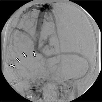
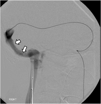
Catheter angiography performed the next day confirmed persistent occlusion of the right lateral sinus (Figure 3), and direct thrombolysis was initiated. The patient was intubated and a guide catheter was placed in the left jugular vein. Two microcatheters were advanced through the left lateral sinus and across into the right lateral sinus to begin direct infusion of tissue plasminogen activator (‘tPA’), supplemented by mechanical disruption with micro-guidewires and balloon angioplasty. Once anterograde flow was established, the microcatheters were left in situ for continued infusion of tissue plasminogen activator, and the patient was transferred to the intensive care unit.
Catheter angiogram (frontal view, venous phase) demonstrating persistent occlusion of the right lateral sinus 1 day after reconstruction of the right sigmoid sinus (arrows demonstrate absent flow).

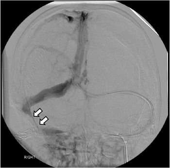
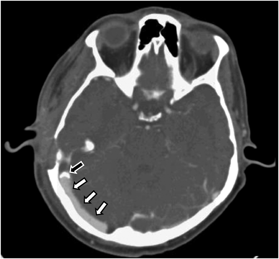
Angiography performed 39 hours later showed complete clearing of thrombus except at the original site of injury, where there was persistent narrowing (Figure 4). This was stented (using a 7 × 38 mm Guidant Omnilink® ‘.018’ stent system) with good expansion of the sinus (Figure 5), and the patient was subsequently extubated. Intracranial venous pressures dropped from 42 mmHg prior to thrombolysis to 15 mmHg after stenting. The CT venography performed 2 days later confirmed a fully patent right lateral sinus and stent (Figure 6). The patient reported resolution of symptoms and the subsequent ophthalmology review confirmed resolution of papilloedema. She was discharged on anticoagulant therapy.
Catheter angiogram (frontal view, venous phase) showing complete clearing of thrombus following direct thrombolysis, except at the site of original injury, where there was persistent narrowing (arrows).

Catheter venogram (frontal view) illustrating expansion of the right sigmoid sinus immediately following stenting (arrows demonstrate patency).

Axial computed tomography venogram demonstrating a fully patent right transverse sinus (white arrows) taken prior to discharge, 2 days after stenting (top of stent indicated by black arrow).

Two months following surgery, the patient reported the return of right-sided headaches, pulsatile tinnitus and increasing imbalance. The CT venography confirmed stent patency, but catheter angiography demonstrated raised venous sinus pressures. Further stenting of the left contralateral venous sinus outflow was performed but without symptomatic control, so ventriculoperitoneal shunting was performed. At the time of writing (14 years after intervention), the patient still has some residual symptoms of vertigo and right-sided conductive deafness, along with occasional headache, but no papilloedema.
Discussion
In the case presented, venous sinus injury occurred as a result of elevating the periosteum over a dehiscent sigmoid venous sinus. Previous reports of unexpected sinus injury suggest that an abnormally anterior course or highly dominant sigmoid sinus makes them susceptible to iatrogenic injury.Reference Zaher Addeen and Al-Mohammad5,Reference Gangopadhyay, McArthur and Larsson6 The incidence of these anomalies has not been reported, but if unexpected venous haemorrhage occurs during mastoid surgery then it is usual to conclude that the sigmoid sinus has been breached and measures to control the bleeding are undertaken. In our unusual case, the referring surgeon believed that bleeding was from a large mastoid emissary vein and so adopted a surgical approach that led to more persistent bleeding. The patient's evolving clinical condition, however, suggested a more complex problem, the nature of which was only resolved after CT venography.
The reported incidence of iatrogenic dural sinus thrombosis during neurosurgical and skull base procedures ranges from 4.8 per cent to 32.4 per cent.Reference Apra, Kotbi, Turc, Corns, Pages and Souillard-Scemama4,Reference Ohata, Haque, Morino, Nagai, Nishio and Nishijima9–Reference Abou-Al-Shaar, Gozal, Alzhrani, Karsy, Shelton and Couldwell11 Reported outcomes following iatrogenic venous sinus thrombosis in the neurosurgical setting range from the asymptomaticReference Zaher Addeen and Al-Mohammad5,Reference Gangopadhyay, McArthur and Larsson6,Reference Abou-Al-Shaar, Gozal, Alzhrani, Karsy, Shelton and Couldwell11 to development of a permanent vegetative state.Reference Ohata, Haque, Morino, Nagai, Nishio and Nishijima9 Although over-packing has been used successfully in some instances,Reference Zaher Addeen and Al-Mohammad5,Reference Gangopadhyay, McArthur and Larsson6 it can lead to post-operative thrombosis,Reference Ohata, Haque, Morino, Nagai, Nishio and Nishijima9 which is likely caused by the ingress of thrombogenic packing material into the lumen of the venous sinus. Iatrogenic dural sinus thrombosis rarely occurs after otological proceduresReference Sim and Lannigan8 and has not been documented to cause raised intracranial pressure. In the case presented, it is probable that packing extending into the lacerated venous sinus contributed to thrombosis and complete occlusion of the dominant venous sinus.
• This paper presents the first known case of cerebral venous sinus thrombosis causing fulminant intracranial hypertension following iatrogenic sinus injury during otological surgery
• Surgeons should be aware of the potential for sinus injury even in routine otological surgery, and manage it appropriately to prevent sinus thrombosis
• Sinus thrombosis in a dominant venous sinus requires urgent intervention to treat raised intracranial pressure
• This case provides an example of successful emergency management of fulminant intracranial hypertension due to iatrogenic sinus thrombosis
Partial or total agenesis of one transverse or sigmoid sinus occurs in 20 per cent of subjects,Reference Zouaoui and Hidden12 and the presence of a dominant sinus thrombosis increases the likelihood of symptoms caused by venous outflow obstruction.Reference Ohata, Haque, Morino, Nagai, Nishio and Nishijima9,Reference Keiper, Sherman, Tomsick and Tew10 Our case highlights that for some, a highly dominant venous sinus is critical for maintaining normal intracranial pressure, and it demonstrates the risk posed if thrombosis occurs within that sinus. Although it is not practical to perform imaging to document venous drainage before all otological procedures, a dominant venous sinus should be presumed if unexpected bleeding occurs that requires substantial packing and further surgery.
There is variation in the management of sinus thrombosis. Some authors advocate the use of thrombolytic therapy alone; others perform endoscopic techniques or open surgery to restore blood flow.Reference Ohata, Haque, Morino, Nagai, Nishio and Nishijima9,Reference Fargen and Hoh13,Reference Al-Sulaiman14 There is insufficient evidence to recommend best practice management in the event of thrombosis following iatrogenic venous sinus injury. We describe an approach that, although it did not result in complete symptom resolution, did prevent major neurological complication. On reviewing the case, we questioned our use of temporalis fascia because it seemed to trigger repeat venous sinus thrombosis. The use of an alternative material, for example an expanded polytetrafluoroethylene graft, may well have been a better choice and will be considered if another similar case presents.
Conclusion
It is our belief that a dominant or dehiscent venous sinus should be considered if unexpected bleeding occurs that requires substantial packing and further surgery. Care should therefore be taken during repair to prevent the ingress of packing material into its lumen or excessive compression applied to the sinus, so as to avoid occlusion and thrombus formation. Thrombosis formation within a dominant sigmoid sinus can cause raised intracranial pressure. Finally, we show that should this occur, urgent repair of the sinus followed by direct thrombolytic techniques can prevent major neurological complications.
Competing interests
None declared