Introduction
As Alzheimer's disease (AD) and vascular dementia (VaD) are the two most common forms of dementia, interest in distinguishing and treating the very early stages of AD and VaD has increased. Mild cognitive impairment (MCI), which is characterized by concerns about cognitive decline but by nearly no interference with activities of daily living (ADL) and an absence of dementia, is considered to be the intermediate stage between normal aging and mild dementia. aMCI, which is identified as memory decline with or without impairment in other cognitive domains, is considered as an important risk factor for developing AD (Petersen et al., Reference Petersen2001). Meanwhile, VCIND specifically refers to MCI subjects whose cognitive deficits are mainly associated with cerebrovascular injuries (Ingles et al., Reference Ingles, Wentzel, Fisk and Rockwood2002). The rates of conversion of aMCI to AD and VCIND to VaD are 35% within 3 years (Zanetti et al., Reference Zanetti, Ballabio, Abbate, Cutaia, Vergani and Bergamaschini2006) and 14.2% per year within 2 years (Marra et al., Reference Marra, Ferraccioli, Vita, Quaranta and Gainotti2011), respectively. Currently, the differentiation of aMCI and VCIND is mainly based on medical history, neuroimaging presentation, and neuropsychological assessment.
Although much research has focused on the differences in cognitive impairment between aMCI and VCIND, the findings of previous studies were partly inconsistent. For example, Nordlund et al. found that MCI was characterized by better memory and executive function than VCIND was (Nordlund et al., Reference Nordlund, Rolstad, Klang, Lind, Hansen and Wallin2007), whereas a meta-analysis conducted by Vasquez and Zakzanis showed that VCIND was characterized by significantly greater executive dysfunction and better memory than MCI (Vasquez and Zakzanis, Reference Vasquez and Zakzanis2015). Furthermore, certain studies tested semantic clustering in subjects with aMCI using the HVLT-R (Price et al., Reference Price2010; Malek-Ahmadi et al., Reference Malek-Ahmadi, Raj and Small2011). Semantic clustering describes the phenomenon of individuals reorganizing items based on a shared semantic feature and then consecutively recalling semantically related words contained in a superordinate category. By “chunking” large amounts of information into smaller units, semantic clustering is an effective strategy for encoding and storing information and more readily retrieving information from memory (Baddeley, Reference Baddeley2001). Although the differences in semantic clustering between aMCI and VCIND patients have not been sufficiently investigated, it has been observed that AD patients have worse semantic clustering capacity, as measured using the HVLT-R, compared with VaD patients (Gaines JJ, Reference Gaines, Shapiro, Alt and Benedict2006).
It is known that the sleep-wake cycle is regulated by complex interactions among brain regions and neurotransmitter systems (Porkka-Heiskanen et al., Reference Porkka-Heiskanen, Zitting and Wigren2013), many of which are also evolved in cognitive function, and especially memory ability (Diekelmann and Born, Reference Diekelmann and Born2010). Mounting evidence has found that patients with aMCI or VCIND have sleep problems. Westerberg et al. specifically observed reduced in sleep efficiency, slow-wave sleep, rapid eye movement, and wake after sleep onset using polysomnography (PSG) in individuals with aMCI. These abnormalities contributed to memory impairments that were considered to be mediated by interfering with sleep-dependent memory consolidation (Westerberg et al., Reference Westerberg2010; Reference Westerberg2012). In addition to PSG assessment, subjective sleep report was demonstrated to detect sleep disturbance in MCI as well (Hita-Yanez et al., Reference Hita-Yanez, Atienza and Cantero2013). Moreover, abnormal sleep was found in VCIND patients using either PSG or the PSQI, and this sleep disturbance was associated with cognitive impairment, as measured using the Montreal Cognitive Assessment (MoCA) (Jiang et al., Reference Jiang2013). Although these studies suggested that both aMCI and VCIND patients have sleep disturbance, the differences in sleep disturbance between aMCI and VCIND patients are still unclear.
In the present study, we compared cognitive impairment between patients with aMCI and VCIND and HCs. We then focused on differences in semantic memory strategy, as measured using the HVLT-R, in patients with aMCI and VCIND. Finally, we assessed the differences in subjective sleep disturbance between aMCI and VCIND patients and further explored the relationship between memory impairment and sleep disturbance. We hypothesized that (1) individuals with aMCI would have more severe impairment in verbal recall, recognition, and semantic memory strategy compared with patients with VCIND, (2) patients with VCIND would have worse performance in executive function compared with individuals with aMCI, and (3) memory impairment would be related to sleep disturbance in both aMCI and VCIND patients.
Methods
Participants
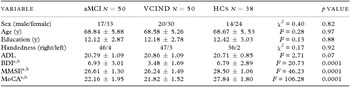
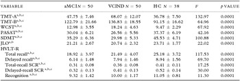
The study was approved by the Ethics Committee of Tianjin Medical University General Hospital. Written informed consent was obtained from the patients and their caregivers. Fifty aMCI (mean age: 68.84 ± 5.88; mean years of education: 12.12 ± 2.87; female: 33), 50 VCIND (mean age: 68.58 ± 5.26; mean years of education: 12.18 ± 2.78; female: 30), and 38 sex- and age-matched HCs (mean age: 68.67 ± 5.53; mean years of education: 12.42 ± 3.03; female: 24) were consecutively enrolled from the memory clinic of Tianjin Medical University General Hospital between April 2014 and July 2015. All candidates completed a comprehensive evaluation, including a full clinical history, a neurologic examination, an informant-based interview, clinical laboratory tests, brain magnetic resonance imaging (MRI), and a full neuropsychological battery. Demographic data are presented in Table 1, and mean neuropsychological test performance values are presented in Table 2. The criteria for participant inclusion were as follows:
Demographic variables for each group

Values represent the mean ± standard deviation (SD) unless otherwise indicated. HCs: healthy controls; aMCI: amnestic mild cognitive impairment; VCIND: vascular cognitive impairment-no dementia; ADL: Activities of Daily Living; BDI: Beck Depression Inventory; MMSE: Mini-Mental State Examination; MoCA: Montreal Cognitive Assessment.
a aMCI compared with HCs, p < 0.05.
b VCIND compared with HCs, p < 0.05.
Neuropsychological and sleep measures for each group

Values represent the mean ± SD. HCs: healthy controls; aMCI: amnestic mild cognitive impairment; VCIND: vascular cognitive impairment-no dementia; TMT-A/B: Trail Making Test-A/B; WCST: Wisconsin Card Sorting Test; PASAT: Paced Auditory Serial Addition Test; SDMT: Symbol Digit Modalities Test; JLO: Benton Judgment of Line Orientation; SCR: semantic clustering ratio.
a aMCI compared with HCs, p < 0.05.
b VCIND compared with HCs, p < 0.05.
c aMCI compared with VCIND, p < 0.05.
Patients with aMCI
The inclusion criteria for subjects with aMCI (including single- and multiple-domain aMCI) were (1) age from 55 to 85 and elementary education or above; (2) fulfillment of Petersen's aMCI criteria (Petersen et al., Reference Petersen2001), including memory complaints, usually corroborated by an informant, and objective memory with or without a decline in other domains (1.5 or more standard deviations (SDs) below the norm) on neuropsychological tests; preserved general cognitive function, as indicated by a Mini-Mental State Examination (MMSE) score ≥24 and a MoCA (Yu et al., Reference Yu, Li and Huang2012) score <26; intact activities of daily living, as indicated by an ADL (Lawton and Brody, Reference Lawton and Brody1969) score ≤22 and a Clinical Dementia Rating (CDR) (Berg, Reference Berg1988) ≤0.5; and not meeting the National Institute of Neurologic and Communicative Disorders and Stroke and the AD and Related Disorders Association (NINCDS-ADRDA) (McKhann et al., Reference McKhann, Drachman, Folstein, Katzman, Price and Stadlan1984) criteria for AD; and (3) the presence of medial temporal lobe atrophy (MTA, visual rating scale score <2) on MRI. The exclusion criteria for aMCI included the following: (1) the presence of cerebral vascular disease or cerebral ischemia on MRI; (2) a history of central nervous system disease or other medical conditions that might affect cognitive function; (3) the presence of psychiatric disorders (e.g. severe depression, a Beck Depression Inventory (BDI) (Zheng and Lin, Reference Zheng and Lin1991) score ≥16 or schizophrenia); (4) pharmacological interventions with cholinesterase inhibitors, memantine, or anti-depressants within 3 months; (5) a history of alcohol or drug abuse; and (6) severe physical, visual, or auditory disability that could influence performance on cognitive assessments.
Patients with VCIND
The inclusion criteria for VCIND were as follows: (1) age from 55 to 85 and elementary education or above; (2) based on the general MCI diagnostic criteria from the international working group on MCI (Winblad et al., Reference Winblad2004), neither normal cognition nor dementia; subjective and/or objective cognitive decline, usually corroborated by an informant; objective cognitive deficits confirmed by neuropsychological tests (1.5 or more SDs below the norm); preserved activities of daily living (ADL ≤22 and CDR ≤0.5); and intact general cognitive function (MMSE ≥24 and MoCA <26); and (3) based on the VCI criteria abstracted from National Institute of Neurological Disorders and Stroke-Canadian Stroke Network (Hachinski et al., Reference Hachinski2006), cognitive decline resulting from vascular risk factors, as well as the presence of multiple lacunar infarctions or white matter lesions in the brain on MRI. The exclusion criteria for VCIND included the presence of obvious brain atrophy (MTA visual rating scale score ≥1) on MRI; the other exclusion criteria were the same as most of the exclusion criteria (from (2) to (6)) for aMCI.
HC subjects
The inclusion criteria for the HCs were as follows: (1) age from 55 to 85 and elementary education or above; (2) an absence of subjective memory complaint; (3) scores not 1.5 SDs below age- and education-adjusted norms for the neuropsychological measures (e.g. the Wechsler Logical Memory Scale); (4) not having notable brain atrophy, ischemia or stroke on MRI; and (5) both MMSE and MoCA scores ≥26. The exclusion criteria for the HCs were (1) the presence of neurological or psychiatric diseases or other clinically significant medical conditions; (2) significant visual and/or auditory impairment; and (3) a history of alcohol or substance abuse.
Neuropsychological assessment
The full neuropsychological battery used in the present study had been validated in the Chinese population in previous studies. Patients with aMCI or VCIND and HCs were administered a neuropsychological battery in the proper order by a trained investigator who was blind to the participants’ clinical conditions. Testing of all participants was conducted in the morning.
-
1. The HVLT-R (Shi et al., Reference Shi, Tian, Wei, Miao and Wang2012) was used to evaluate verbal memory. This test consisted of 3 learning trials utilizing a list of 12 words divided into three semantic categories (jewels, animals, and dwellings) that were read aloud one by one, with one word per second, to an individual. Once the 12 words had been read, the individual was asked to recall as many words as he or she could. The individual was again asked to complete the delayed recall after 20 minutes. Immediately following the delayed-recall trial, a yes–no recognition task was administered, including 12 target and 12 (6 semantically related, 6 semantically unrelated) non-target words. A semantic cluster included consecutive recall of two categorically related words (i.e. sapphire and emerald) contained in a larger word list (Gaines et al., Reference Gaines, Shapiro, Alt and Benedict2006). SCRs were calculated by dividing the number of semantic clusters by the total number of words correctly recalled during the trial. The correct word scores in the immediate- and delayed-recall trials and the SCRs were then used to evaluate the patients’ learning and memory abilities and strategies. The word recall scores and SCRs were thus the outcomes.
-
2. The TMT-A/B (Rossini and Karl, Reference Rossini and Karl1994) was used to test executive function. The TMT-A consisted of a relatively simple numerical sequencing task, and the TMT-B was an alphanumeric sequencing task with required ongoing shifts between each overlearned series. The outcome was the time to finish each task.
-
3. The WCST (Mok et al., Reference Mok2004) was used to test higher executive function. The participants placed cards, one by one, under four stimulus cards according to a principle that they had to deduce from the pattern of a computer's responses of “right” or “wrong”. The score for perseverative errors was the outcome.
-
4. The PASAT (Gronwall, Reference Gronwall1977) was used to evaluate auditory processing speed, attention, and working memory. A series of 61 numbers from 1 to 9 were randomly delivered at presentation rates of one number every two seconds, and participants were instructed to add each digit to the one immediately preceding it. Performance was assessed based on the number of correct responses.
-
5. The SDMT (Smith, Reference Smith1982) was used to assess visual processing speed and working memory. The test consisted of single digits paired with abstract symbols, and the participants were expected to write the number that corresponded with a specific symbol. Ninety seconds were allowed for each trial, and performance was assessed based on the number of correct responses.
-
6. The Benton JLO test (Benton, Reference Benton1994) was used to assess spatial processing. The test consisted of 30 items, each showing a different pair of angled lines to be matched to display cards. The two test forms, or H and V, present the same items, but in different order. The outcome was the score for the number of items on which judgments for both lines were correct.
Subjective sleep assessment
Subjective sleep assessment, which was performed using the PSQI, ESS, and ISI, was conducted after neuropsychological assessment. The PSQI (Sohn et al., Reference Sohn, Kim do, Lee and Cho2012) consists of seven subscales, including subjective sleep quality, sleep-onset latency, sleep duration, sleep medication use, sleep efficiency, sleep disturbances, and daytime dysfunction. The total score possible on this test is 21, and lower scores indicate higher sleep quality. The ESS (Cho et al., Reference Cho, Lee, Son, Lee, Shin and Johns2011) and the ISI (Bastien et al., Reference Bastien, Vallieres and Morin2001) were used to evaluate daytime sleepiness and insomnia, respectively. Higher scores on the ESS and ISI indicate more severe daytime sleepiness and insomnia, respectively.
Statistical analyses
Statistical analyses were carried out with SPSS 18.0 (SPSS Inc., USA). Values of p < 0.05 were regarded as statistically significant. Demographic, neuropsychological and sleep data from the three groups were analyzed using χ2 tests for categorical data and analysis of variance for measuring materials. Most data in our study presented a normal distribution, and there were no missing values. To examine differences in the cognitive and sleep measures between the aMCI, VCIND, and HC groups, the data were analyzed using the analysis of covariance (ANCOVA) procedure followed by post-hoc least significant difference (LSD) tests, with cognitive and sleep measures as the dependent variable; the different groups (aMCI, VCIND, and HCs) as the between subjects variable; and age, gender, and years of education as covariates. Eta squared (ŋ2), which is an index of effect size that varies between 0 and 1, was calculated in the parametric statistical tests. In addition to ANCOVA, partial correlation analyses controlling for age were performed to analyze the relationship between memory and sleep variables. The assumptions underlying the parametric statistical tests were not violated, and no data transformations were performed during the analyses.
Results
Demographic characteristics are shown in Table 1. No significant differences were found between the aMCI, VCIND, and HC groups in terms of gender, age, years of education, handedness, or ADL score. Compared with the HC group, aMCI, and VCIND patients had higher BDI scores but lower MMSE and MoCA scores (p < 0.001). Table 2 displays neuropsychological test performance values for the three groups. ANCOVA followed by LSD, with sex, age, and education as covariates, was repeatedly used to evaluate cognitive function. In general, the HCs showed better performance than the patients with aMCI and VCIND did on the following cognitive tests: the TMT-A (F(2,132) = 132.97, p < 0.001, ŋ2 = 0.67), the TMT-B (F(2,132) = 64.96, p < 0.001, ŋ2 = 0.50), the WCST (F(2,132) = 67.92, p < 0.001, ŋ2 = 0.51), the PASAT (F(2,132) = 41.16, p < 0.001, ŋ2 = 0.39), the SDMT (F(2,132) = 100.88, p < 0.001, ŋ2 = 0.49), and JLO (F(2,134) = 22.02, p < 0.001, ŋ2 = 0.35). Compared with aMCI patients, VCIND patients showed more significant impairments on the following cognitive tests: the TMT-A (p < 0.001), the TMT-B (p < 0.001), the WCST (p < 0.001), the PASAT (p = 0.002), and the SDMT (p < 0.001). Meanwhile, JLO scores were similar between aMCI and VCIND patients.
Memory assessment
Overall, the HC group performed better than either the aMCI or the VCIND group in terms of recall, recognition, and SCR values (Table 2). ANCOVA followed by LSD, with sex, age, and education as covariates, demonstrated that the HCs had higher scores for total recall (F(2,132) = 117.53, p < 0.001, ŋ2 = 0.64), delayed recall (F(2,132) = 69.70, p < 0.001, ŋ2 = 0.51), and recognition (F(2,132) = 11.30, p < 0.001, ŋ2 = 0.34) and higher total-recall (F(2,132) = 17.25, p < 0.001, ŋ2 = 0.34) and delayed-recall (F(2,132) = 30.00, p < 0.001, ŋ2 = 0.21) SCRs. Compared with VCIND patients, aMCI patients showed poorer performances in terms of total recall (p < 0.001), delayed recall (p < 0.001) and recognition (p = 0.001) as well as lower total-recall (p = 0.002) and delayed-recall (p < 0.001) SCRs. Differences in SCRs are displayed graphically in Figure 1.
Comparisons of the semantic clustering ratios between the three groups (HCs, aMCI, and VCIND). SCR: semantic clustering ratio, #p < 0.05.

Subjective sleep assessment
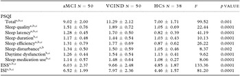
Subjective sleep variables are shown in Table 3. ANCOVA followed by LSD, with age as a covariate, was used in the sleep measures analysis. In general, the aMCI group had moderate PSQI, ESS, and ISI scores, which were higher than those in the HC group and lower than those in the VCIND group. For the PSQI subtests, the aMCI and VCIND patients showed higher scores for sleep quality (F(2,134) = 24.06, p < 0.001, ŋ2 = 0.26), sleep latency (F(2,134) = 44.21, p < 0.001, ŋ2 = 0.40), sleep efficiency (F(2,134) = 25.06, p < 0.001, ŋ2 = 0.27), and sleep disturbance (F(2,134) = 8.37, p < 0.001, ŋ2 = 0.10) compared with HCs. The HCs and aMCI groups had similar sleep duration, daytime dysfunction, and sleep medication use scores, which were lower than the scores for the VCIND group. Compared with the aMCI group, the VCIND group had higher scores for all PSQI subtests, except that these groups had similar sleep disturbance scores.
Sleep measures for each group

Values represent the mean ± SD. PSQI: Pittsburgh Sleep Quality Index; ESS: Epworth Sleepiness Scale; ISI: Insomnia Severity Index Scale.
a aMCI compared with HCs, p < 0.05.
b VCIND compared with HCs, p < 0.05.
c aMCI compared with VCIND, p < 0.05.
Correlation between HVLT-R and sleep measures
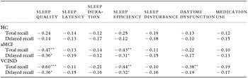
Partial correlation analyses controlling for age were performed for the learning and delayed-recall scores for the HVLT-R and variables of sleep measures (Table 4). For the HC group, there was no relationship between learning and delayed-recall scores for the HVLT-R and variables of sleep measures. For the aMCI and VCIND groups, sleep quality and sleep efficiency showed negative correlations with total recall and delayed recall (all r values ranged from −0.31 to −0.60, p < 0.05). Moreover, daytime dysfunction negatively correlated with total recall (r = −0.38, p = 0.007) for the VCIND group.
Correlation values between HVLT-R and sleep measures

Values represent the r values.
* p < 0.05; **p < 0.001; ***p < 0.0001.
Discussion
To our knowledge, this is the first study to compare verbal memory and semantic strategy, as assessed using the HVLT-R, and sleep characteristics, as assessed using the PSQI, between aMCI and VCIND patients. We found that individuals with aMCI had more significant difficulties in memory and semantic strategies but relatively better executive functioning and sleep than individuals with VCIND did. In addition, our results indicated that there are relationships between memory deficits and sleep disturbance in both aMCI and VCIND patients.
In this study, we found that individuals with aMCI exhibited poorer acquisition (immediate recall) and retrieval (delayed recall and recognition) than subjects with VCIND did, which indicates that memory impairment is more severe in aMCI than in VCIND. These results are consistent with the findings of most previous studies (Frisoni et al., Reference Frisoni, Galluzzi, Bresciani, Zanetti and Geroldi2002; Meyer et al., Reference Meyer, Xu, Thornby, Chowdhury and Quach2002; Villeneuve et al., Reference Villeneuve, Massoud, Bocti, Gauthier and Belleville2011). However, a disparate observation that has been reported is that individuals with VCIND exhibit slightly worse memory performance than MCI patients do. The discrepancy between that study and our present study may be mainly attributed to the inclusion of participants with aMCI or VCIND. In particular, in the previous study, the inclusion of MCI patients was not focused on aMCI, and the authors presumed that VCIND patients might have both vascular disease and AD pathology. In our study, we only focused on the amnestic type of MCI and tried our best to include the purely vascular-induced cognitive impairment in the VCIND group, using an MTA score <1 as an inclusion criterion. However, the inconsistent findings for the distinction in memory ability between aMCI and VCIND suggest that this topic deserves further investigation focusing on memory impairment features, such as semantic strategy.
It has been demonstrated that poor memory performance could be related to the diminished use of active encoding strategies (e.g. semantic clustering) in aMCI patients (Ribeiro et al., Reference Ribeiro, Guerreiro and De Mendonca2007; Price et al., Reference Price2010). However, whether there are any differences between aMCI and VCIND patients in terms of semantic strategy in the process of memory remains unknown. In the current study, we found that aMCI patients showed less semantic clustering than VCIND patients did. Although no other studies have specifically investigated the differences in semantic clustering between aMCI and VCIND patients, our results support the findings of Gaines et al., who showed that reduced memory and semantic clustering are more prominent in AD than in VaD (Gaines et al., Reference Gaines, Shapiro, Alt and Benedict2006). Ribeiro et al. noted that the poor semantic clustering in aMCI might be related to executive dysfunction (Ribeiro et al., Reference Ribeiro, Guerreiro and De Mendonca2007). However, the relationship between semantic clustering and executive function remains controversial. Malek-Ahmadi et al. found that executive functioning was highly correlated with verbal recall, but not semantic clustering (Malek-Ahmadi et al., Reference Malek-Ahmadi, Raj and Small2011), whereas McLaughlin et al. suggested that executive dysfunction contributed to poor semantic clustering to a certain extent (McLaughlin et al., Reference McLaughlin2014). Interestingly, in our study, the aMCI patients showed better executive function than the VCIND patients did, as in most previous studies (O'Brien et al., Reference O'Brien2003; Nordlund et al., Reference Nordlund, Rolstad, Klang, Lind, Hansen and Wallin2007), but the use of semantic strategy in aMCI patients was strongly diminished compared with that in VCIND patients. Our results support the view that semantic memory may be based on a separate brain network, independent of the executive system. Further neuropsychological studies combined with brain function assessment, such as functional MRI (fMRI), have the potential to elucidate the anatomic and physiologic mechanisms of semantic strategy in memory.
Sleep disturbances often coexist with cognitive disorders and appear in the early stages of dementia, such as AD and VaD. These disturbances are assumed to be caused by two mechanisms: (1) neural structures involved in sleep regulation, affecting the normal sleep-wake system, are directly damaged by neurodegeneration (Westerberg et al., Reference Westerberg2012) and/or cerebrovascular diseases (Schwartz and Roth, Reference Schwartz and Roth2008), and (2) a central imbalance of neurotransmitters, and especially acetylcholine, occurs (Bassetti and Hermann, Reference Bassetti and Hermann2011). Recently, studies suggested that damage of basal forebrain nuclei, and especially the nucleus basalis of Meynert, contributes to sleep disturbance, even in MCI (Grothe et al., Reference Grothe2010). In line with previous studies (Westerberg et al., Reference Westerberg2012), we found that both aMCI and VCIND patients had more severe sleep disturbance compared with HCs. We also found that VCIND patients showed poorer sleep quality, lower sleep efficiency, prolonged sleep latency, decreased sleep duration, and prominent daytime dysfunction compared with aMCI patients. These findings may be supported by a study on dementia patients, in which patients with VaD experienced more severe symptoms of disrupted sleep compared with patients with AD (Fuh et al., Reference Fuh, Wang and Cummings2005). It is conceivable that the common lesion areas in VCIND, such as the frontal lobes and thalamus, are involved in the projection of sleep-wake regulation (Hirshkowitz, Reference Hirshkowitz2004). It was observed that white matter hyperintensity was associated with sleep disturbances in VCIND (Cheng et al., Reference Cheng, Tsai, Wang, Hsu and Fuh2013). Moreover, vascular risk factors such as hypertension and diabetes mellitus, which frequently exist in VCIND, also contribute to sleep disturbance (Joo et al., Reference Joo2011). Therefore, VCIND patients may experience more severe sleep disturbances than aMCI patients do.
Sleep is considered to be important for memory consolidation. Because the specific patterns of neuromodulatory and electrical activities in the brain during sleep facilitate hippocampal-neocortical interaction, quantitative, and qualitative changes in new memory occur subsequently (Diekelmann and Born, Reference Diekelmann and Born2010). The relationship between sleep disturbance and memory deficits has been demonstrated in cognitively healthy individuals (Kloepfer et al., Reference Kloepfer2009; Takeuchi et al., Reference Takeuchi2014). In addition, a functional neuroimaging study suggested that memory and cognition could be influenced by sleep deprivation (Chee and Chuah, Reference Chee and Chuah2008). Moreover, several studies demonstrated that sleep changes contribute to cognitive dysfunction, and especially memory deficit, in patients with aMCI or VCIND (Westerberg et al., Reference Westerberg2010; Reference Westerberg2012; Jiang et al., Reference Jiang2013). Our study found that several sleep characteristics, including sleep quality and sleep efficiency, showed significant correlations with learning and delayed recall during memory assessment in both aMCI and VCIND patients. These findings suggest that sleep disturbances may be a comorbid condition at the very early stage of cognitive impairment. The complex interaction between cognitive decline and sleep disturbance may ultimately lead to severe consequences, such as dementia.
Limitations
There are certain limitations to our study. First, although the ages and education of our patients were matched with those of the HCs, our participants were relatively young (mean age: 68.70 ± 5.53) and highly educated (mean years of education: 12.22 ± 3.62); therefore, the results may not be generalizable to all individuals with aMCI or VCIND. Second, the diagnosis of aMCI was based upon neuropsychological assessments and brain MRI; however, cerebrospinal fluid biomarkers and positron emission tomography (PET) scans could improve the specificity of diagnoses of MCI due to AD. Finally, we did not follow all patients until several of them converted from aMCI or VCIND to AD or VaD. Which cognitive domain, memory strategy, or sleep measures can predict the conversion from MCI to dementia will be investigated in our future work.
Conclusion
Our findings support the idea that aMCI and VCIND differ in their characteristics of impairment in cognitive function, memory strategy and sleep, which are helpful to identify and distinguish patients with cognitive impairment at a very early stage. In addition, our results suggest that memory deficits are associated with sleep disturbance in both aMCI and VCIND patients.
Conflict of interest
None.
Description of authors’ roles
Nan Zhang formulated the research questions. Nan Zhang and Qingna Sun designed the study. All authors contributed to conducting the study and acquiring or interpreting the data. Qingna Sun carried out the statistical analysis. Qingna Sun and Nan Zhang wrote the paper. Yan Cheng supervised the study.
Acknowledgments
This work was supported in part by the Science and Technology Development Project of the Colleges and Universities of Tianjin Municipality, China (grant No. 20110116); the Key Project of Tianjin Municipal Science and Technology Commission (grant No. 13ZCZDSY01900); the National Natural Science Foundation of China (grant No. 81500926); and the National Key Clinical Specialty Construction Project of China.