Introduction
Much attention has been paid to the adiponectin gene, since it was first isolated and cloned from mouse fatty tissue by Scherer et al. (Reference Scherer, Williams, Fgliano, Baldini and Lodish1995). The adiponectin gene coding regions in many species have been cloned and published, including human (NC-000003), rat (NM-144744), mouse (NM-009605), pig (AY627882), dog (AB110099), cow (NM-174742), chicken (AY786316) and duck (DQ452618). Mammalian adiponectin genes, which contain three exons and two introns, are highly conserved between species (Hu et al., Reference Hu, Liang and Spieglman1996; Maeda et al., Reference Maeda, Okubo, Shimomura, Funahashi, Matsuzawa and Matsubara1996; Jacobi et al., Reference Jacobi, Ajuwon, Weber, Kuske, Dyer and Spurlock2004). The distinction between poultry and mammalian adiponectin is obvious: chicken and duck adiponectin only contain two exons and one intron (Yuan et al., Reference Yuan, Liu, Liu and Li2006; Dong, Reference Dong, Gong and Meng2007, whereas those of mammals have three exons and two introns); the homology between these two species is 86%, and they only have 66% homology with mammals.
Adiponectin, which is secreted by fatty tissue, has many physiological functions, like some other fatty cytokines such as leptin and interleukins (ILs) (Berg and Scherer, Reference Berg and Scherer2005). It has been proved that adiponectin can regulate endothelium function, glucolipid metabolism and enhance insulin sensitivity in energy metabolism and lipogenesis (Xu et al., Reference Xu, Wang, Keshaw, Xu, Lam and Cooper2003; Han et al., Reference Han, Quon, Kim and Koh2007; Saely et al., Reference Saely, Risch and Hoefle2007). However, the mechanism of action is not clear. The sequences of the adiponectin genes or their coding region have been submitted to GenBank and there have been many achievements in research into their physiological function (Jacobi et al., Reference Jacobi, Ajuwon, Weber, Kuske, Dyer and Spurlock2004; Devaraj et al., Reference Devaraj, Siegel and Jialal2007; Higurashi et al., Reference Higurashi, Kunieda, Matsuyama and Kawakami2007). In contrast, little information is available on goose adiponectin. As a foundation for further research into goose adiponectin, we cloned and sequenced the goose adiponectin gene and studied its expression profile in various tissues by semi-quantitative reverse transcription-polymerase chain reaction (RT-PCR).
Materials and methods
Experimental animals and DNA/RNA isolation
Three 11-week-old female geese were euthanized by decapitation and tissues, including adipose, liver, skeletal muscle, heart, spleen, small intestine, glandular stomach, muscular stomach, diencephalon, ovary, lung and kidney, were collected immediately. Genomic DNA was extracted from goose blood using phenol, chloroform and ethanol precipitation. Total RNA was extracted from different tissues using Trizol (Invitrogen, Carlsbad, California, USA), and digested in DNase I (Takara, Tokyo, Japan) at 37°C for 60 min. The concentrations of RNA samples were measured and diluted to 1 μg/μl. Samples were stored at −70°C until use.
Cloning of the adiponectin gene
The adipose tissue RNA samples were reverse transcribed using an RNA PCR kit (Takara) following the manufacturer's instructions. The reaction mixture (10 μl) consisted of 2 μl MgCl2 (25 mmol/l), 1 μl 10×RT buffer, 3.75 μl RNase-free distilled water, 1 μl dNTP mixture (10 mmol/l each), 0.25 μl RNase inhibitor (40 U/μl), 0.5 μl avian myeloblastosis virus (AMV) reverse transcriptase (5 U/μl), 0.5 μl oligo dT-adaptor primer (2.5 pmol/l), 1 μl sample RNA (about 1 μg total RNA). The reaction mixture was incubated at 42°C for 30 min, followed by 99°C for 5 min and 5°C for 5 min.
The PCR reaction was performed in the thermal cycler in a mixture reaction of 25 μl containing 2.5 μl 10×PCR buffer (without Mg2+), 1.5 μl MgCl2 (25 mmol/l), 2 μl dNTP (2.5 mmol/l each), 1 μl (10 pmol/l) of each primer, 0.2 μl rTaq enzyme (5 U/μl; Takara) and 1 μl RT product as template. The PCR programme was as follows: initial denaturation at 95°C for 5 min, followed by 30 cycles at 95°C for 30 s, 50–55°C for 45 s, 72°C for 60 s, and then a final extension at 72°C for 10 min. The primers used were as follows:
F1: 5′-CACTTCAGGAACGCACCATG-3′;
R1: 5′-CGGAGCGGTAAACGTAGGC-3′;
F2: 5′-TCAGTAGGCTTCCTCCTTTG-3′;
R2: 5′-TGTCTGGGTACAGGAGGAAG-3′;
F3: 5′-ACAAGAAGGACAAGGCGGTC-3′;
R3: 5′-TAGTGCCAGAGCCCCAGA-3′.
The PCR products were purified by Takara Gel Extraction Kit (Takara), followed by cloning into pMD 18-T vector (Takara). Sequences were determined by Shanghai Sangon Biological Engineering Technology Co., Ltd, China.
Semi-quantitative RT-PCR
Semi-quantitative RT-PCR was performed to examine the expression of adiponectin mRNAs in 12 different tissues. The RNA samples were reverse transcribed as above. PCR was performed in the thermal cycler in a reaction mixture of 25 μl containing 0.25 μl Taq polymerase (5 U/μl, Takara), 2.5 μl 10×PCR buffer (without Mg2+), 2 μl dNTP (2.5 mmol/l each), 2 μl RT-F (5 pmol/l), 2 μl RT-R (5 pmol/l), 1 μl cDNA and 15.25 μl double-distilled water. The cycle parameters were subjected to denaturation at 95°C for 5 min, 32 cycles of denaturation at 95°C for 30 s, annealing at 60°C for 30 s, and extension at 72°C for 30 s, and a final extension at 72°C for 10 min. β-Actin was used for standardization for each adiponectin. The annealing temperature and cycles of β-actin were 45°C and 26 cycles, respectively. PCR products were separated by electrophoresis on 2% (w/v) agarose gel containing ethidium bromide (0.5 mg/ml) and photographed under UV illumination. Densitometry was performed using a Gel-Pro analyser (Ultra-Violet Products Ltd, Cambridge, UK), and the ratio of the adiponectin to β-actin mRNA was obtained. The primers used were as follows:
RT-F: 5′-CCTCCTTTGCTCACTGCTGCTGGTG-3′;
RT-R: 5′-CCCTGGGTATCCTGGAAATCCTC-3′.
β-actin:
F: 5′- ACGAGCGGTTCAGGTGTC-3′;
R: 5′-GAAAAGAAGGCTTATTCCAGTTT-3′.
Statistical analysis and software
Primer 5.0 software was used to design primers. DNAMAN 5.2.2 software (Lynnon Corp., Quebec, Canada) was applied to sequence alignment, splicing the whole gene and for translation. The predicted isoelectric point and molecular weight of the goose adiponectin were determined by Sequence Analyzer at http://www.expasy.org/tools/protparam.html. The results of semi-quantitative RT-PCR were analysed by Lab Works 4.0 software (Ultra-Violet Products Ltd). Applying SPSS 12.0 software (SPSS Inc., Chicago, Illinois, USA), the results were expressed as the actual mean ± SEM of the ratio of the adiponectin to β-actin mRNA.
Results
The adiponectin gene clone
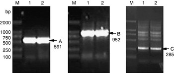
Three fragments (A, B and C) were obtained by PCR amplification using three pairs of primers (Fig. 1), which had short homologous sequences at the 3′ and 5′ ends. Three adiponectin segments were joined using DNAMAN software. A sequence of 1062 bp, which was composed of two exons and one intron (GenBank accession No. EU370686), was obtained by splicing using DNAMAN software. This sequence had a 738 bp integrity coding region, which encoded a protein of 245 amino acids.
Identification results by PCR for goose adiponectin gene recombinant plasmid. A, B and C, amplification fragments of F1/R1, F2/R2 and F3/R3, respectively. Lanes: M, DL2000 DNA marker; 1 and 2, different single colonies chosen at random.

Sequence analysis and homology comparison
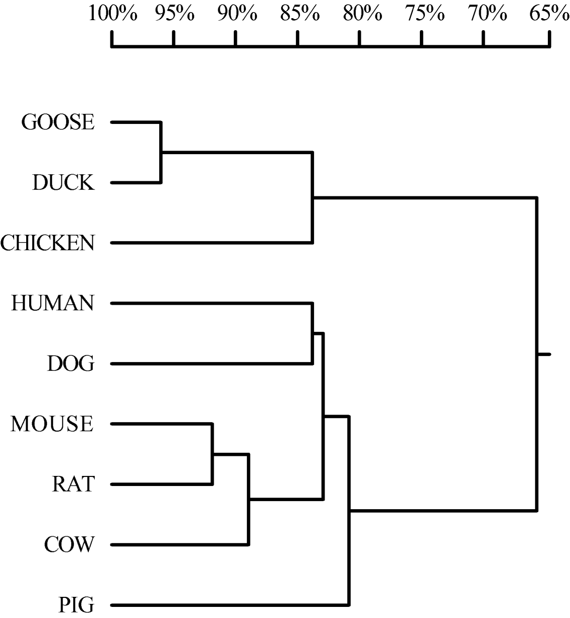
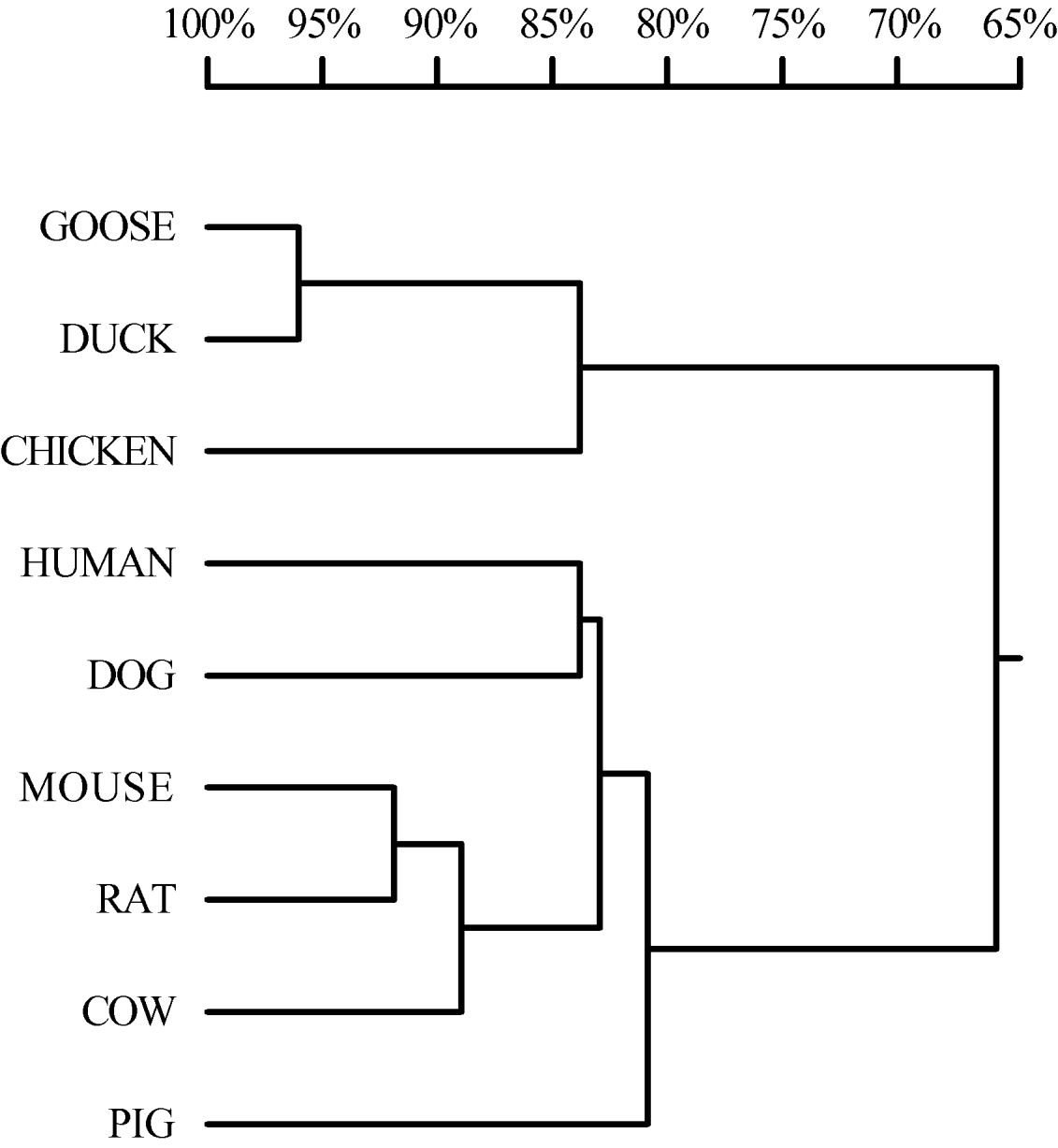
The results of homology comparison showed that the goose adiponectin gene coding sequence (CDS) had a high homology with those of the chicken and duck, which was above 86.9%, and that the homology with mammals was 62.4–64.2%, while the corresponding amino acid sequence shared a 95.5% correspondence with that of ducks, 84.1% with the chicken protein, and 63.3–65.9% with that of mammals (Fig. 2 and Table 1).
Amino acid sequence homology tree of partial adiponectin of animals.
Homologous comparison of nucleotide and amino acid sequences of the goose adiponectin with that of other animals

The molecular weight of the goose adiponectin protein was 26,603.3 Da, similar to that of chicken, duck, human and mouse adiponectin. The predicted isoelectric point (pI) was 5.19, which was highly similar to that of the above animals. Goose adiponectin protein contained 22 Gly–X–Y collagen repeated sequence structures (Gly represents aminoacetic acid, X, Y represent arbitrary amino acids).
mRNA expression level in various tissues
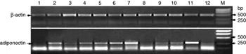
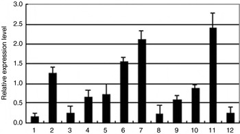
The results of semi-quantitative RT-PCR revealed that the goose adiponectin mRNA was highly expressed in skeletal muscle, adipose, heart and muscular stomach, moderately expressed in the small intestine, glandular stomach, kidney, lung, and weakly expressed in the liver, spleen, diencephalon and ovary (Figs 3 and 4).
Agarose gel electrophoresis of goose adiponectin gene expression in various tissues. Lanes: 1, liver; 2, heart; 3, spleen; 4, small intestine; 5, glandular stomach; 6, muscular stomach; 7, adipose tissue; 8, ovary; 9, kidney; 10, lung; 11, skeletal muscle; 12, diencephalon; M, DL2000 DNA marker.

The histogram of the expression of the goose adiponectin mRNA in various tissues by the semi-quantitative RT-PCR. The vertical axis indicates the value of the adiponectin mRNA quantity relative to β-actin mRNA quantity, whereas the horizontal axis indicates the various tissues. 1, liver; 2, heart; 3, spleen; 4, small intestine; 5, glandular stomach; 6, muscular stomach; 7, adipose tissue; 8, ovary; 9, kidney; 10, lung; 11, skeletal muscle; 12, diencephalon. Bars represent the standard deviation (SD).

Discussion
According to the known sequences of the adiponectin gene of chicken, duck, human, mouse, etc., we designed three pairs of primers and amplified geese genomic DNA. The obtained sequence had complete ORF and encoded 245 amino acids. The avian adiponectin gene, which is composed of two exons and one intron, is different from that of mammals, which has three exons and two introns. The coding sequence and the corresponding protein of the goose adiponectin shared the highest homology with duck adiponectin, which were 94.85% and 95.51%, respectively. The next highest homology was with chicken adiponectin, and the lowest homology was with that of mammals. The predicted molecular weight and pI of goose adiponectin protein were highly similar to those of other species. The adiponectin gene contained an N-signal peptide, a negative homology region, collagen repeated sequence and globular carboxy-terminus (Berg et al., Reference Berg, Combs and Scherer2002). In the negative homology region, the goose adiponectin amino acids from the N-terminus were lower in homology to those of other species, except duck adiponectin which is a waterfowl too. In the collagen repeated sequence region, 22 Gly-X-Y structures, which were present in the adiponectin of mammals, chicken and duck, were also found in the goose adiponectin. The analysis results confirmed that the DNA obtained in the present study was a partial goose adiponectin gene.
Maddineni et al. (Reference Maddineni, Metzger, Ocon, Hendricks and Ramachandran2005) found that chicken adiponectin mRNA was present in adipose tissue, liver, antehypophysis, diencephalons, skeletal muscle, kidney, ovaries and spleen, but not in blood. They also confirmed that chicken adiponectin mRNA displayed the highest expression levels in adipose tissue, then in the liver, antehypophysis, diencephalons, kidney and skeletal muscle. Semi-quantitative RT-PCR results from Yuan et al. (Reference Yuan, Liu, Liu and Li2006) indicated that chicken adiponectin mRNA displayed high expression levels in adipose, heart, stomach and skin, and weak expression in skeletal muscle. Our semi-quantitative RT-PCR results revealed the goose adiponectin was expressed in all tissues and its expression profile conformed to the duck adiponectin mRNA expression profile (Dong et al., Reference Dong, Gong and Meng2007). This reveals that adiponectin has a broad function in life.
By semi-quantitative RT-PCR detection, the result revealed that the goose adiponectin mRNA was highly expressed in skeletal muscle, adipose tissue, heart and muscular stomach, moderately expressed in the small intestine, glandular stomach, kidney and lung, and weakly expressed in the liver, spleen, diencephalons and ovary, which had a few differences compared to the previous results gained by Maddineni et al. (Reference Maddineni, Metzger, Ocon, Hendricks and Ramachandran2005), Yuan et al. (Reference Yuan, Liu, Liu and Li2006) and Dong et al. (Reference Dong, Gong and Meng2007). Genetic variance among species is considered to be the main reason for the differential expression of the adiponectin gene in these species.
Adiponectin displays high expression in adipose tissue, as a cytokine secreted by this tissue, suggesting that adiponectin is implicated in adipose cell differentiation (Ye et al., Reference Ye, Zhang, Wang and Lin2005). Another tissue with high adiponectin mRNA expression is skeletal muscle. Previous research indicated that this gene is expressed in human, porcine and chicken skeletal muscle (Ding et al., Reference Ding, Liu and Ko2004; Maddineni et al., Reference Maddineni, Metzger, Ocon, Hendricks and Ramachandran2005; Punyadeera et al., Reference Punyadeera, Zorenc and Koopman2005). It has also been predicted that adiponectin is concerned with glucose parabiosis in muscle and avian cold endurance (Hirabayashi et al., Reference Hirabayashi, Ijiri, Kamei, Tajima and Kanai2005). Many previous results show that adiponectin plays an important role in fat metabolism, carbohydrate metabolism and inflammation, and that it is involved in regulating energy metabolism (Qi et al., Reference Qi, Takahashi and Hileman2004) and the regulation of several hormones, such as growth hormone (GH), via the thalamic neuron (Berryman et al., Reference Berryman, List, Coschigano, Behar, Kim and Kopchick2004). In addition, adiponectin can regulate steroid synthesis by animal ovaries (Tosca et al., Reference Tosca, Froment, Solnais, Ferre, Foufelle and Dupont2005; Chabrolle et al., Reference Chabrolle, Tosca, Crochet, Tesseraud and Dupont2007). However, the mechanism of action in these processes is not clear. In order to reveal the function and mechanism of action of adiponectin in life, more considerable and penetrating work should be pursued.
In conclusion, we have cloned a partial sequence of the goose adiponectin gene. The gene homology comparison indicated that the goose shared its highest similarity with the duck and lower similarity with mammals. Marked regional differences in the adiponectin expression level observed in different tissues suggest that the adiponectin gene has broad functions in the metabolism process.
Acknowledgements
This work was supported by the National Natural Science Foundation of China (No. 30771541).