Introduction
Although it is known that schizophrenia is associated with neurocognitive dysfunction, the extent to which this is related to the disorder, as opposed to vulnerability to schizophrenia, is unclear. There is also increasing evidence that neuroimaging abnormalities may change over the course of psychotic disorders (Lieberman, Reference Lieberman1999; Rapoport et al. Reference Rapoport, Giedd, Blumenthal, Hamburger, Jeffries, Fernandez, Nicolson, Bedwell, Lenane, Zijdenbos, Paus and Evans1999; Lieberman et al. Reference Lieberman, Chakos, Wu, Alvir, Hoffman, Robinson and Bilder2001; Pantelis et al. Reference Pantelis, Velakoulis, McGorry, Wood, Suckling, Phillips, Yung, Bullmore, Brewer, Soulsby, Desmond and McGuire2003) and can be affected by treatment (Chakos et al. Reference Chakos, Schobel, Gu, Gerig, Bradford, Charles and Lieberman2005; Dazzan et al. Reference Dazzan, Morgan, Orr, Hutchinson, Chitnis, Suckling, Fearon, McGuire, Mallett, Jones, Leff and Murray2005). Determining variables that are linked to vulnerability to schizophrenia, rather than to the disorder itself, is important for identifying those who may benefit from interventions that may prevent the onset of the disorder, and also allows understanding of how the disorder develops and progresses. One way of clarifying the relative contribution of these factors is to compare individuals who are at very high risk of psychosis, patients who have just developed schizophrenia and have had minimal treatment, and healthy volunteers.
People with ‘prodromal’ symptoms of psychosis have a 25–40% risk of developing a psychotic disorder in the next 12 months (Yung et al. Reference Yung, Phillips, Yuen, Francey, McFarlane, Hallgren and McGorry2003) and thus have an ‘at-risk mental state’ (ARMS). However, this rate of transition has not remained the same in other centres or, indeed, over time. Other groups have reported higher rates of transition (Miller et al. Reference Miller, McGlashan, Rosen, Somjee, Markovich, Stein and Woods2002), and the Melbourne Personal Assessment and Crisis Evaluation (PACE) service has recently reported a transition rate of less than 10% (Yung et al. Reference Yung, Yuen, Berger, Francey, Hung and McGorry2007). Within our own service, OASIS, current transition rates are at approximately 21% (Valmaggia et al. Reference Valmaggia, McCrone, Knapp, Woolley, Broome, Tabraham, Johns, Prescott, Bramon, Lappin, Power and McGuire2009). Knowledge of neurocognitive function in this group is growing rapidly. Neuropsychological studies point to an impairment of executive and memory functions (Brewer et al. Reference Brewer, Francey, Wood, Jackson, Pantelis, Phillips, Yung, Anderson and McGorry2005) with some deficits only evident when the task demands are relatively high (Broome et al. Reference Broome, Johns, Valli, Woolley, Tabraham, Brett, Valmaggia, Peters, Garety and McGuire2007). In general, neuropsychological performance in ARMS subjects has been found to be at an intermediate level relative to patients with schizophrenia and controls (Wood et al. Reference Wood, Pantelis, Proffitt, Phillips, Stuart, Buchanan, Mahony, Brewer, Smith and McGorry2003; Brewer et al. Reference Brewer, Francey, Wood, Jackson, Pantelis, Phillips, Yung, Anderson and McGorry2005; Lencz et al. Reference Lencz, Smith, McLaughlin, Auther, Nakayama, Hovey and Cornblatt2006; Wagner et al. Reference Wagner, Frommann, Jessen, Pukrop, Bechdolf, Ruhrmann, Klosterkotter, Brinkmeyer, Woelwer, Decker and Maier2006; Pukrop et al. Reference Pukrop, Ruhrmann, Schultze-Lutter, Bechdolf, Brockhaus-Dumke and Klosterkotter2007), with evidence suggesting that spatial working memory (SWM) is impaired. Structural magnetic resonance imaging (MRI) studies suggest that the ARMS is associated with reduced grey matter volumes in the prefrontal, cingulate and temporal cortex (Pantelis et al. Reference Pantelis, Velakoulis, McGorry, Wood, Suckling, Phillips, Yung, Bullmore, Brewer, Soulsby, Desmond and McGuire2003) whereas functional MRI (fMRI) studies have reported differential prefrontal activation in ARMS subjects relative to controls and patients with schizophrenia during a visual oddball paradigm (Morey et al. Reference Morey, Inan, Mitchell, Perkins, Lieberman and Belger2005) and during verbal fluency and the N-back verbal working memory tasks (Broome et al. Reference Broome, Matthiasson, Fusar-Poli, Woolley, Johns, Tabraham, Bramon, Valmaggia, Williams, Brammer, Chitnis and McGuire2009). In both these studies, the clinical high-risk group demonstrated activations intermediate between those with schizophrenia and healthy controls, with the control subjects typically showing greatest activation and those with psychosis, the least.
Working memory refers to the retention of information in conscious awareness when it is not present in the environment. Working memory has been implicated as an important contributor to language processing, learning, planning, reasoning and general fluid intelligence (Postle, Reference Postle2006). It can be subdivided into a memory component (holding information ‘online’) and a manipulation component (working on the information being held). It has been further subdivided according to the form of the information involved (verbal versus non-verbal; spatial versus non-spatial; verbal versus object memory) (Pollmann & von Cramon, Reference Pollmann and von Cramon2000). In our previous imaging work with the ARMS groups we studied verbal working memory (Broome et al. Reference Broome, Matthiasson, Fusar-Poli, Woolley, Johns, Tabraham, Bramon, Valmaggia, Williams, Brammer, Chitnis and McGuire2009). In the present study our focus is on SWM. SWM impairments have been well documented in schizophrenia (Park & Holzman, Reference Park and Holzman1992; Fleming et al. Reference Fleming, Goldberg, Binks, Randolph, Gold and Weinberger1997) and have been highlighted as a neuropsychological dysfunction that is core to the disorder (Silver et al. Reference Silver, Feldman, Bilker and Gur2003; Joyce & Huddy, Reference Joyce and Huddy2004). Impairments in visuospatial working memory are evident early in the course of schizophrenia (Wood et al. Reference Wood, Proffitt, Mahony, Smith, Buchanan, Brewer, Stuart, Velakoulis, McGorry and Pantelis2002, Reference Wood, Pantelis, Proffitt, Phillips, Stuart, Buchanan, Mahony, Brewer, Smith and McGorry2003; Smith et al. Reference Smith, Park and Cornblatt2006; Vance et al. Reference Vance, Hall, Bellgrove, Casey, Karsz and Maruff2006), but it is unclear whether impairments in SWM predate the onset of psychosis. Studies of monozygotic and dizygotic twins pairs discordant for schizophrenia (Cannon et al. Reference Cannon, Huttunen, Lonnqvist, Tuulio-Henriksson, Pirkola, Glahn, Finkelstein, Hietanen, Kaprio and Koskenvuo2000; Glahn et al. Reference Glahn, Ragland, Abramoff, Barrett, Laird, Bearden and Velligan2005) indicate that SWM deficits are associated with increased genetic risk for schizophrenia, and it has been suggested that a higher genetic loading for disease-related traits is linked to greater cognitive impairment (Saperstein et al. Reference Saperstein, Fuller, Avila, Adami, McMahon, Thaker and Gold2006). Impaired spatial memory performance has also been reported in subjects with high levels of schizotypy (Park et al. Reference Park, Holzman and Lenzenweger1995) or schizotypal personality disorder (Farmer et al. Reference Farmer, O'Donnell, Niznikiewicz, Voglmaier, McCarley and Shenton2000), and in those with a history of very preterm birth (Narberhaus et al. Reference Narberhaus, Lawrence, Allin, Walshe, McGuire, Rifkin, Murray and Nosarti2009).
Several studies have reported impaired memory performance in the ARMS (Wood et al. Reference Wood, Pantelis, Proffitt, Phillips, Stuart, Buchanan, Mahony, Brewer, Smith and McGorry2003; Brewer et al. Reference Brewer, Francey, Wood, Jackson, Pantelis, Phillips, Yung, Anderson and McGorry2005; Francey et al. Reference Francey, Jackson, Phillips, Wood, Yung and McGorry2005; Lencz et al. Reference Lencz, Smith, McLaughlin, Auther, Nakayama, Hovey and Cornblatt2006; Pukrop et al. Reference Pukrop, Ruhrmann, Schultze-Lutter, Bechdolf, Brockhaus-Dumke and Klosterkotter2007). Brewer et al. (Reference Brewer, Francey, Wood, Jackson, Pantelis, Phillips, Yung, Anderson and McGorry2005) found that ARMS subjects showed impairments on measures of visual reproduction and verbal memory, and that this deficit was specific to the subgroup that went on to develop psychosis. Brewer and colleagues performed a paired-associate task, but one that assessed verbal, rather than spatial, memory. To date, functional neuroimaging studies of working memory in the ARMS have been limited to the verbal domain (Broome et al. Reference Broome, Matthiasson, Fusar-Poli, Woolley, Johns, Tabraham, Bramon, Valmaggia, Williams, Brammer, Chitnis and McGuire2009). However, SWM has been studied in the offspring of people with schizophrenia using a memory-guided saccade task; this genetically high-risk group showed decreased activation in the dorsolateral prefrontal and inferior parietal cortex while performing the task relative to controls (Keshavan et al. Reference Keshavan, Diwadkar, Spencer, Harenski, Luna and Sweeney2002).
In the present study, we used fMRI to assess cortical activation during an object–location paired-associate memory task. This task is complex, comprising elements of encoding, recognition, learning and discrimination (Narberhaus et al. Reference Narberhaus, Lawrence, Allin, Walshe, McGuire, Rifkin, Murray and Nosarti2009). Interpretation of data from non-verbal associative learning tests can be compromised if the stimuli are easy to verbalize (Goldstein et al. Reference Goldstein, Canavan and Polkey1988). Paired-associate learning (PAL) tasks attempt to overcome this problem by pairing abstract designs with spatial locations (Brewer et al. Reference Brewer, Francey, Wood, Jackson, Pantelis, Phillips, Yung, Anderson and McGorry2005). The paradigm we used also incorporated different levels of mnemonic load, which allowed us to examine whether functional deficits were related to the demands on working memory. In addition, the repetition of trials over the course of the study enables us to examine whether abnormalities were related to the ability to learn the relationship between the pairs of stimuli and their spatial location. We studied three groups: (1) patients with a first episode of schizophrenia, (2) subjects with an ARMS, and (3) healthy controls. We hypothesized that, relative to controls, individuals with an ARMS would show qualitatively similar functional abnormalities to patients with first-episode psychosis (FEP) but that the magnitude of these abnormalities would be less severe. More specifically, we predicted that group differences in activation would be evident in the frontal and parietal cortex (Curtis, Reference Curtis2006), with the superior frontal cortex implicated in the maintenance of spatial information and the dorsolateral cortex implicated in its manipulation (Postle et al. Reference Postle, Berger, Taich and D'Esposito2000), and that these differences would become more apparent as the mnemonic demands of the task were increased (Gould et al. Reference Gould, Brown, Owen, ffytche and Howard2003). A further prediction was that differential frontal and parietal activation would be evident in association with differential learning across repeated trials of the task (Brewer et al. Reference Brewer, Francey, Wood, Jackson, Pantelis, Phillips, Yung, Anderson and McGorry2005; Lencz et al. Reference Lencz, Smith, McLaughlin, Auther, Nakayama, Hovey and Cornblatt2006).
Method
Subjects
ARMS group
Individuals meeting PACE criteria for the ARMS were recruited from Outreach and Support in South London (OASIS; Broome et al. Reference Broome, Woolley, Johns, Valmaggia, Tabraham, Gafoor, Bramon and McGuire2005a). The diagnosis was based on assessment by two experienced clinicians using the Comprehensive Assessment for the ARMS (CAARMS; Yung et al. Reference Yung, Phillips, McGorry, McFarlane, Francey, Harrigan, Patton and Jackson1998, Reference Yung, Phillips, Yuen, Francey, McFarlane, Hallgren and McGorry2003) and a consensus meeting with the clinical team. None of the subjects had ever received antipsychotic medication. An individual can meet criteria for the ARMS in one or more of three ways: first, a recent decline in function coupled with either schizotypal personality disorder or a first-degree relative with psychosis; second, ‘attenuated’ positive symptoms; and third, a brief psychotic episode of less than 1 week's duration that resolves without antipsychotic medication.
FEP group
Patients who had recently presented with a first episode of psychosis (n=10) were recruited from Lambeth Early Onset (LEO) Services (www.slam.nhs.uk/services/). All met ICD-10 criteria for schizophreniform psychosis at the time of scanning and met OPCRIT criteria (McGuffin et al. Reference McGuffin, Farmer and Harvey1991) for schizophrenia when subsequently reassessed 12 months after first presentation. Three of these patients were unmedicated. The other seven had been treated with either oral risperidone or quetiapine for a mean of 10 days [95% confidence interval (CI) 4.7–16.3] at mean doses of 1.7 and 63.75 mg respectively.
Controls
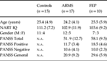
Healthy volunteers (n=15) were recruited through advertisements in the local media. All subjects lived in the borough of Lambeth (London), were native speakers of English and were right-handed. The groups were matched on sociodemographic variables (Table 1), including age (F=0.35, p=0.71) and handedness. Subjects were excluded if there was a history of neurological disorder or they met DSM-IV criteria for a substance misuse disorder. General intellectual function was estimated in all subjects using the National Adult Reading Test (NART). The severity of symptoms in the clinical groups was assessed with the Positive and Negative Syndrome Scale (PANSS; Kay, Reference Kay1990) on the day of scanning by a psychiatrist (M.R.B. or P.M.) trained in its use.
Age, IQ, gender and psychopathology ratings

NART, National Adult Reading Test; M, male; F, female; PANSS, Positive and Negative Syndrome Scale; ARMS, at-risk mental state; FEP, first-episode psychosis; n.a., not applicable.
Values given as mean (standard deviation).
Experimental task
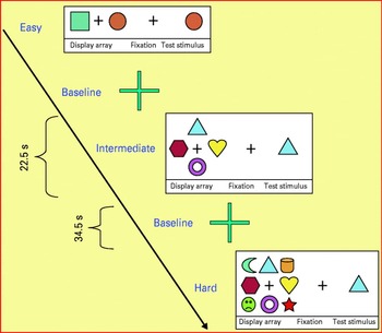
Stimuli were presented in 22.5 s epochs, alternating with 34.5 s epochs of cross-hair fixation; this cycle was repeated 12 times (for a total of 24 epochs) so the total duration of the experiment was 686 s or 343 images [repetition time (TR)=2 s]. Cognitive load was manipulated by presenting trials at one of three levels of difficulty (easy, intermediate, and hard) in a block design, with four blocks of each level of difficulty. Thus, there were a total of 12 blocks of trials alternating with 12 blocks of cross-hair fixation. The blocks of trials were always presented in the same sequence with respect to level of difficulty: easy, intermediate, and then hard. Each block comprised seven trials.
In an easy trial, two stimuli (highly discriminable coloured shapes) were shown either side (left and right) of a central cross-hair, followed by the central cross-hair alone, then the central presentation of one of the two original stimuli. Subjects had been trained to move a joystick with their right hand in the direction of the location originally occupied by the central stimulus. Intermediate and hard trials were the same except that four and eight stimuli were presented around the central cross-hair respectively. The speed and accuracy of the joystick movements were recorded during scanning. To avoid habituation of the subject, every stimulus had a randomly varied time of presentation, either between stimuli or before the presentation of the probe. As the stimuli were jittered randomly in every block, we did not need to take account of this in the block design analysis (Fig. 1).
The paired-associate learning task.

Behavioural data
All behavioural data, response accuracy and response latency, were recorded on a personal computer using Visual Basic (Microsoft Corp., USA) and analysed in SPSS version 11.0 (SPSS Inc., USA).
Image acquisition
Images were acquired on a 1.5-T Signa (GE) system at the Maudsley Hospital, London. T2*-weighted images were acquired in 38×3 mm slices, with a 0.3 mm gap in 14 axial planes, and a TR of 2 s, echo time (TE) 40 ms, and flip angle 90°. To facilitate anatomical localization of activation, a high-resolution inversion recovery image dataset was also acquired, with 3 mm contiguous slices and an in-plane resolution of 3 mm [TR 1600 ms, inversion time (TI) 180 ms, TE 80 ms].
Image analysis
Individual brain activation maps
The data were analysed with software developed at the Institute of Psychiatry, using a non-parametric approach. Data were realigned (Bullmore et al. Reference Bullmore, Brammer, Rabe-Hesketh, Curtis, Morris, Williams, Sharma and McGuire1999b) and then smoothed using a Gaussian filter [full-width at half-maximum (FWHM) 7.2 mm]. Responses to the experimental paradigms were detected by convolving each component of the design with each of two gamma variate functions (peak responses at 4 and 8 s respectively). The best fit between the weighted sum of these convolutions and the time series at each voxel was computed using the constrained blood oxygen level-dependent (BOLD) effect model (Friman et al. Reference Friman, Borga and Lundberg2003). A goodness-of-fit statistic comprising the ratio of the sum of squares of deviations from the mean image intensity (over the whole time series) divided by the sum of squares of deviations due to the residuals (SSQratio) was then computed at each voxel.
The data were then permuted by a wavelet-based method (Bullmore et al. Reference Bullmore, Long, Suckling and Fadili2001) to calculate the null distribution of SSQratios under the assumption of no experimentally determined response. This was used to calculate the critical value of SSQratio needed to threshold the maps at a type I error rate of <1. The detection of activated voxels was then extended from voxel to cluster level (Bullmore et al. Reference Bullmore, Suckling, Overmeyer, Rabe-Hesketh, Taylor and Brammer1999a). To minimize the potential confounding effects of between-group and between-condition variation in task performance, in the analysis of data from the task the BOLD response in each subject was modelled using only trials associated with correct responses. In addition to the SSQratio, the size of the BOLD response to each experimental condition was computed for each individual at each voxel as a percentage of the mean resting image intensity level. To calculate the BOLD effect size, the difference between the maximum and minimum values of the fitted model for each condition was expressed as a percentage of the mean image intensity level over the whole time series.
Group maps
The SSQratio maps for each individual were transformed into the standard space of Talairach & Tournoux (Reference Talairach and Tournoux1988) using a two-stage warping procedure (Brammer et al. Reference Brammer, Bullmore, Simmons, Williams, Grasby, Howard, Woodruff and Rabe-Hesketh1997). Group activation maps were computed by determining the median SSQratio at each voxel (across all individuals) in the observed and permuted data maps. The distribution of median SSQratios from the permuted data was used to derive the null distribution of SSQratios and the critical SSQratio to threshold group activation maps at a cluster level threshold of <1 expected type I error cluster per brain.
Linear trend analysis
Two different types of linear trend analysis were performed to assess linear change in neural activation dependent on group (analysis 1) and on mnemonic load (analysis 2). In the first analysis, for each mnemonic load (easy, intermediate, hard), control, ARMS and FEP subjects were respectively coded with dummy variables −1, 0 and 1. A linear model was selected to test the hypothesis that activation in the ARMS group would be intermediate between that in the controls and FEP subjects. In the second analysis, for each group (ARMS, control, FEP), the easy, intermediate and difficult levels of the task were respectively coded with dummy variables −1, 0 and 1. In this case a linear model was selected to test the hypothesis that activation at the intermediate level would be intermediate between that during the easy and hard levels. To minimize the potential confounding effects of between-group and between-condition variation in task performance, in each subject the BOLD response was covaried with the performance score. To ensure that we examined whether the middle group was intermediate in both the linear trend analyses (ARMS group in comparison to FEP and controls; medium mnemonic load in comparison to easy or hard load), an additional quadratic trend analysis was performed using the dummy variables (−1, 2, −1). Data that failed to fit this model, but that fitted the linear model, would hence have a middle group that was indeed intermediate between the other groups.
ANOVA was carried out on the effect size maps representing percentage change in BOLD response in standard space by first computing the difference in median SSQratio between groups at each voxel. Subsequent inference of the probability of this difference under the null hypothesis was made by reference to the null distribution obtained by repeated random permutation of group membership and recomputation of the difference in median SSQratios between the two groups obtained from the resampling process. Cluster-level maps were then obtained as described above. We set a voxel-wise p value of 0.05 and a cluster-wise p value of 0.01. This method ensured a total number of false-positive clusters of <1. Corrections for multiple comparisons were not required, as thresholds were set on an image-wide, not a voxel-wise, basis.
Group comparison
Given the possible limitations of the linear trend analysis described above, and to identify more precisely the relationship between groups (ARMS, FEP, control), task loads (two objects, four object, eight objects), or the effects of task repetition (comparing the first half of the run with the latter half), post-hoc comparisons were made between the respective conditions. Comparison of responses between groups or experimental conditions was performed by fitting the data at each intracerebral voxel at which all subjects had non-zero data using a linear model of the type Y=a+bX+e, where Y is the vector of BOLD effect sizes for each individual, X is the contrast matrix for the particular inter-condition/group contrasts required, a is the mean effect across all individuals in the various conditions/groups, b is the computed group/condition difference and e is a vector of residual errors. The model was fitted by minimizing the sum of absolute deviations rather than the sums of squares to reduce outlier effects. The null distribution of b was computed by permuting data between conditions/groups (assuming the null hypothesis of no effect of experimental condition or group membership) and refitting the above model. Group difference maps were computed using BOLD effect maps rather than standardized measures such as SSQratio, F or t as these contain explicit noise components (error SSQ or error variance), raising the possibility that group differences resulting from F, SSQratio or t comparisons could reflect differences in noise rather than signal.
Statistical significance
We have consistently adopted stringent levels of statistical significance for all the hypothesis tests reported on imaging data. For all between-group ANOVAs we set a voxel-wise p value of 0.05 and a cluster-wise p value of 0.01. For trend analysis conducted at cluster level, we set a voxel-wise p value of 0.05 and a cluster-wise probability p value of 0.01. This method ensured a total number of false-positive clusters of <1. Corrections for multiple comparisons were not required, as thresholds were set on an image-wide, not a voxel-wise, basis.
The method of analysis we used (XBAM) makes use of median statistics to control outlier effects and permutation rather than normal theory-based inference. The main test statistic is computed by standardizing for individual differences in residual noise before embarking on second-level, multi-subject testing using robust permutation-based methods. Approaches using a mixed effects analysis and permutation-based and cluster-level inference seem to be more valid than analyses involving simple random effects and voxel-level inference (Thirion et al. Reference Thirion, Pinel, Meriaux, Roche, Dehaene and Poline2007).
Results
Behavioural data
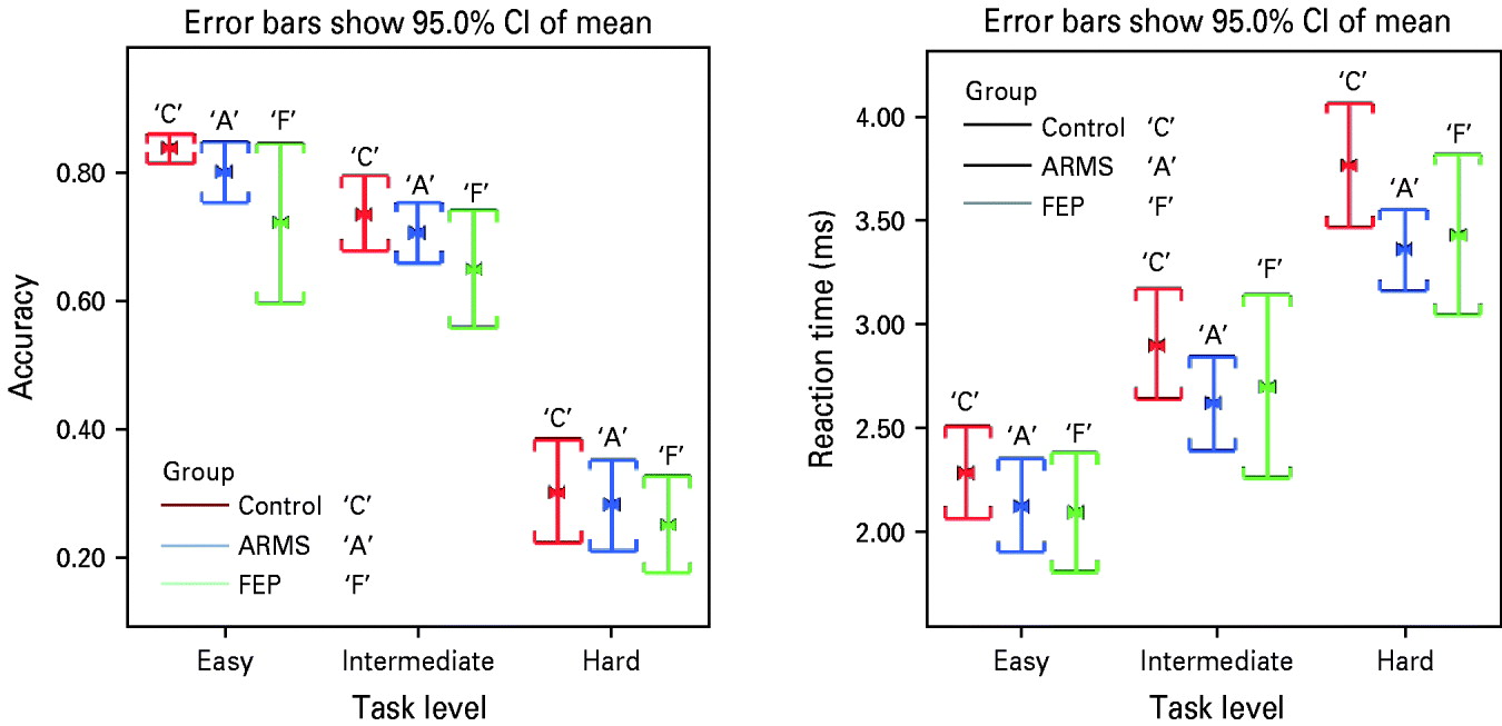
Repeated-measures ANCOVA showed a main effect for task difficulty with respect to accuracy (F=154.29, p<0.00) and latency (F=229.47, p<0.00). As the mnemonic load increased, response latency increased and response accuracy decreased in an approximately linear fashion (Fig. 2). No main effect for group was observed with respect to accuracy (F=1.59, p=0.271) or latency (F=1.7, p=0.247). However, post-hoc analysis revealed that, compared to controls, FEP showed impaired accuracy during the intermediate and hardest level of the task (t tests<0.05). No significant interactions of group by task load were observed with respect to accuracy (F=0.226, p=0.013) or latency (F=1.19, p=0.415) (Fig. 2).
Performance during the paired-associate learning (PAL) task. ARMS, ‘At-risk mental state’; FEP, first-episode psychosis.

To explore the effect for learning, we compared the accuracy in the first half of the run with accuracy in the second half of the run. During the easiest level of the task, there were no significant differences in accuracy between the first and the last blocks (paired t tests>0.05). Conversely, when performing the more demanding levels of the task (intermediate plus hard level), subjects performed better during the second half of the run than the first. This effect was evident for all three groups, FEP, ARMS and controls (all t tests<0.05).
fMRI results
Main effect of task (independent of group and mnemonic load): group analysis
Across all groups and levels of task demand, relative to baseline (cross-hair fixation), the task was associated with activation in a wide region spanning the cerebellum and occipital cortex bilaterally (precuneus x=−25, y=−70, z=37). Conversely, cross-hair fixation was associated with activation in the left posterior cingulate gyrus (x=−7, y=−63, z=15) (voxel p=0.05, cluster p=0.001, type I error p<1).
Activation associated with mnemonic load: linear trend analysis
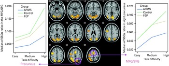
Independent of group, increasing the number of stimuli (from two to four to eight) was associated with activation in the left medial frontal/superior frontal and precentral gyrus, the cerebellum bilaterally and the right cuneus (voxel p=0.05, cluster p=0.01, type I error, p<1). After controlling for response accuracy, the activation in the left medial frontal/superior frontal gyrus and right precuneus remained significant (voxel p=0.05, cluster p=0.01). In both these regions the intermediate level of the task showed less activation than the most demanding level but greater activation than the easy level (all t tests<0.05) (Fig. 3).
Main effect for task difficulty across all groups. Areas shown in yellow revealed increasing activation as task demand was increased (high level>medium level>easy level). MFG, medial frontal gyrus; SFG, superior frontal gyrus; ARMS, ‘at-risk mental state’; FEP, first-episode psychosis.

Changes in activation dependent upon group: linear trend analyses
Easy level (two objects)
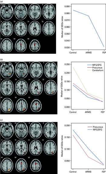
While performing the easiest level of the PAL task, there was differential activation across the three groups in a cluster spanning the medial frontal and anterior cingulate gyrus (voxel p<0.05, cluster p<0.01) (Fig. 4 a). In this region the magnitude of activation in the ARMS group was similar to that in controls, whereas the FEP group showed less activation than both other groups. Post-hoc paired comparisons confirmed that the FEP group showed significantly less activation than both the ARMS and control groups (t tests<0.05), with no significant difference between the latter two groups (t tests>0.05) (Fig. 4 a). These differences remained significant after covarying for accuracy (voxel p<0.05, cluster p<0.01).
(a) Easy level: between-group difference in activation (controls>ARMS>FEP) in the medial frontal gyrus (voxel p=0.05, cluster p=0.01). The left side of the brain is shown on the left of the picture. (b) Intermediate level: between-group differences in activation (controls>ARMS>FEP) in the middle frontal gyrus, right precuneus and right cerebellum (voxel p=0.05, cluster p=0.01). (c) Hard level: between-group differences in activation (controls>ARMS>FEP) in the medial frontal gyrus and right precuneus (voxel p=0.05, cluster p=0.01). MFG, medial frontal gyrus; SFG, superior frontal gyrus; ARMS, ‘at-risk mental state’; FEP, first-episode psychosis.

Intermediate level (four objects)
While performing the intermediate level of the PAL task, we detected differential activation across the three groups in the right cerebellum, right precuneus [Brodmann area (BA) 19] and medial frontal/superior frontal gyrus (BA 6/32) (voxel p<0.05, cluster p<0.025; Fig. 4 b). In these regions the magnitude of activation in the ARMS group was intermediate between that of the controls and FEP subjects, with the FEP group showing less activation than both other groups, and control subjects the greatest activation.
Hard level (eight objects)
While subjects were performing the most demanding level of the PAL task, there was differential activation across the three groups in the medial frontal gyrus/superior frontal gyrus (BA 32/6) and right precuneus (19) (voxel p<0.05, cluster p<0.01; Fig. 4c). In these regions the magnitude of activation in the ARMS group was intermediate between that of both controls and FEP subjects, with the FEP group showing less activation than both other groups and the control subjects the greatest (Fig. 5). These differences remained significant after covarying for accuracy (voxel p<0.05, cluster p<0.01).
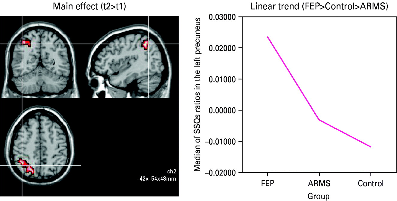
Effect of learning on activation. There was a main effect across all groups in the right precuneus, with greater activation during the second half of the experiment (voxel p=0.05, cluster p=0.01). t1=intermediate and hard level of the paired-associate learning (PAL) task during the first half of the run; t2=intermediate and hard level of the task during the second half of the run. ARMS, ‘At-risk mental state’; FEP, first-episode psychosis.

Main effect of task repetition (independent of group): group analysis
Across all groups, processing the most demanding levels of the task (intermediate plus hard) was associated with a greater activation in the right precuneus (x=21, y=−59, z=36) during the second half of the run than that in the first half (voxel p=0.01, cluster p=0.0075). There were no brain areas that showed greater activation in the first half of the run compared with the second (Fig. 5).
Group differences in effect of task repetition
There was also a difference between the groups in the effect of repetition of the most demanding levels of the task in a region spanning the left cuneus/precuneus (BA 7/19, x=−10, y=−74, z=31). In this region, there was a greater response during the second half of the run relative to the first, with the magnitude of the within-group difference greatest in the FEP group, smallest in the controls and intermediate in the ARMS (all t tests<0.05).
Effects of antipsychotic medication: correlational analysis
Within the FEP group (which was the only group that included subjects on antipsychotic medication), for all levels of task demand, there was no correlation (Pearson's r, n=10, p>0.05) between activation in brain areas that were differentially engaged in the FEP group relative to the other two groups and measures of antipsychotic treatment (daily and cumulative dose in chlorpromazine equivalents, or exposure).
Discussion
The present study used fMRI to study the neural substrate of SWM in subjects with an ARMS. In line with our hypothesis, there was a consistent pattern of differential activation across the groups on all levels of difficulty of the task, with an additional differential response across the groups on the analysis to examine the effects of learning.
The differential activation was not attributable to impairments in task performance, as there were no significant differences in the speed or accuracy of responses across groups, and the analysis selectively modelled the BOLD response to those trials associated with correct responses. Hence, any remaining difference in activation is likely to be due to the disorder of interest.
Similarly, the findings are unlikely to be related to effects of antipsychotic medication as both the ARMS subjects and controls were antipsychotic medication naive, and in the first-episode group there was no relationship between medication exposure and activation in the regions that were differentially engaged across groups.
When quadratic trend analysis was carried out, there were no significant clusters activated differentially across the groups, indicating that there was a predominantly linear relationship in activation across the groups on all the tasks.
Neural network underlying object and SWM
Compared to cross-hair fixation, PAL engaged a wide area spanning the cerebellum and visual cortex bilaterally. We also manipulated task difficulty by parametrically varying memory load in the task. We found that the medial frontal/superior frontal gyrus and the right precuneus showed a linear response. These brain areas have been extensively implicated in spatial and object working memory (McCarthy et al. Reference McCarthy, Puce, Constable, Krystal, Gore and Goldman-Rakic1996; LaBar et al. Reference LaBar, Gitelman, Parrish and Mesulam1999; Rypma et al. Reference Rypma, Prabhakaran, Desmond, Glover and Gabrieli1999).
A medial superior frontal gyrus region, centred on the supplementary motor area (SMA), has been traditionally associated with motor control necessary in the selection of action sets and in monitoring of response conflict (Rushworth et al. Reference Rushworth, Walton, Kennerley and Bannerman2004) and also speech expression and memory (Chung et al. Reference Chung, Han, Jeong and Jack2005). The anterior part of the SMA is known to interconnect with the prefrontal cortex and to play a role in the more complex components of movement (Exner et al. Reference Exner, Weniger, Schmidt-Samoa and Irle2006). As such, the role of the SMA is likely to be key in performing the SWM task. The absence of hippocampal activation may suggest that such activation does not distinguish the groups or the effect of mnemonic load. However, imaging data have demonstrated both over- and underactivation of the hippocampus in memory tasks in schizophrenia (Boyer et al. Reference Boyer, Phillips, Rousseau and Ilivitsky2007).
Functional imaging findings in healthy subjects evidenced a central role for the precuneus, in visuospatial imagery, episodic memory retrieval (Krause et al. Reference Krause, Schmidt, Mottaghy, Taylor, Halsband, Herzog, Tellmann and Muller-Gartner1999; Wagner et al. Reference Wagner, Shannon, Kahn and Buckner2005) and self-processing operations (Cavanna & Trimble, Reference Cavanna and Trimble2006). Nagahama et al. (Reference Nagahama, Okada, Katsumi, Hayashi, Yamauchi, Sawamoto, Toma, Nakamura, Hanakawa, Konishi, Fukuyama and Shibasaki1999) showed that the precuneus may process spatial attention and attention shift between object features. Lesion studies suggest that the dorsal stream of the ‘visuospatial sketchpad’ involves the precuneus, and this may enable spatial operations (Muller & Knight, Reference Muller and Knight2006). Hence, our finding implicating differential precuneus activation may be due to its probable role in visual imagery, spatial behaviour and spatial attention, all functions relevant for the SWM task.
Group differences in activation
Consistent with our first hypothesis, during both the intermediate and the hard versions of the task, activation in the medial frontal cortex and precuneus in the ARMS group was intermediate relative to that in the FEP group and controls. The localization of the group differences in activation in the medial frontal cortex and precuneus is consistent with data from previous neuroimaging studies of SWM in schizophrenia (Gould et al. Reference Gould, Brown, Owen, ffytche and Howard2003; Curtis, Reference Curtis2006). A different pattern of activation across groups was evident during the least demanding (‘easy’) version of the task. In this case there were no significant differences in activation between the ARMS and the control subjects, and differences in activation were limited to the medial frontal cortex, which responded more weakly in the FEP than the other groups. This suggests that functional abnormalities in the ARMS group became more evident as the task demands were increased, as has been reported in behavioural studies of other paradigms (Broome et al. Reference Broome, Johns, Valli, Woolley, Tabraham, Brett, Valmaggia, Peters, Garety and McGuire2007).
SWM as a neurocognitive vulnerability marker for psychosis
Kuperberg et al. (Reference Kuperberg, Broome, McGuire, David, Eddy, Ozawa, Goff, West, Williams, van der Kouwe, Salat, Dale and Fischl2003) found significant cortical thinning in the medial frontal areas of adult schizophrenic patients, and grey matter reductions have been reported in the medial premotor cortex (Honey et al. Reference Honey, Sharma, Suckling, Giampietro, Soni, Williams and Bullmore2003). Imaging studies suggest that the medial frontal gyrus (Stevens et al. Reference Stevens, Goldman-Rakic, Gore, Fulbright and Wexler1998; Paillere-Martinot et al. Reference Paillere-Martinot, Caclin, Artiges, Poline, Joliot, Mallet, Recasens, Attar-Levy and Martinot2001) and the medial prefrontal cortex (Ananth et al. Reference Ananth, Popescu, Critchley, Good, Frackowiak and Dolan2002) are affected early in psychosis. Suzuki et al. (Reference Suzuki, Zhou, Takahashi, Hagino, Kawasaki, Niu, Matsui, Seto and Kurachi2005) found the SMA to be reduced in subjects with recent-onset schizophrenia whereas Exner et al. (Reference Exner, Weniger, Schmidt-Samoa and Irle2006) showed that reduced volume of the SMA in FEP subjects was related to impaired implicit learning. Medial frontal cortex dysfunction is compatible with the hypothesis of a core deficit in early stages of psychosis involving a failure to monitor actions generated internally (Exner et al. Reference Exner, Weniger, Schmidt-Samoa and Irle2006). Activation in the cuneus and precuneus increased in the second half of the run, during the harder versions of the task, in all three groups. However, this increase itself was greatest in the FEP group, least in the controls, and intermediate in the ARMS cohort. Given that behavioural performance improved, it is likely that this differential activation of the precuneus may underpin such a learning effect, and, furthermore, that greater activation in the FEP group is required than in the control group (with the ARMS group intermediate) to enable the same degree of learning. Barnett et al. (Reference Barnett, Sahakian, Werners, Hill, Brazil, Gallagher, Bullmore and Jones2005) suggested that visuospatial PAL failure may be a marker of clinical severity, in a first-episode cohort, whereas executive dysfunction may reflect more stable, trait-like impairment. However, in our data there seems to be some relationship between executive function and clinical course in the at-risk sample (Fusar-Poli et al. Reference Fusar-Poli, Broome, Matthiasson, Woolley, Mechelli, Johns, Tabraham, Bramon, Valmaggia, Williams and McGuire2009) and we are currently undertaking longitudinal studies of the PAL task in the at-risk cohort.
Conclusions
This study suggests that the ARMS is associated with a dysfunction in the neural substrate for SWM. This is not attributable to an effect of psychotic illness, as none of the subjects were psychotic, nor an effect of antipsychotic treatment, as all of the ARMS subjects were medication naive. These observations are consistent with independent neuroimaging and neuropsychological evidence that the ARMS is associated with neurofunctional abnormalities that are qualitatively similar to, but less severe than, those seen in patients with schizophrenia (Broome et al. Reference Broome, Woolley, Tabraham, Johns, Bramon, Murray, Pariante, McGuire and Murray2005b, Reference Broome, Johns, Valli, Woolley, Tabraham, Brett, Valmaggia, Peters, Garety and McGuire2007, Reference Broome, Matthiasson, Fusar-Poli, Woolley, Johns, Tabraham, Bramon, Valmaggia, Williams, Brammer, Chitnis and McGuire2009; Fusar-Poli et al. Reference Fusar-Poli, Perez, Broome, Borgwardt, Placentino, Caverzasi, Cortesi, Veggiotti, Politi, Barale and McGuire2007). As those in the ARMS group had a high risk of developing a psychotic disorder but were not psychotic, the functional abnormalities they displayed can be seen as a correlate of their increased vulnerability to psychosis.
Limitations of the study
This study reports cross-sectional data, from a modestly sized sample, on ARMS, FEP and control subjects. The findings in the ARMS group may be a correlate of the subjects' increased vulnerability to psychosis. However, to determine this formally will require a longitudinal study, a study informed by the findings presented here and, in particular, whether the pattern and degree of activation during visuospatial working memory tasks predict transition to psychosis in a clinical high-risk group. We hope to report longitudinal fMRI data, and particularly the relationship to clinical outcomes such as transition to psychosis, in the future.
Acknowledgements
OASIS is supported by the Guy's and St Thomas' Charitable Foundation and the South London and Maudsley National Health Service (NHS) Trust. We thank all the clients, staff and referrers of both OASIS and LEO.
Declaration of Interest
None.