Introduction
Several grafting materials are available for chronic tympanic membrane perforation repair and ossicular reconstruction. Most of these materials have generally acceptable closure rates and functional benefits post-tympanoplasty.
To date, temporalis fascia has been (and may still continue to be) the most common graft material used for the closure of tympanic membrane perforation. Nevertheless, 25–30 per cent of cases of type I tympanoplasty present short-term problems of re-perforation.Reference Ozbek, Ciftçi, Tuna, Yazkan and Ozdem1, Reference Kazikdas, Onal, Boyraz and Karabulut2 Moreover, it is a well known factReference Buckingham3, Reference Cayé-Thomasen, Andersen, Uzun, Hansen and Tos4 (supported by our own clinical experience) that if temporalis fascia is used, there is a very high prevalence of retraction and/or atrophy of the neotympanum over long-term follow up.
Notably, in Buckingham's studyReference Buckingham3 of 89 tympanic membrane defects repaired with either fascia or perichondrium, of 63 ears available for 10-year follow up, 54 had cholesteatomas and 9 had perforations. Moreover, this author reported graft atrophy in 35 of 43 ears (80 per cent) receiving fascial grafts and in 8 of 20 ears (40 per cent) with perichondrial grafts. Grafts maintained their relatively thick and fibrous character in only 20 of 63 ears (32 per cent).
Fascial and perichondrial grafts have also been used to correct atelectasis due to an atrophic tympanic membrane. However, if such grafts failed at the rate indicated by Buckingham's results, atelectasis would be expected to recur.
Modelled autologous cartilage has also been used for ossicular reconstruction during tympanoplasty. However, of those studies assessing histological findings from cartilage explants retrieved at revision surgery, most concluded that such cartilage structures may lose their rigidity over time, rendering them unfit as sound conductors.Reference Steinbach and Pusalkar5–Reference Smyth, Kerr, Pahor and Law7 Yung's literature review on cartilage tympanoplastyReference Yung8 concluded that few surgeons have used cartilage for ossicular reconstruction because of the risk that the cartilage will become softer with time and may even be reabsorbed.
In recent years, many authors have considered cartilage to be the best choice for the reconstruction of the tympanic membrane in ‘at risk’ cases, such as those with chronic tubal dysfunction, adhesive processes, recurrent perforation, sub-total or total perforation, in children, and in patients with nasal respiratory problems.Reference Dornhoffer9–Reference Ulkü12 According to these same authors, cartilage has better results than temporalis fascia (in the cited conditions) as its mechanical characteristics enable superior resistance to retraction and re-perforation and also permit better acoustic performance.
Two main techniques have been described for cartilage reconstruction of the tympanic membrane: the cartilage and perichondrium island flap,Reference Ulkü12–Reference Beutner, Huttenbrink, Stumpf, Beleites, Zahnert and Luers14 which utilises tragal cartilage, and the palisade technique, which uses cartilage from the tragusReference Ozbek, Ciftçi, Tuna, Yazkan and Ozdem1, Reference Anderson, Cayé-Thomasen and Tos15 or cymba.Reference Kazikdas, Onal, Boyraz and Karabulut2 Autologous cartilage may better withstand negative middle-ear pressures due to its rigidity and convexity, and also seems to offer high resistance to infection.Reference Bernal-Sprekelsen, Romaguera Lliso and Sanz Gonzalo16
The aim of the current study was to compare our surgical experience and anatomical and functional results, one year following the use of either an island-type autologous chondro-perichondral tragal graft or temporalis fascia graft, in a group of adults undergoing type I tympanoplasty for subtotal perforation (with no other selection criteria). We also employed various surgical techniques in order to save time and improve standardisation,Reference De Seta, Covelli, De Seta, Mancini and Filipo17 which favoured the use of cartilage type I tympanoplasty as the treatment of choice.
Materials and methods
During the previous six years, our department had used different types of cartilage and composite cartilage grafts as alternatives to the classical temporalis fascia graft for tympanoplasty.
Over the last three years, 106 consecutive adults with near-total pars tensa perforation for simple chronic otitis with normal middle-ear mucosa were selected for this comparative, retrospective study.
In order to reduce variability, we excluded patients with a history of any previous otological procedure, cholesteatoma or ossicular involvement, as well as those younger than 18 years.
Of the 106 patients, a temporalis fascia graft was used in 53 patients (male:female ratio, 1.2; mean age, 44 years) and a composite chondro-perichondral tragal graft in the other 53 patients (male:female ratio, 1.1; mean age, 41 years).
Surgical technique
In both groups, an incision was performed 0.5 cm from the retroauricular fold using a post-auricular approach.
In the fascia graft group, a 1.5 × 2 cm piece of temporalis fascia was harvested. A tympano-meatal flap was then raised, the fibrous margin of the perforation was removed, and an underlay graft technique was utilised. The mesotympanic space was filled with gelfoam (Gelita Tampon; Aesculap, Tuttlingen, Germany).
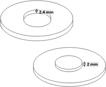
In the cartilage graft group, all patients underwent the same procedure for harvesting and preparation of the composite graft, as follows. A skin incision was made on the medial aspect of the tragus, followed by elevation of the whole medial cutaneous surface of the tragus (leaving perichondrium attached to the cartilage). An incision was made along the external margin of the tragal cartilage, leaving a 1 mm strip on the dome. The tragal cartilage was elevated from the side of the perichondrium, enabling the removal of a large chondro-perichondral graft. The tympanic membrane perforation size was measured using squared micro-hooks, in order to determine the dimensions of the cartilage island. The island graft was created using an auricular speculum (generally 8 or 9 mm in diameter), by applying gentle pressure to isolate a round segment of cartilage, which remained attached to its perichondrium. Then, holding the speculum in situ, the excess cartilage was removed from the perichondrium with a spatula or a sickle micro-knife. The graft was placed on a cartilage cutter device composed of two steel discs (Figure 1) and trimmed to the required shape (Figure 1). It was then manually thinned to 0.3 mm using a size 11 scalpel blade. A notch for the malleus handle was created using a sickle micro-knife. Finally, the graft was placed in an underlay fashion, with the perichondrium on the lateral aspect.
The cartilage cutter device. A knife on the surface of the second disc permits the removal of excess cartilage and the creation of the required graft thickness.

All patients underwent otomicroscopy one month, six months and one year post-operatively to evaluate the state of the neotympanum, especially as regards graft re-perforation and retraction, following the classification of Sade.Reference Sade18
Pure tone audiometry was also performed six months and one year post-operatively to assess air and bone conduction thresholds, enabling comparison of the mean air–bone gap (ABG) for the two groups.
Statistical analysis
Audiological findings in the two groups were compared using the Mann–Whitney U test. A p value of less than 0.05 was considered statistically significant. All statistical analyses were performed using SPSS software (version 14.0; SPSS Ltd, Chicago, Illinois, USA).
Results
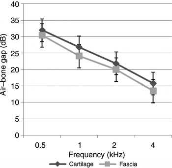
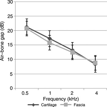
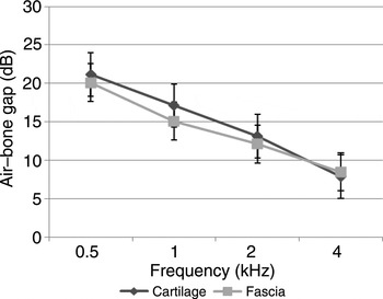
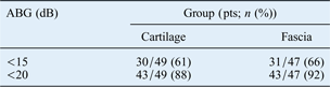
The mean pre-operative ABG values for the fascia and cartilage groups were similar, as shown in Figure 2. At six months and one year after surgery, ABG results for the two groups were also similar, with no statistically significant differences (Figures 3 and 4); at one year, p values for results at 0.5 and 1 kHz were 0.167 and 0.991, respectively. Table I reports the functional success in the two groups, in terms of ABG closure to within 15 dB and within 20 dB, one year after surgery. One year post-operatively, four patients had been lost to follow up: two in the cartilage group and two in the fascia group (see Table I). Six patients presented with surgical failure (i.e. anterior graft failure or re-perforation) one month post-operatively; these patients were not included in the long-term follow-up analysis.
Pre-operative values for mean air–bone gap on pure tone audiometry, for cartilage and fascia groups. Differences between the two groups were not statistically significant.

Six month follow-up values for mean air–bone gap on pure tone audiometry, for cartilage and fascia groups. Differences between the two groups were not statistically significant.

One year follow-up values for mean air–bone gap on pure tone audiometry, for cartilage and fascia groups. Differences between the two groups were not statistically significant.

Air–bone gap at 1 year
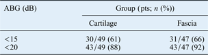
ABG = air–bone gap; pts = patients
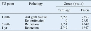
The anatomical outcome of the operation, in terms of otomicroscopically determined perforation and clinically evaluated retraction (following Sade's classification system)Reference Sade18 was as follows. In the cartilage group, at six-month follow up there were two cases of anterior graft failure (3.7 per cent) and one of graft retraction (1.8 per cent); at one-year follow up, there were two cases of grade I retraction (5.5 per cent) and no cases of graft perforation. In the fascia group, at six-month follow up there were two cases of anterior graft failure and two of graft perforation (i.e. a 7.4 per cent prevalence of re-perforation) and four cases of grade I graft retraction (7.4 per cent); at one-year follow up, there were six cases of retraction (11 per cent; four grade I cases and two grade II cases) (Table II).
Otomicroscopic results

FU = follow up; pts = patients
The patients with anterior fascia graft failure and re-perforation underwent revision of cartilage tympanoplasty; cartilage was taken from the tragus using the technique previously described. The two patients in the cartilage group with anterior graft failure underwent revision of tympanoplasty with autologous composite cartilage graft from the cymba.
Discussion
These findings indicate a satisfactory improvement in auditory threshold at one year follow up, with no statistically significant differences in ABG between the two groups. The graft ‘take’ rate at one month was 96.3 per cent in the cartilage group and 92.6 per cent in the fascia group.
In most studies, the indications for cartilage tympanoplasty are limited to particular categories of patients and pathology. In contrast, our study groups included all adult patients with subtotal tympanic membrane perforation, regardless of clinical risk category. In the literature and also in our own experience, so-called ‘at risk’ patients actually constitute a very large proportion of the total, and include almost all type I tympanoplasty candidates (i.e. children, patients requiring reintervention, and those with nasal pathology or total perforation). Moreover, conditions such as nasal respiratory problems are almost always present in these patients, even if scarcely evident or subclinical. The proposed cartilage technique saves operating time and should be considered as an alternative to the use of temporalis fascia in all type I tympanoplasty procedures.
Many types of cartilage tympanoplasty have been described. TosReference Tos19 proposed a classification of 23 different techniques divided into 6 groups. These techniques vary in the tragal or conchal cartilage arrangements used: thin plates or thick foils, palisades or rods, overlapping or close to each other, and positioned using underlay, inlay or onlay techniques. GoodhillReference Goodhill20 described the use of a perichondral graft with a circumferential cartilage batten in cases with a shallow middle-ear cavity, in order to avoid sagging of perichondrium onto the promontory. Other surgeons have trimmed the cartilage part of the chondro-perichondral composite graft into the shape of a shield,Reference Duckert, Müller, Makielski and Helms21 double island,Reference Dornhoffer22 ‘Mercedes-Benz sign’,Reference Spielmann and Mills23 wheel,Reference Shin, Lee, Kim and Lee11 coin with butterfly edges,Reference Eavey24 crown corkReference Hartwein, Leuwer and Kehrl25 or lamellae.Reference Neumann and Jahnke26 The use of a composite graft, generally with perichondrium, is more common than the use of cartilage alone as a reinforcement for the tympanic membrane or a fascia graft. The use of a thin, composite, chondro-perichondral island graft in an underlay fashion, as previously reported,Reference Ulkü12, Reference Kirazli, Bilgen, Midilli and Ogüt13, Reference Onal, Arslanoglu, Oncel, Songu, Kopar and Demiray27 seems to be the best choice for type I tympanoplasty for subtotal perforation.
Regarding the optimal thickness of a cartilage graft, the majority of authors agree that full thickness (i.e. approximately 1 mm) tragal or conchal cartilage should not be used. From an acoustic point of view, Mürbe et al. Reference Mürbe, Zahnert, Bornitz and Hüttenbrink28 reported that a 0.5 mm cartilage plate seems to be the best choice. In contrast, Lee et al. Reference Lee, Chen, Chou, Hsu, Chen and Liu29 have stated that the optimal thickness of a cartilage graft is between 0.1 and 0.2 mm, for myringoplasty for medium or large tympanic membrane perforations, on the basis of biomechanical analysis. In our study, based upon our preliminary experience of different thicknesses of cartilage graft, we chose a 0.3 mm graft thickness.Reference De Seta, Covelli, De Seta, Mancini and Filipo17
Another important issue, little emphasised by most authors, is the cosmetic appearance after the removal of tragal cartilage. In our experience, there are three relevant steps contributing to cosmesis. Firstly, the site of the skin incision, which we placed along the medial surface of the tragus to prevent an unaesthetic scar. Secondly, it is important to leave a 1 mm strip of cartilage along the dome of the tragus in order to prevent post-operative deformity (including an amputated tragus). Thirdly, the surgeon should spare the lateral perichondrium in order to assist regrowth of cartilaginous tragal tissue. In our cartilage group, we encountered one haematoma in the tragal region, but no patients with noticeable post-operative alteration in the shape of the tragus; six months after surgery, we found cartilaginous consistency of the tragus on palpation in 73 per cent of patients.
One of the obvious disadvantages of the cartilage tympanoplasty technique is its longer operative time compared with the use of temporalis fascia. However, we used two techniques which saved time and guaranteed greater precision and consistency, compared with other surgical techniques: the cartilage cutter device and the use of an ear speculum to create the cartilage island graft. In our cartilage group, the mean operative time was only 15 minutes longer than in the fascia group. This comparative time saving was due in large part to the use of the cartilage cutter device, which enabled the creation of cartilage slices of a uniform 0.3 mm thickness, and which was specifically designed to cut composite cartilage–perichondrium grafts (in contrast to other, similar devices designed to cut only cartilage tissue).
• Cartilage tympanoplasty is the leading surgery for ‘at risk’ patients with tympanic membrane perforation due to chronic otitis media
• This study compared chondro-perichondral tragal vs temporalis fascia grafts
• During graft creation, use of an ear speculum and cartilage cutter device saved time and improved standardisation
• At one year, the cartilage group had fewer retractions and perforations, with comparable audiological outcomes
The use of these surgical tools and methods conferred two important advantages. Firstly, we gained enhanced operating speed and precision, in comparison with other cartilage techniques. Secondly, we benefited from improved technical standardisation and consequently gained better, more reliable anatomical and functional results which were less dependent on the individual surgeon's skill and experience; this had the additional academic benefit of making our data more homogeneous and easy to compare.
Conclusion
This study's functional results confirm that the reported island chondro-perichondral graft technique was of similar value to temporalis fascia grafting over a one-year follow-up period. Furthermore, anatomical results were better in the cartilage graft group, in terms of perforation and retraction. However, the number of study participants was limited and the study lacked statistical power as a result. Longer follow up of a greater number of patients would enable better evaluation of long-term complications, including graft retraction and atrophy (the commonest long-term complication of fascia tympanoplasty). If the present study results were confirmed in a longer-term study of a larger patient group, then island chondro-perichondral tragal graft tympanoplasty should be considered as the preferred surgical technique for type I tympanoplasty.