Introduction
The ability to detect surface-enhanced Raman scattering (SERS)Reference Moskovits1,Reference Le Ru and Etchegoin2 from single moleculesReference Le Ru, Meyer and Etchegoin3–Reference Weiss and Haran6 located in the vicinity of noble metal plasmonic nanostructures has opened unique opportunities for probing the nanoscale environment and has provided the basis for ultrasensitive diagnostics and nano-optical applications.Reference Haran7–Reference Le Ru and Etchegoin13 From a basic science point of view, single-molecule SERS (SM-SERS) is an excellent tool for probing local dynamics at metallic surfaces, including phenomena such as charge transferReference Shegai, Vaskevich, Rubinstein and Haran14 and surface diffusion.Reference Weiss and Haran6,Reference Haran15 The strong electromagnetic field localization required for the observation of SM-SERS is readily achieved when a molecule is adsorbed inside the nanometer gap between closely adjacent nanoparticles, especially when these particles possess a localized plasmon resonance of frequency close to that of the excitation source and the Raman band.Reference Michaels, Nirmal and Brus4,Reference Hao and Schatz16–Reference Michaels, Jiang and Brus20
State-of-the-art strategies for nanoparticle synthesis have rendered possible precise control of nanoparticle size and shape,Reference Xia, Zeng, Zhang, Moran and Xia21,Reference Xia, Xiong, Lim and Skrabalak22 the inter-particle gap, and even molecular binding within the gap.Reference Lim, Jeon, Kim, Nam and Suh23 When a gap-bound molecule is excited with the enhanced local electric field at the laser frequency, it is the scattering of its Raman-shifted emission by the localized surface plasmon excitations on the nanoparticles that determines the light characteristics, for example, its directivity and polarization.Reference Shegai, Li, Dadosh, Zhang, Xu and Haran24–Reference Etchegoin, Galloway and Le Ru28 These excitations depend on two factors: The metal dielectric functionReference Bohren and Huffman29,Reference Kreibig30 and the way the nanoparticles are arranged.Reference Romero, Aizpurua, Bryant and García de Abajo17,Reference Li, Shegai, Haran and Xu18,Reference Quinten and Kreibig31–Reference Brandl, Mirin and Nordlander38 It has been shown that by manipulating these properties, one can build new materials with improved optical characteristics.Reference Yu, Aieta, Genevet, Kats, Gaburro and Capasso39–Reference Zhao, Belkin and Alù43
SM-SERS is a new field of spectroscopy that has developed rapidly in the last decade. Our interest in better understanding the mechanisms behind the interactions of molecules with localized surface plasmons has motivated a series of spectroscopic and theoretical studies that are summarized in this review.Reference Shegai, Li, Dadosh, Zhang, Xu and Haran24,Reference Mock, Barbic, Smith, Schultz and Schultz44–Reference Chuntonov and Haran46 In particular, we examined the relation between the polarization of Raman-scattered light and the plasmon mode structure of nanoparticle clusters, an aspect that has not received much attention.Reference Etchegoin, Galloway and Le Ru28 We focused on trimers, which are the smallest nanoparticle clusters that may show Raman optical activity (ROA) (i.e., conversion of linearly polarized light into circularly polarized light at the Raman frequency) in SM-SERS.
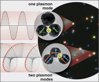
We start our discussion with the question of how the plasmon mode structure of nanoparticle trimers depends on their symmetry. We then demonstrate a relation between mode structure and the SM-SERS light polarization using a simple physical model of harmonic oscillators. Figure 1 shows the main take-home message of this review: When a molecule trapped within a nanoparticle cluster emits light (e.g., Raman scattering), it may excite either one or multiple plasmon modes, depending on the symmetry of the cluster. In the case of multiple-mode excitation, the interference between modes can lead to elliptical and even circular polarization of the Raman-scattered light in the far field. Understanding the connection between plasmon modes and the characteristics of emitted light may facilitate the design of novel plasmonic devices based on well-defined polarization states of light.
Plasmonic clusters and Raman optical activity. A dark-field image of silver plasmonic nanoparticle clusters is shown on the right; nanoparticle monomers appear in blue and clusters (e.g., trimers), in more orange color as determined by their dominant plasmon resonances. Transmission electron microscope images taken from the same clusters studied optically are also shown (insets). The arrows drawn on the nanoparticles represent excited plasmon normal modes of the clusters. The single-molecule surface-enhanced Raman spectroscopy reporter occupying the interparticle gap is shown in yellow. Upper trimer: only one plasmon mode is excited in a cluster of D3h symmetry, leading to the linear polarization of the emitted light. Lower trimer: two orthogonal plasmon modes are excited in a cluster of C2v symmetry, which generally results in elliptical polarization of the emitted light.

Plasmon mode structure of nanoparticle trimers
The plasmon modes of metal nanoparticle clusters can be studied with single-particle dark-field optical spectroscopy, sometimes called plasmon spectroscopy in the current context.Reference Mock, Barbic, Smith, Schultz and Schultz44 Spectra of single nanoparticle clusters obtained in this manner can be analyzed based on their structural symmetry using symmetry-adapted linear combinations (SALCs) of the individual nanoparticle plasmon modes.Reference Prodan, Radloff, Halas and Nordlander35,Reference Chuntonov and Haran45–Reference Dadosh, Sperling, Bryant, Breslow, Shegai, Dyshel, Haran and Bar-Joseph47 When the symmetry of the cluster is lowered due to the change in its geometry, the irreducible representations of the symmetry group are transformed, as summarized in relevant correlation tables, in a similar way to molecular vibrations.Reference Bunker and Jensen48,Reference Cotton49 However, correlation tables alone provide only information on the symmetry changes and are not sufficient to track the plasmon mode transformation in detail. The explicit shapes and energies of the plasmon modes for arbitrary cluster geometries can be calculated using plasmon hybridization theory.Reference Prodan, Radloff, Halas and Nordlander35–Reference Brandl, Mirin and Nordlander38 For simplicity, here we focus only on the dipolar modes of the multipole expansion. The dipolar model is valid in the long wavelength limit (ka << 1, where k is the scattering wave vector, and a is the radius of a nanoparticle), which holds approximately for silver nanoparticles of ca. 50 nm excited with visible light.
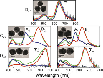
Different clusters of three silver nanoparticles are shown in Figure 2 together with their plasmon scattering spectra, corresponding to two orthogonal orientations of the linearly polarized excitation light: Longitudinal and transverse. In the former orientation, the polarization is parallel to one of the bases of the triangle formed by the nanoparticles, while in the latter, it is perpendicular to this base. The equilateral triangle, which belongs to symmetry point group D3h, possesses two degenerate low-energy plasmon resonances. They correspond to the doubly degenerate irreducible representation of symmetry E′, as dictated by the presence of the rotational subgroup C3.Reference Bunker and Jensen48,Reference Cotton49 This assignment was made based on the correlation between surface charge distributions calculated numerically for each of the cluster resonances and dipolar SALCs constructed based on the D3h group character table. The corresponding dipolar SALCs, which also agree with the results of plasmon hybridization theory,Reference Brandl, Mirin and Nordlander38 are illustrated in Figure 3 (left column). In addition to the degenerate E′ plasmon modes, it is important to consider the ring-like  ${{\rm{A'}}_2}$ mode of the D3h trimer. This mode is dark owing to its zero net dipole moment. Nevertheless, when the symmetry of this trimer is broken, it plays an important role in the interaction with light.
Scattering spectra of silver nanoparticle trimers showing mode splitting due to gradual symmetry breaking. Thin black lines, raw experimental data; color lines, low pass filter applied. The clusters are excited by either nonpolarized light (green) or light linearly polarized along the longitudinal (red) and transverse (blue) directions. The polarized spectra are normalized to the corresponding peaks of the nonpolarized spectra. Plasmon mode symmetries of the bonding modes are marked. Transmission electron microscopy images of the clusters are shown in the insets (scale bars are 50 nm). Adapted with permission from Reference 44. © 2011 American Chemical Society.

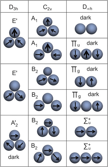
Correlation table of bonding dipolar plasmon eigenmodes of trimers obtained using plasmon hybridization theory. The symbols of the corresponding irreducible representations are shown. Adapted with permission from Reference 45. © 2011 American Chemical Society.

The symmetry of the D3h cluster can be broken in several ways, for example, by opening one of the vertex angles of the triangle or by “re-sizing” one of the nanoparticles, such that the resulting clusters have C2v symmetry. Gradual opening of the vertex angle eventually leads to a linear chain of three nanoparticles, while reducing the size of one of the nanoparticles leads to a dimer (linear chain of two nanoparticles). Both linear clusters have D∞h symmetry. Representative examples of symmetry-broken trimers are shown in Figure 2 together with their plasmon spectra. It is evident from the figure that breaking the symmetry of the trimer lifts the degeneracy of the E′ modes and facilitates the appearance of new peaks in the plasmon spectrum.
Using the group theoretical framework and plasmon hybridization theory, we constructed a correlation table for the plasmon modes in the sequence of shapes, as shown in Figure 3. As the C3 rotation operation becomes invalid, two new plasmon bands, selectively excited by light linearly polarized along the cluster’s longitudinal and transverse directions, appear. As shown in the correlation table, the transverse and longitudinal in-plane E′ modes transform to modes A1 and B2 of C2v, respectively, which are symmetric and anti-symmetric with respect to the σv reflection plane. The net dipoles of the A1 and B2 modes are oriented along the cluster’s transverse and longitudinal directions.
As the symmetry breaking further increases, the cluster attains D∞h symmetry corresponding to a linear chain of nanoparticles. Here, only the single bright bonding mode  $\Sigma _{\rm{u}}^ +$ exists, corresponding to a parallel alignment of the individual dipoles. All other modes of the C2v cluster transform to various dark modes (with no net dipole) in the D∞h limit (see Figure 3). The interchange of the mode dipole moments and the associated energy shifts obtained from hybridization theory are in full agreement, with detailed electromagnetic numerical simulations that were performed for all possible cluster geometries.
Effect of plasmon modes on the polarization of SM-SERS
As we already noted, single molecules can provide valuable information on their immediate environment within the interparticle gaps through their enhanced Raman spectra. SM-SERS is being used to probe an ever-increasing list of nanoscale phenomena, and we cite only a few examples here. Thus, blinking and spectral jumps of single molecules were studied in order to understand molecular diffusion on metallic surfacesReference Kitahama, Tanaka, Itoh and Ozaki50 and were also correlated with electrical conductance through a molecular junction.Reference Ward, Halas, Ciszek, Tour, Wu, Norlander and Natelson51 Single molecules were also used to probe charge transfer phenomenaReference Shegai, Vaskevich, Rubinstein and Haran14,Reference Park and Kim52 and to study chemical reactions on the nanoscale.Reference Park and Kim52 Finally, the structure of electromagnetic hot spots was mapped by super-resolution imaging of Raman scattering from individual moleculesReference Wu, Liu, Huang, Ren, Xu and Tian53 (see the Introductory and Jokerst et al. articles in this issue).
One important observation that can provide information on the interaction of the electromagnetic field generated by a molecule and the plasmon modes of the cluster on which it resides is the polarization state of Raman-scattered light.Reference Weber and Willets54 We studied SM-SERS from single Rhodamine 6G molecules embedded within nanoparticle clusters.Reference Shegai, Li, Dadosh, Zhang, Xu and Haran24 The polarization function of the Raman-scattered light,  ${\rm{\rho }}\left( {\rm{\theta }} \right) = \left[ {{I_\parallel }\left( {\rm{\theta }} \right) - {I_ \bot }\left( {\rm{\theta }} \right)} \right]/\left[ {{I_\parallel }\left( {\rm{\theta }} \right) + {I_ \bot }\left( {\rm{\theta }} \right)} \right]$, was measured in our lab for different orientations of each cluster in the laboratory frame defined by the angle
 ${\rm{\theta }}$. Here
 ${I_\parallel }$ and
 ${I_ \bot }$ are two orthogonally polarized intensity components of the SERS spectrum that were measured simultaneously.Reference Shegai, Li, Dadosh, Zhang, Xu and Haran24 Note that the emitting molecule, excited within the interparticle gap by the plasmon-enhanced electric field normal to the surface, is the only source of light observed at the Raman-scattered wavelength.Reference Kerker, Wang and Chew56 Therefore, only the interaction of the Raman emission with the plasmonic cluster affects the polarization of the far-field signal, as can be formally shown.Reference Shegai, Li, Dadosh, Zhang, Xu and Haran24
Consider that a molecule adsorbed within one of the interparticle gaps in a cluster excites the cluster’s plasmon modes. Therefore, the far field measured in SM-SERS experiments can be modeled as a superposition of electromagnetic fields emitted by each plasmon mode excited by the molecule. The electric field of light observed at the zenith point can be written simply as a linear combination of complex amplitudes of these modes. Each plasmon mode can be represented as a harmonic oscillator, with the amplitude and phase determined by the resonance frequency, spectral width, and its net dipole moment. To a good approximation, these parameters can be obtained directly from the plasmon spectrum of the cluster. The linear combination of the electric field components includes all possible transverse or longitudinal modes of the cluster.Reference Chuntonov and Haran57
In general, the interference between excited plasmon modes implies that light emitted by the plasmonic cluster has elliptical polarization even though linearly polarized light was used for the excitation. This phenomenon is similar to ROA, a well-known phenomenon in molecular spectroscopy.Reference Barron58 ROA is defined as  $\Delta = {{{I_R} - {I_L}} \over {{I_R} + {I_L}}}$, where
 ${I_{R,\;L}}$ is the intensity of the right- (or left-)handed circularly polarized light at the Raman-scattered frequency. It is typically observed in chiral molecules, due to the interference between the molecular polarizability and optical activity tensors, the latter involving both induced electric quadrupole and magnetic dipole moments of the molecule. ROA serves as the basis for an emerging powerful characterization tool for structural biology. However, the effect in chiral molecules is weak, with typical
 $\Delta$ values of several tenths of a percent only.Reference Barron58,Reference Barron, Zhu, Hecht, Tranter and Isaacs59
We found that single molecules adsorbed on symmetry-broken trimers such as those described previously show giant ROA, with values as high as the maximal Δ =  $\pm$1, which corresponds to pure circular polarization at the Raman-scattered wavelength.Reference Chuntonov and Haran57 Notably, the ROA observed in the present case originates from the interaction between the molecular electric dipole and multiple electric dipole resonances of the nanoparticle cluster, as opposed to the case of optical activity observed in bare plasmonic nanostructures that originates from the interaction between electric and magnetic modes.Reference Plum, Liu, Fedotov, Chen, Tsai and Zheludev60–Reference Valev, Baumberg, Sibilia and Verbiest62 No magnetic dipole of the molecule is involved, which is unique to the present effect when compared to Raman optically active chiral molecules.Reference Barron58
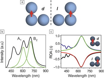
To understand how such a giant ROA is obtained, consider C2v trimers of the shape shown in Figure 4a, with a dipolar emitter (a molecule) residing in one of the interparticle gaps, marked by an arrow.Reference Chuntonov and Haran57 The two possible geometries shown in the figure are related to each other as mirror images, and therefore serve as an example for a plane-chiral pair.Reference Fedotov, Mladyonov, Prosvirnin, Rogacheva, Chen and Zheludev63,Reference Kuwata-Gonokami, Saito, Ino, Kauranen, Jefimovs, Vallius, Turunen and Svirko64 One of the structures will convert linear polarization preferentially into right-handed circular polarization and will therefore be called dextrorotatory (d), while the other will generate left-handed circular polarization (levorotatory, 1). This behavior arises from the interference between the two plasmon normal modes of A1 and B2 symmetry excited by the emitting molecule, as shown in the numerically simulated emission spectrum in Figure 4b. At around 650 nm, the electric fields emitted by the cluster along two perpendicular axes are equal in amplitude and in quadrature phase. This corresponds to purely circularly polarized light, with values of Δ = +1 and –1 obtained for the d and 1 clusters, respectively. Modeling the cluster emission using two harmonic oscillators (whose parameters are found from fitting the spectral resonances of the cluster’s emission spectrum in Figure 4b to Lorentzian line shapes), we calculated the ROA and found it to be in very good agreement with the numerical results, as compared in Figure 4c.Reference Chuntonov and Haran57 This approach can be readily generalized for multiple plasmon resonances.
Raman optical activity (ROA) in a silver trimer of C2v symmetry. (a) Two plane-chiral configurations of the cluster, related by mirror symmetry and corresponding to dextrorotatory (d ) and levorotatory (l ) behavior (see text). Molecular dipoles residing within the interparticle gaps are depicted by the red arrows. (b) Plasmon spectrum of the trimer in either configuration, with the symmetries of the excited plasmon modes marked. Green lines show Lorentzian fits of the spectral peaks. (c) ROA of the d and l configurations. Black and blue lines, electromagnetic simulation; red and green lines, harmonic oscillator model. Adapted with permission from Reference 56. © 2013 American Chemical Society.

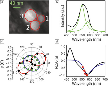
We also tested the harmonic oscillator model of plasmonic ROA by analysis of experimental SM-SERS results reported in Reference 24. As an example, we show an SEM image of a cluster in Figure 5a. The parameters of the harmonic oscillators used in the model were obtained from a fit to the simulated emission spectrum shown in Figure 5b. The experimentally measured  ${\rm{\rho }}\left( {\rm{\theta }} \right)$ is shown in Figure 5c for two specific choices of wavelength in black and green dots. While the pattern measured at 555 nm (black) shows that the polarization is very close to linear, the polarization at 583 nm (green) is essentially circular. The ROA values calculated from the experimental data are plotted for several wavelengths in Figure 5d as red dots. The ROA spectrum calculated by electromagnetic simulation (black line) and the harmonic oscillator model results (blue line) are in a very good agreement with the experimental data. While for excitation at 550 nm, one plasmon resonance clearly dominates the spectrum and results in an almost linear polarization of the scattered light; for excitation at 583 nm, the contributions of both resonances are of the same magnitude, and their spectral distance from the excitation results in amplitudes and phases of the electric field components corresponding to circularly polarized scattered light.
Experimental observation of the effect of a symmetry broken trimer on the polarization of surface-enhanced Raman spectroscopy from a single molecule. (a) Scanning electron microscopy image of a silver trimer on which a single Rhodamine 6G molecule was adsorbed. Our calculations show that the molecule resided in the gap between particles 1 and 2. (b) Simulated emission spectrum (black) using nanoparticle dimensions and gap sizes estimated from the image (see text). (c) Experimental polarization functions ρ(θ) of the Raman-scattered light measured at 555 nm (black dots) and 583 nm (green dots). Red and blue lines show a fit. (d) Raman optical activity (ROA) calculated from the experimental data (red dots), from the electromagnetic simulation (black line), and from the harmonic oscillator model (blue line). Adapted with permission from Reference 56. © 2013 American Chemical Society.

Conclusions
The symmetry of a noble metal nanoparticle cluster defines the shapes and general structure of its plasmon normal modes, which, along with the metallic dielectric function, define the cluster’s optical response. Raman light emitted by a molecule trapped within the interparticle gap of the plasmonic cluster is shaped by the interaction with the plasmon modes. In this review, we have shown how plasmon modes of small clusters can be studied and analyzed using concepts from the theory of symmetry. This analysis helps in explaining the formation of these modes, their evolution with changes in cluster geometry, and the impact they have on the properties of SM-SERS light. The broad spectrum of analytical tools used in the present study, including group theoretical analysis, plasmon hybridization theory, full electrodynamic simulations, and harmonic oscillator models, enabled an in-depth understanding of the physical systems studied here. Although we have discussed clusters of three nanoparticles, this approach can, in principle, be applied to more complex clusters. However, increasing the number of nanoparticles in a cluster leads to a more complex plasmon mode structure and congested plasmon spectra, and will therefore make the application of harmonic oscillator models more difficult, requiring detailed case-specific analysis.
The importance of the novel insights gained from this study is twofold. First, the availability of a simple model of molecule-cluster interaction, based on the concept of plasmon modes and their interference, will substantially assist the correct interpretation of SM-SERS experiments. Second, the present approach paves the way for designing future subwavelength devices, in which control over light characteristics will be crucial for the efficient coupling of electromagnetic fields into plasmon modes and back.
Acknowledgments
The authors acknowledge Ady Stern (Weizmann Institute) and Peter Nordlander (Rice University) for valuable discussions. Timur Shegai (Chalmers Institute of Technology) is thanked for help with the data presentation and for his crucial involvement in early stages of this project, while Takumi Sannomiya (Tokyo Institute of Technology) is thanked for help with MMP simulations. This research was made possible by a grant to G.H. from the Israel Science Foundation (450/10).