Introduction
Cognitive dysfunction in attention, memory and executive function is a core symptom of unipolar disorder (UD) and bipolar disorder (BD) that contributes to impaired workforce capacity (Jaeger et al. Reference Jaeger, Berns, Uzelac and Davis-Conway2006; Depp et al. Reference Depp, Mausbach, Harmell, Savla, Bowie, Harvey and Patterson2012). Executive dysfunction persists after remission and is seen in the domains of working memory (WM) and inhibitory control (Bora et al. Reference Bora, Harrison, Yucel and Pantelis2013; Bourne et al. Reference Bourne, Aydemir, Balanza-Martinez, Bora, Brissos, Cavanagh, Clark, Cubukcuoglu, Dias, Dittmann, Ferrier, Fleck, Frangou, Gallagher, Jones, Kieseppa, Martinez-Aran, Melle, Moore, Mur, Pfennig, Raust, Senturk, Simonsen, Smith, Bio, Soeiro-de-Souza, Stoddart, Sundet, Szoke, Thompson, Torrent, Zalla, Craddock, Andreassen, Leboyer, Vieta, Bauer, Worhunsky, Tzagarakis, Rogers, Geddes and Goodwin2013). Executive dysfunction across UD and BD is associated with WM-associated hypoactivity in the dorsolateral prefrontal cortex (dlPFC) during symptomatic phases (Siegle et al. Reference Siegle, Thompson, Carter, Steinhauer and Thase2007; Townsend et al. Reference Townsend, Bookheimer, Foland-Ross, Sugar and Altshuler2010; Garrett et al. Reference Garrett, Kelly, Gomez, Keller, Schatzberg and Reiss2011; Fernandez-Corcuera et al. Reference Fernandez-Corcuera, Salvador, Monte, Salvador, Goikolea, Amann, Moro, Sans-Sansa, Ortiz-Gil, Vieta, Maristany, McKenna and Pomarol-Clotet2013) and remission (Townsend et al. Reference Townsend, Bookheimer, Foland-Ross, Sugar and Altshuler2010; Pomarol-Clotet et al. Reference Pomarol-Clotet, Alonso-Lana, Moro, Sarro, Bonnin, Goikolea, Fernandez-Corcuera, Amann, Romaguera, Vieta, Blanch, McKenna and Salvador2015), although hyperactivity has also been observed (Harvey et al. Reference Harvey, Fossati, Pochon, Levy, Lebastard, Lehericy, Allilaire and Dubois2005; Matsuo et al. Reference Matsuo, Glahn, Peluso, Hatch, Monkul, Najt, Sanches, Zamarripa, Li, Lancaster, Fox, Gao and Soares2007). This is accompanied by a failure to suppress activity in the default mode network (DMN) (Sheline et al. Reference Sheline, Barch, Price, Rundle, Vaishnavi, Snyder, Mintun, Wang, Coalson and Raichle2009; Fernandez-Corcuera et al. Reference Fernandez-Corcuera, Salvador, Monte, Salvador, Goikolea, Amann, Moro, Sans-Sansa, Ortiz-Gil, Vieta, Maristany, McKenna and Pomarol-Clotet2013), a network of medial brain regions that includes the hippocampus, medial PFC and inferior parietal cortex and is implicated in self-referential thoughts (Raichle et al. Reference Raichle, MacLeod, Snyder, Powers, Gusnard and Shulman2001).
There is no effective treatment for cognitive dysfunction in mood disorders. Current pharmacological treatments may in fact impair cognition because of anticholinergic, extrapyramidal, sedative, and/or blunting effects (Dias et al. Reference Dias, Balanza-Martinez, Soeiro-de-Souza, Moreno, Figueira, Machado-Vieira and Vieta2012). Preliminary evidence indicates that psychological interventions like cognitive remediation (CR) may have beneficial effects on cognition in mood disorder (Demant et al. Reference Demant, Almer, Vinberg, Kessing and Miskowiak2013; Porter et al. Reference Porter, Bowie, Jordan and Malhi2013). However, in a recent randomized, controlled study we found no overall cognitive benefits of 12 weeks CR in 44 remitted BD patients (Demant et al. Reference Demant, Vinberg, Kessing and Miskowiak2015).
The discovery of effective treatments for cognitive dysfunction is hampered by the absence of rapid methods to screen novel candidate treatments. Identification of biomarker models for cognitive enhancement is therefore a priority for psychiatric research. Blood-oxygen-level dependent (BOLD) functional magnetic resonance imaging (fMRI) is a sensitive biomarker of abnormal brain function (Nathan et al. Reference Nathan, Phan, Harmer, Mehta and Bullmore2014). Characterization of post-interventional change in task-related BOLD response within key neural circuits may therefore reveal neurobiological targets associated with cognitive enhancement (Nathan et al. Reference Nathan, Phan, Harmer, Mehta and Bullmore2014).
Erythropoietin (EPO) is a promising candidate treatment for cognitive dysfunction in mood disorders. EPO and its receptor are expressed in the brain and are important for neurodevelopment, neuroprotection and cognition (Siren et al. Reference Siren, Fasshauer, Bartels and Ehrenreich2009; Kastner et al. Reference Kastner, Grube, El-Kordi, Stepniak, Friedrichs, Sargin, Schwitulla, Begemann, Giegling, Miskowiak, Sperling, Hannke, Ramin, Heinrich, Gefeller, Nave, Rujescu and Ehrenreich2012). Systemically administered EPO crosses the blood–brain barrier and enhances neuroplasticity and cognitive function in animal models of brain disease and in healthy animals (Miskowiak et al. Reference Miskowiak, Vinberg, Harmer, Ehrenreich and Kessing2012). In healthy volunteers, we showed that EPO modulates WM-associated fronto-parietal activation and improves executive function independent of red blood cell changes (Miskowiak et al. Reference Miskowiak, Inkster, O'Sullivan, Selvaraj, Goodwin and Harmer2008). Further, 8 weeks EPO v. saline treatment improved some aspects of cognition in treatment-resistant depression (TRD) and BD in recent studies from our group (Miskowiak et al. Reference Miskowiak, Ehrenreich, Christensen, Kessing and Vinberg2014a , Reference Miskowiak, Vinberg, Christensen, Bukh, Harmer, Ehrenreich and Kessing c ). Prospective volumetric MRI assessments revealed a reversal of hippocampal cornu ammonis 1–3 and subiculum volume loss as a potential neural mechanism of patients’ memory improvement (Miskowiak et al. Reference Miskowiak, Vinberg, Macoveanu, Ehrenreich, Koster, Inkster, Paulson, Kessing, Skimminge and Siebner2015).
Here we report the results of a concurrent prospective fMRI investigation in 56 patients from the above trials (Miskowiak et al. Reference Miskowiak, Ehrenreich, Christensen, Kessing and Vinberg2014a , Reference Miskowiak, Vinberg, Christensen, Bukh, Harmer, Ehrenreich and Kessing c ), which aimed to elucidate the changes in BOLD fMRI underlying the EPO-associated improvement in executive function. Given WM-related hypoactivity of dlPFC and deficient task-related suppression of DMN activity across BD and UD and the increase in WM-related right dlPFC activation after a single EPO administration in healthy volunteers, we hypothesized that EPO would (i) reinforce task-related activation of right dlPFC and (ii) suppress hippocampal activity during a spatial WM task.
Materials and method
Study design and participants
This report is based on our double-blind, placebo-controlled efficacy studies (Miskowiak et al. Reference Miskowiak, Ehrenreich, Christensen, Kessing and Vinberg2014a , Reference Miskowiak, Vinberg, Christensen, Bukh, Harmer, Ehrenreich and Kessing c ). Patients aged 18–65 years were screened with Schedules for Clinical Assessment in Neuropsychiatry to confirm ICD-10 diagnosis. Eligible patients had unipolar TRD with moderate depression [17-item Hamilton Depression Rating Scale (HAMD-17) score ⩾17] or BD in partial remission [HAMD-17 and Young Mania Rating Scale (YMRS) scores ⩽14] with subjective cognitive difficulties. Patients’ medication remained stable from ⩾2 weeks before inclusion and throughout the study. For a detailed description of the screening, exclusion criteria and safety precautions, see Miskowiak et al. Reference Miskowiak, Ehrenreich, Christensen, Kessing and Vinberg2014a , Reference Miskowiak, Vinberg, Christensen, Bukh, Harmer, Ehrenreich and Kessing c . The study was approved by the local ethics committee, Danish Medicines Agency, and Danish Data Agency, and registered at clinicaltrials.gov (no. NCT00916552). After complete description of the study, written informed consent was obtained from all participants.
Randomization and masking
Block randomization was stratified for age (<35 or ⩾35 years) and gender. Outcome assessors were blinded to patients’ treatment allocation throughout the study period and data analysis (see Miskowiak et al. Reference Miskowiak, Ehrenreich, Christensen, Kessing and Vinberg2014a , Reference Miskowiak, Vinberg, Christensen, Bukh, Harmer, Ehrenreich and Kessing c ).
Procedures
Patients received eight weekly intravenous infusions of EPO (Eprex; 40 000 IU; Janssen-Cilag) or saline (NaCl 0.9%). The dose and treatment schedule were chosen because under conditions of an intact blood–brain barrier, around 1% of peripherally injected EPO enters the brain and leads to concentrations in brain tissue comparable to the optimal concentrations for neuroprotection in neuron cultures (Brines et al. Reference Brines, Ghezzi, Keenan, Agnello, de Lanerolle, Cerami, Itri and Cerami2000; Ehrenreich et al. Reference Ehrenreich, Degner, Meller, Brines, Béhé, Hasselblatt, Woldt, Falkai, Knerlich, Jacob, von Ahsen, Maier, Brück, Rüther, Cerami, Becker and Sirén2004). Whole-brain fMRI was performed at baseline and week 14, 6 weeks after treatment completion at which time point the haematological parameters were expected to have normalized in the EPO group (Miskowiak et al. Reference Miskowiak, Vinberg, Macoveanu, Ehrenreich, Koster, Inkster, Paulson, Kessing, Skimminge and Siebner2015). Blood tests and blood pressure were taken weekly during the treatment period and in week 14 with the examiners being blind to the results. Mood symptoms were assessed with the HAMD-17, Beck Depression Inventory (BDI) and YMRS at weeks 1, 5, 9, and 14.
Spatial n-back WM task
n-back WM tasks are widely used to probe memory aspects of executive function (Wager & Smith, Reference Wager and Smith2003). Here we used a spatial n-back WM task during fMRI (Supplementary Fig. S2). The task consisted of blocks of three types of conditions that differed in terms of WM load (0-back, 1-back and 2-back). In each condition, a yellow circle appeared in a sequence of 14 appearances at random locations in a 5 × 5 grid. The circle was displayed for 300 ms followed by an empty grid for 1200 ms. In 1-back and 2-back conditions, patients indicated with a button press when the circle appeared at the same location as one trial and two trials back, respectively. During the 0-back condition, patients pressed a button when the circle appeared in one of the four grid corners. The blocks had an average of three target trials and were presented successively five times (15 blocks in total) interleaved with 8 s fixation crosses. The total task length was 7 min 35 s.
Control task
To investigate whether WM-related neural responses were confounded by global effects of EPO on cerebral blood flow, neural activation in the occipital cortex was assessed with a control visual stimulation paradigm: a flashing checkerboard (8 Hz) presented in blocks of 14 s alternating with 14 s of a fixation cross for a total of six cycles (duration 3 min 8 s). Patients were instructed to lie with their eyes open during this time.
Other fMRI paradigms
We also investigated the effects of EPO on picture encoding and emotional face processing in the same scan session using fMRI paradigms from our previous studies (Miskowiak et al. Reference Miskowiak, O'Sullivan and Harmer2007, Reference Miskowiak, Glerup, Vestbo, Harmer, Reinecke, Macoveanu, Siebner, Kessing and Vinberg2014b ). For clarity reasons, the results of these tasks will be reported elsewhere.
MRI
Whole-brain MRI data were collected at the Danish Research Centre for Magnetic Resonance with a 3-T Siemens Trio MR scanner using an eight-channel head array coil. BOLD-sensitive fMRI used a T2*-weighted echo-planar imaging (EPI) sequence with an echo time (TE) of 30 ms, repetition time (TR) of 2.49 ms and a low flip angle of 20° to minimize physiological noise (Gonzalez-Castillo et al. Reference Gonzalez-Castillo, Roopchansingh, Bandettini and Bodurka2011). For the WM task, a total of 184 brain volumes were acquired in a single fMRI session, each consisting of 42 slices with a slice thickness of 3 mm and a field of view (FOV) of 192 × 192 mm using a 64 × 64 grid. The control task employed an identical MR sequence as the WM task with 68 brain volumes acquired. High-resolution 3D structural T1-weighted spin echo images were obtained after the first session of BOLD fMRI (TI = 800, TE = 3.93, TR = 1540 ms, flip angle 9°; 256 × 256 FOV; 192 slices).
fMRI data analysis
Our analysis approach was to investigate our a priori hypotheses that EPO would (i) increase WM-related right dlPFC activity and (ii) suppress hippocampal activity by comparing the task-related mean percent signal change within predefined regions of interest (ROIs) between groups (details below). We explored additional effects of EPO v. saline on changes in neural response with whole-brain exploratory analysis.
fMRI data processing was performed with the fMRI Expert Analysis Tool (FEAT; version 6.00) part of FMRIB's Software Library (FSL; http://www.fmrib.ox.ac.uk/fsl). Pre-processing included image realignment, non-brain removal, spatial normalization to an MNI (Montreal Neurologic Institute) template and spatially smoothing (Gaussian kernel, 5 mm full-width-half-maximum). The time series in each session were high pass-filtered (to max 0.008 Hz). Three conditions, ‘1-back’, ‘2-back’ and ‘0-back’ were modelled as blocks convolved with a canonical haemodynamic response function.
To investigate hypothesis (i), we defined a spherical ROI with a 10 mm radius around the peak right dlPFC region (x = 40, y = 34, z = 29) involved in active spatial WM processes as reported in a meta-analysis (Wager & Smith, Reference Wager and Smith2003). The ROI was constructed on the Montreal Neurological Institute template using the WFU PickAtlas toolbox (Maldjian et al. Reference Maldjian, Laurienti, Kraft and Burdette2003). Given the absence of meta-analyses of WM-associated DMN deactivation, we tested hypothesis (ii) by extracting mean percent signal change at baseline in a functional mask in the left and right hippocampus (i.e. clusters showing negative linear relation with increasing load obtained by using the hippocampal structural masks for small volume correction in FSL (thresholded at Z > 2.0 and p ≤ 0.05, corrected for multiple comparisons at a cluster level). We extracted the mean percent BOLD signal change in the dlPFC and hippocampal ROIs during 1-, 2- and 0-back and computed the differential signal change for the 2-back > 0-back and 2-back > 1-back contrasts to assess the effects of EPO on neural response during high-load WM and high-load specific WM, respectively.
For analysis of our a priori ROIs, we conducted repeated-measures analysis of variance (ANOVA) with time and group to examine differential change in WM-related activity between groups from baseline to follow-up. Because of high within-subject variation in BOLD fMRI signal change across repeated measurements (Zandbelt et al. Reference Zandbelt, Gladwin, Raemaekers, van, Neggers, Kahn, Ramsey and Vink2008), we also conducted exploratory between-group comparisons at the post-treatment session given the randomized controlled trial design with well-matched groups. Activity in these ROIs was also compared between groups at baseline. We used residual plots to check the model assumptions and excluded any outliers with standardized residual values ⩾5. Signal change analyses were performed in Statistical Package for Social Sciences (SPSS v. 22; IBM Corporation, USA).
For the whole-brain exploratory analysis, a fixed-effects analysis was conducted for each participant to identify regions of activity increase and decrease from baseline to follow-up. These ‘difference images’ were entered into group-level analyses, which employed a full mixed-effects approach (Woolrich et al. Reference Woolrich, Behrens, Beckmann, Jenkinson and Smith2004). Z (Gaussianized T/F) statistic images were thresholded using clusters determined by an extent threshold of Z > 2.0 and a (corrected) cluster significance threshold of p ⩽ 0.05 (Worsley, Reference Worsley, Jezzars, Matthews and Smith2001). We used the high-load WM measure (2-back > 0-back) and the high-load specific WM measure (2-back > 1-back) in the ROI analyses. A standard anatomical atlas (Talairach & Tournoux, Reference Talairach and Tournoux1988) was used to localize peak cluster activation. For clusters showing either a group × time interaction or change in the EPO group over time, we extracted mean percent BOLD signal change for each participant and examined this with ANOVA to identify what was driving the interaction/activity change.
For the control paradigm, we calculated the mean percent BOLD signal change at baseline and follow-up within an ROI within the occipital (calcarine) cortex for each participant, consistent with the approach in our previous studies (Miskowiak et al. Reference Miskowiak, O'Sullivan and Harmer2007, Reference Miskowiak, Inkster, O'Sullivan, Selvaraj, Goodwin and Harmer2008, Reference Miskowiak, Favaron, Hafizi, Inkster, Goodwin, Cowen and Harmer2009). Repeated-measures ANOVA with group and time was conducted to examine differential change between groups in neural response to photic stimuli in this ROI (Maldjian et al. Reference Maldjian, Laurienti, Kraft and Burdette2003).
Statistical analyses of behavioural and mood data
The effect of EPO on n-back WM performance was examined with repeated-measures ANOVA with time (weeks 1, 14) and WM load (1, 2) as within-subject factors and group as the between-subject factor (all tests were two-tailed). Significant interactions were followed up by simple main effect analyses. We corrected for non-sphericity when appropriate using the Greenhouse-Geisser correction. Signal detection theory was applied to obtain a measure of WM accuracy corrected for participants’ response tendency (d’) (Grier, Reference Grier1971). Changes between groups in mood symptoms were analysed in repeated-measures ANOVA. Associations between changes in BOLD response and WM accuracy, and the correlations between these variables and changes in mood, haemoglobin and blood pressure, were examined with Pearson's correlations. Medication status in EPO v. saline groups was examined with Pearson's χ2 and Fisher's exact tests. Haemoglobin and blood pressure at follow-up were examined with simple main effect analyses (t tests). Statistical analyses were performed in SPSS. Significance level was set at p ⩽ 0.05.
Results
Patient flow and characteristics
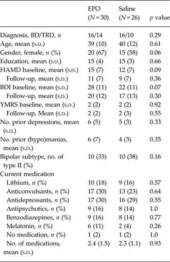
Table 1 and the CONSORT chart (Supplementary Fig. S1) display patients’ characteristics and flow, respectively. Of the 84 patients included in the trials, one patient withdrew at baseline, MRI data collection was incomplete for 14 and behavioural data was missing for 10 because of technical difficulties with the response box. Of the remaining 59 patients, three outliers with WM accuracy of ⩾2 s.d. below the mean were excluded. Data was thus analysed for n = 56 patients (EPO: n = 30; Saline: n = 26). Groups were well-matched on baseline characteristics and medication (p > 0.05, see Table 1).
Patients’ characteristics

BD, Bipolar; TRD, treatment-resistant depression; EPO, erythropoietin; HAMD, Hamilton Depression Rating Scale; BDI, Beck Depression Inventory; YMRS, Young Mania Rating Scale.
Safety of EPO treatment
EPO treatment was well-tolerated (Miskowiak et al. Reference Miskowiak, Ehrenreich, Christensen, Kessing and Vinberg2014a , Reference Miskowiak, Vinberg, Christensen, Bukh, Harmer, Ehrenreich and Kessing c ). Seven (17%) of the 41 EPO-treated patients had to discontinue treatment after weeks 5–7 because of increased thrombocytes but completed all assessments. Six patients [EPO: n = 5 (12%); saline: n = 1 (2%)] reached the criteria for bloodletting (haematocrit levels >50% and >48% for men and women, respectively, on two consecutive measurements) in weeks 3–7; three were bled once, two were bled twice and one was bled three times. All haematological parameters were normalized 2–6 weeks after treatment completion (for further details see Miskowiak et al. Reference Miskowiak, Ehrenreich, Christensen, Kessing and Vinberg2014a , Reference Miskowiak, Vinberg, Christensen, Bukh, Harmer, Ehrenreich and Kessing c ).
Behavioural results
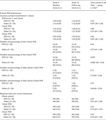
The WM accuracy (d’) scores data showed a negative skew due to near-ceiling accuracy rates. As d’ scores are in the range 0–1, we arcsine-transformed the data to approach normal distribution prior to statistical analysis (McDonald, Reference McDonald2014). At baseline, there was no difference in WM accuracy between groups (p ⩽ 0.31). EPO improved WM accuracy from baseline to follow-up compared with saline (F 1,54 = 4.23, p = 0.045; post-hoc t tests non-significant) (see Table 2). Speed of WM was not influenced by EPO (p ⩽ 0.70). For more details, see Table 2.
N-back working memory (WM) performance at baseline (week 0) and follow-up (week 14) and effects of EPO v. saline

s.d. Standard deviation; EPO, erythropoietin; WM, working memory.
a Degrees of freedom (df) are identical for time and time × group.
fMRI results
dlPFC
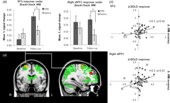
In line with previous studies (Wager & Smith, Reference Wager and Smith2003), the right dlPFC was significantly activated by high-load WM (2-back > 0-back) (t = 12.40, df = 55, p < 0.001) and showed greater response specifically under high v. low loads (2-back > 1-back) (t = −4.79, df = 55, p < 0.001). Baseline dlPFC response showed no differences between groups during high-load WM or high-load specific WM (p⩾0.50). One outlier (saline) was excluded because of a standardized residual value ⩾5 for WM-related dlPFC response at follow-up and data was thus analysed for n = 55 (EPO: n = 30, saline: n = 25). There was no differential change in WM-related dlPFC activity between groups over time (p ⩾0.36). However, exploratory post-treatment comparisons revealed greater right dlPFC response in EPO- v. saline groups during 2-back > 1-back WM (t = 2.21, df = 53, p = 0.032) (see Fig. 1) in the absence of differences during 2-back > 0-back WM (p⩾0.12).
(a) Neural network activated during high working memory (WM) load (2-back > 1-back) across all patients at baseline (green). Region in the superior frontal gyrus (SFG) (BA 6) showing increased response specifically during this high WM load in the erythropoietin (EPO) group in the exploratory whole-brain analysis (yellow); a priori ROI in the right dorsolateral prefrontal cortex (dlPFC) (red). Images are thresholded at Z > 2.0 and p < 0.05, corrected for multiple comparisons at a cluster level. (b) Plot of change from baseline to follow-up in mean % blood oxygen level-dependent (BOLD) signal change during high WM load (2-back > 1-back) within SFG and dlPFC in EPO (red bars) and saline (blue bars) groups at baseline and follow-up. Bars show the mean; error bars show the s.e. In the a priori ROI, dlPFC, between-group comparison of mean percent signal change at the follow-up scan with adjustment for baseline activity showed that EPO produced greater activity compared with saline during high WM loads (p = 0.03). Notably, this post-treatment group difference occurred in the absence of significant activity changes from baseline to follow-up within each group separately (p ⩾0.40). EPO also increased high-load specific BOLD response in the SFG from baseline to follow-up compared with saline (group × time interaction for extracted mean percent signal change: F 2,54 = 6.78, p = 0.01; t test at follow-up: t = 2.34, df = 54, p = 0.02). Note that the ROIs were non-independent and that the effect sizes of the extracted mean percent signal change may therefore be exaggerated. (c) Linear relation across the entire sample between change in WM performance and change in BOLD response in the SFG during high-load specific WM and in the dlPFC during high-load WM. There was a positive correlation between change in WM accuracy and WM-related BOLD signal in these regions across the entire cohort (r = 0.30–0.35, p < 0.05).

Functional cluster in the hippocampus
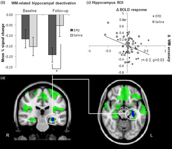
WM was associated with deactivation in regions of the DMN including the hippocampus, medial frontal and inferior parietal regions (see Fig. 2). There were no differences between groups at baseline in WM-associated deactivation of the hippocampus ROIs (p ⩾0.46). EPO enhanced left hippocampal deactivation during 2-back > 0-back WM from baseline to follow-up compared with saline (F 1,54 = 5.24, p = 0.026), which was driven by greater deactivation in EPO v. saline groups at follow-up (t = 2.28, df = 54, p = 0.027) (see Fig. 2). There was also a greater hippocampal deactivation in EPO v. saline groups over time during 2-back > 1-back WM (F 1,54 = 5.02, p = 0.029), which reflected reversal of greater baseline activity in EPO group (baseline: t = 2.44, df = 54, p = 0.018; follow-up: p ⩾0.65). In contrast, there were no WM-related activity differences between groups in the right hippocampal ROI (p ⩾0.38).
(a) Default mode network (DMN) showing deactivation during working memory (WM) (i.e. regions showing a negative linear relation with increasing WM load) across all participants at baseline (green). The (overlapping) functional cluster within the left hippocampus identified as showing WM-associated hippocampal deactivation (blue). Images are thresholded at Z > 2.0 and p < 0.05, corrected for multiple comparisons at a cluster level. (b) Plot of mean % blood oxygen level dependent (BOLD) signal change during high-load WM (2-back > 0-back) within the left hippocampal ROI in the erythropoietin (EPO) (red bars) and saline (blue bars) groups at follow-up. Bars show the mean; error bars show the s.e. EPO produced greater WM-related deactivation compared with saline at follow-up (p = 0.02). Note that the ROIs were non-independent and that the effect sizes of the extracted mean percent signal change may therefore be exaggerated. (c) Linear relation across the entire sample between change in WM performance and in WM-related BOLD response in the left hippocampal ROI. There was a negative correlation between change in WM accuracy and WM-associated hippocampal activity across the entire cohort, indicating increase in WM accuracy in patients with greater hippocampal deactivation (r = −0.30, p = 0.03).

Whole-brain exploratory analyses
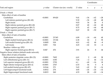
High-load (2-back > 0-back) and high-load specific (2-back > 1-back) WM activated a broad network of overlapping regions including the dlPFC, SFG, cerebellum, temporo-parietal and occipital regions consistent with previous findings (Wager & Smith, Reference Wager and Smith2003) (for peak cluster activations, see Table 3). Exploratory whole-brain analysis revealed no differences between groups in neural activity changes during high-load WM or high-load specific WM over time. However, in the EPO group there was an activity increase over time in the right caudal SFG specifically under high v. low WM loads (at Z > 2.0, cluster corrected p = 0.05) (see Fig. 1 a; for peak cluster activation see Table 3). Extraction and between-group comparison of mean percent signal change showed that this effect was driven by increased high-load specific (2-back > 1-back) activity in the EPO v. saline groups at follow-up (see Fig. 1 b). In contrast, the SFG showed no activity changes during 2-back > 0-back WM in EPO v. saline groups (p ⩾0.25).
Peak cluster activation in regions identified in whole-brain analyses (with Z = 2.0, p = 0.05, cluster-corrected) as showing (i) increased BOLD response during general n-back working memory (2-back > 0-back), (ii) greater response during high WM load (2-back > 1-back) and (iii) functional deactivation during working memory (2-back < 1-back < 0-back) across all participants at baseline, and (iv) regions showing activity change from baseline to follow-up in EPO-treated patients

BA, Brodmann area; EPO, erythropoietin.
MNI coordinates (x, y, z) refer to the point of peak activation within each cluster identified using this threshold.
Visual stimulation control experiment
Mean percent BOLD signal change to visual stimulation within the occipital (calcarine) cortex ROI showed no baseline differences between groups or any effects of EPO v. saline (p ⩾0.39).
Associations between BOLD fMRI and WM performance
To investigate the functional relevance of the EPO-associated effects on neural activity, we correlated baseline WM accuracy with baseline WM-related BOLD response in the right dlPFC, the left hippocampal functional mask and the SFG cluster identified in the whole-brain analysis. We further correlated the change in WM accuracy with the change in WM-related activity in these regions across the entire sample. At baseline, patients displayed a significant correlation between WM accuracy and (i) high-load WM-related right dlPFC activity (r 54 = 0.27, p = 0.044) and (ii) high-load specific activity in the SFG (r 54 = 0.35, p = 0.009), as well as (iii) a trend towards correlation between WM accuracy and hippocampal deactivation during high-load WM (r 54 = −0.25, p = 0.066). Again using the entire sample, there was a correlation between WM improvement and increase in (i) high-load WM-related right dlPFC response (r 54 = 0.29, p = 0.030), (ii) increase in high-load specific SFG activity (r 54= 0.28, p = 0.035), and (iii) greater high-load WM-related left hippocampal deactivation (r 54 = −0.30, p = 0.026) (see Figs 1 c and 2 c). For exploratory purposes we also performed these correlation analyses for EPO-treated patients only but found no significant correlations (p ⩾0.36), possibly because of reduced statistical power due to the reduction in numbers.
No influence of changes in haematocrit, blood pressure, mood or medication
EPO increased haemoglobin during the active treatment phase (for details see Miskowiak et al. Reference Miskowiak, Ehrenreich, Christensen, Kessing and Vinberg2014a , Reference Miskowiak, Vinberg, Christensen, Bukh, Harmer, Ehrenreich and Kessing c ), but this effect had tapered off before the follow-up fMRI scan at which time there was no difference between groups (t = −0.39, df = 54, p = 0.70). There was also no difference between groups in blood pressure (p ⩾0.39). EPO-treated patients showed no correlation between changes in haemoglobin and neural response within the identified ROIs or WM improvement (p ⩾ 0.21). There were no significant correlation between changes in blood pressure and neural response in these regions (p ⩾0.08). EPO had no significant effect on mood symptoms (HDRS: p ⩾ 0.13; BDI: p ⩾0.08) and there was no significant correlation between changes in mood symptoms and changes in (i) WM accuracy in EPO or saline groups (p ⩾ 0.69 and p ⩾ 0.12, respectively) or (ii) high-load specific activity in the dlPFC or SFG (EPO: p ⩾ 0.38; saline: p ⩾ 0.43) or hippocampal ROIs (EPO: p ⩾ 0.09; saline: p ⩾ 0.42). Finally, there was no correlation between age and neural response or WM accuracy (p ⩾ 0.18). Post hoc comparisons of patients with or without lithium, antipsychotic and antidepressant treatment, respectively, revealed no effects of these medications on post-interventional activity in the right dlPFC (p ⩾ 0.5) or in the hippocampus ROI (p ⩾ 0.4) or SFG (p ⩾ 0.14).
Discussion
This fMRI study provides a cerebral basis for the EPO-associated improvement of executive function in patients with mood disorders. Our data show that 8 weeks of EPO treatment of patients with TRD or BD increased right caudal SFG response specifically during high WM loads and enhanced WM-related deactivation of the left hippocampus compared with saline. Post-treatment comparisons also revealed greater high-load specific activity in the a priori dlPFC ROI in EPO v. saline groups, although notably dlPFC activity change over time did not differ between groups. EPO treatment also improved patients’ WM accuracy. Across the entire sample, WM improvement correlated with increase in WM-related dlPFC and SFG activity and hippocampal deactivation. The present effects of EPO were not associated with changes in mood, haemoglobin or blood pressure.
The effects of EPO on WM-related neural activity may counteract hypoactivity in dorsal PFC regions and failure to suppress DMN activity in the hippocampus across mood disorders (Siegle et al. Reference Siegle, Thompson, Carter, Steinhauer and Thase2007; Sheline et al. Reference Sheline, Barch, Price, Rundle, Vaishnavi, Snyder, Mintun, Wang, Coalson and Raichle2009; Garrett et al. Reference Garrett, Kelly, Gomez, Keller, Schatzberg and Reiss2011; Fernandez-Corcuera et al. Reference Fernandez-Corcuera, Salvador, Monte, Salvador, Goikolea, Amann, Moro, Sans-Sansa, Ortiz-Gil, Vieta, Maristany, McKenna and Pomarol-Clotet2013; Pomarol-Clotet et al. Reference Pomarol-Clotet, Alonso-Lana, Moro, Sarro, Bonnin, Goikolea, Fernandez-Corcuera, Amann, Romaguera, Vieta, Blanch, McKenna and Salvador2015). Greater task-related suppression of hippocampal activity in the EPO group may be an indirect marker of improved WM (increased disengagement from task-irrelevant thoughts), consistent with the observed correlation between hippocampal activity suppression and WM performance. The PFC shows low activity at low load, increased activity at high load, and decreased activity when the load exceeds WM capacity (Callicott et al. Reference Callicott, Mattay, Bertolino, Finn, Coppola, Frank, Goldberg and Weinberger1999). Given this, the greater prefrontal response specifically during high WM loads in EPO v. saline-treated patients at follow-up suggests that EPO may prevent breakdown of WM capacity. These results are in accordance with increased dorsal PFC activity during executive function tests after cognitive training in multiple sclerosis (Filippi et al. Reference Filippi, Riccitelli, Mattioli, Capra, Stampatori, Pagani, Valsasina, Copetti, Falini, Comi and Rocca2012) and schizophrenia (Ramsay & MacDonald, Reference Ramsay and MacDonald2015). The observed effects on WM-related neural circuit activity may therefore not be specific to EPO but also occur in response to other compounds with pro-cognitive effects. This would highlight normalization of hypoactivity in dorsal PFC regions and DNM hyperactivity during executive control tasks as key neurobiological targets associated with pro-cognitive effects of both biological and psychological treatments across a range of neuropsychiatric disorders.
Several neurobiological mechanisms may underlie the observed effects of EPO. Although EPO increased haemoglobin during the active treatment phase, this was normalized at follow-up and there were no correlations between changes in haemoglobin and BOLD response within the identified ROIs. Blood pressure has been shown to correlate positively with BOLD response (Wang et al. Reference Wang, Foniok, Wamsteeker, Qiao, Tomanek, Vivanco and Tuor2006). However, we found no significant differences in blood pressure between groups or correlations between EPO-associated change in BOLD response and blood pressure. The visual control task also showed no difference between groups in occipital activation to photic stimuli, suggesting that the observed effects of EPO did not result from any global haemodynamic changes. Finally, there were no significant effects of EPO on mood symptoms across the UD and BD groups or correlations between changes in mood and neural response or WM accuracy. Taken together, this suggests that direct neurobiological actions mediate the observed effects of EPO. Impaired neuroplasticity may be a central cause of patients’ executive dysfunction and aberrant prefrontal and hippocampal activity (Carlson et al. Reference Carlson, Singh, Zarate, Drevets and Manji2006; Marsden, Reference Marsden2013). The ability of EPO to increase neuroplasticity through activation of anti-inflammatory, anti-apoptotic and anti-oxidant signalling cascades (Siren et al. Reference Siren, Fasshauer, Bartels and Ehrenreich2009) and inhibition of glycogen synthase kinase 3 beta (GSK3β) (Ge et al. Reference Ge, Zhu, Geng, Zhang and Liu2012) may hence be important for the observed effects of EPO on neural and behavioural measures of WM. These effects may be generalizable to other neuropsychiatric disorders, consistent with the findings of EPO-associated improvement of executive function in multiple sclerosis, schizophrenia (Ehrenreich et al. Reference Ehrenreich, Fischer, Norra, Schellenberger, Stender, Stiefel, Siren, Paulus, Nave, Gold and Bartels2007a , Reference Ehrenreich, Hinze-Selch, Stawicki, Aust, Knolle-Veentjer, Wilms, Heinz, Erdag, Jahn, Degner, Ritzen, Mohr, Wagner, Schneider, Bohn, Huber, Czernik, Pollmacher, Maier, Siren, Klosterkotter, Falkai, Ruther, Aldenhoff and Krampe b ) and in healthy individuals (Miskowiak et al. Reference Miskowiak, Inkster, O'Sullivan, Selvaraj, Goodwin and Harmer2008).
A strength of the study was the relatively large sample size of N = 56 patients with complete pre- and post-treatment data. In comparison, previous prospective fMRI investigations of treatments targeting cognition involved n = 20–28 patients (Filippi et al. Reference Filippi, Riccitelli, Mattioli, Capra, Stampatori, Pagani, Valsasina, Copetti, Falini, Comi and Rocca2012; Meusel et al. Reference Meusel, Hall, Fougere, McKinnon and MacQueen2013; Subramaniam et al. Reference Subramaniam, Luks, Garrett, Chung, Fisher, Nagarajan and Vinogradov2014). In addition, the randomized, placebo-controlled design could accommodate for any practice effects and effects of repeated scanning. It is a potential limitation that patients were medicated since this could have produced non-specific changes in global brain activation. However, such confounding effects were unlikely since EPO and saline groups showed no differences in medication status or baseline neural response. In addition, we observed no effects of lithium, antipsychotic or antidepressant treatment on neural activity in the identified ROIs. Indeed, it is noteworthy that the effects of EPO on WM-related neural activity occurred over and above any non-specific effects of patients’ medication. Another limitation is that our cohort included both TRD and BD patient groups who may be characterized by distinct although overlapping pathogenic processes. Assessment of the effects of EPO for the TRD and BD groups separately was not possible due to insufficient statistical power with the small number of participants for such subgroup analyses. Nevertheless, executive dysfunction and aberrant WM-related neural responses in the dlPFC and DMN occur across both disorders, and EPO has been shown to improve cognition across several brain disorders (Ehrenreich et al. Reference Ehrenreich, Degner, Meller, Brines, Béhé, Hasselblatt, Woldt, Falkai, Knerlich, Jacob, von Ahsen, Maier, Brück, Rüther, Cerami, Becker and Sirén2004, Reference Ehrenreich, Hinze-Selch, Stawicki, Aust, Knolle-Veentjer, Wilms, Heinz, Erdag, Jahn, Degner, Ritzen, Mohr, Wagner, Schneider, Bohn, Huber, Czernik, Pollmacher, Maier, Siren, Klosterkotter, Falkai, Ruther, Aldenhoff and Krampe2007b ; Miskowiak et al. Reference Miskowiak, Ehrenreich, Christensen, Kessing and Vinberg2014a , Reference Miskowiak, Vinberg, Christensen, Bukh, Harmer, Ehrenreich and Kessing c ). Since the study did not include a healthy control arm, it cannot be determined if our patients displayed WM-related prefrontal hypoactivity at baseline or if the EPO-associated change in BOLD signal represents normalization. While we found a significantly higher dlPFC response in the EPO group post-treatment compared with saline, the effect of treatment on change in dlPFC activity was not significant. Consequently, caution should be applied when interpreting the effects of EPO on WM-related dlPFC activity. Given the evidence for abnormal task-related prefrontal and DMN activity in mood disorders and our demonstration of correlations between BOLD signal and WM performance, we suggest that our findings indicate an improved neural network functioning in EPO-treated patients. Notably, this post-interventional fMRI investigation of the effects of EPO on neural circuitry activity was exploratory in nature according to our original published trial protocol (Miskowiak et al. Reference Miskowiak, Vinberg, Harmer, Ehrenreich, Knudsen, Macoveanu, Hansen, Paulson, Siebner and Kessing2010). Bonferroni correction for the three employed fMRI paradigms was therefore not applied. From a clinical perspective, the therapeutic use of EPO in patients with mood disorders is limited by its haematological effects. Although EPO was well-tolerated in our studies, it is a labor-intensive treatment because of the necessity to perform careful screening and regular medical examinations and blood tests. A clinically interesting alternative is therefore non-haemapoietic EPO molecules such as carbamylated EPO that cross the blood–brain barrier and enhance neuroplasticity and cognition in preclinical models (Siren et al. Reference Siren, Fasshauer, Bartels and Ehrenreich2009). If pharmacokinetic studies indicate that such EPO derivatives can be safely administered to humans, it would be a logical next step to investigate if they produce target engagement in the PFC and DMN.
In conclusion, our data links EPO-related improvement of executive function in mood disorders with increased SFG response specifically during high-load WM, and WM-related deactivation of DMN activity. This is consistent with preclinical evidence for the ability of EPO to enhance neuroplasticity and cognition and highlights EPO as a candidate treatment for executive dysfunction in mood disorders.
Supplementary material
For supplementary material accompanying this paper visit http://dx.doi.org/10.1017/S0033291716000209.
Acknowledgements
The study was funded by the Danish Council for Independent Research, Novo Nordisk Foundation, Beckett Fonden, and Savværksejer Juhl's Mindefond. The sponsors had no role in the planning or conduct of the study or in the interpretation of the results. The authors thank the study nurses, Susanne Sander and Hanne Nikolajsen for their work with logistical planning and running the trial. We also thank Dr Jeanette Mumford, Department of Psychology, University of Texas at Austin, for her valuable advice on the design of the fMRI analyses and Dr Lene T. Skovgaard, Department of Biostatistics at University of Copenhagen, for her valued input on the statistical analyses of the fMRI data. Professor Siebner was supported by a Grant of Excellence on the control of actions ‘ContAct’ from Lundbeck Foundation (R59 A5399) and the Novo Nordisk Foundation Interdisciplinary Synergy Program 2014 (NNF14OC0011413). The Simon Spies Foundation is acknowledged for donation of the Siemens Trio Scanner. The Lundbeck Foundation provided half of Dr Miskowiak's post-doctorate salary at the Psychiatric Center Copenhagen (2012–2015) to allow her to do full-time research in this period.
Declaration of Interest
K.W.M. reports having received consultancy fees from Lundbeck. M.V. discloses consultancy fees from Eli Lilly, Lundbeck; Servier and AstraZeneca. H.E. has submitted/holds user patents for EPO in stroke, schizophrenia, and MS. O.B.P. is a member of the board of directors of the Elsass Foundation. C.J.H. has received consultancy fees from P1vital Ltd, Lundbeck, Servier, Eli-Lilly and is a company director of Oxford Psychologists Ltd. C.J.H. has also received grant income from GSK, UCB, Janssen Inc., Lundbeck, Servier and AstraZeneca. G.M.K. received within the last 3 years honoraria as a field editor for International Journal of Neuropsychopharmacology and as scientific advisor for Lundbeck. H.R.S. discloses honoraria as reviewing editor for Neuroimage, as speaker for Biogen Idec Denmark A/S, and scientific advisor for Lundbeck within the past 3 years. L.V.K. reports having been a consultant for Lundbeck and AstraZeneca within the last 3 years. All other authors report no biomedical financial interests or potential conflicts of interest.