Introduction
Older patients, who account for more than half of intensive care unit (ICU) admissions and 60% of all ICU days in the USA (Angus et al., Reference Angus, Kelley, Schmitz, White and Popovich2000), frequently experience delirium during critical illness. This form of acute brain dysfunction affects up to 80% of mechanically ventilated ICU patients (Girard et al., Reference Girard, Pandharipande and Ely2008b) and is associated with numerous adverse outcomes in this vulnerable population, including prolonged hospitalization (Girard et al., Reference Girard, Pandharipande and Ely2008b), increased costs (Girard et al., Reference Girard, Pandharipande and Ely2008b), higher short- and long-term mortality (Girard et al., Reference Girard, Pandharipande and Ely2008b; Pisani et al., Reference Pisani, Kong, Kasl, Murphy, Araujo and van Ness2009), and persistent brain dysfunction in the form of long-term cognitive impairment (Girard et al., Reference Girard2010).
A variety of mechanisms have been proposed to explain the pathophysiology of delirium; these include inflammation, abnormalities in neurotransmission, and direct effects of medications, e.g. sedatives and analgesics (Inouye and Ferrucci, Reference Inouye and Ferrucci2006; Gunther et al., Reference Gunther, Morandi and Ely2008). In particular, several studies have examined the association of delirium with markers of inflammation, such as inflammatory and anti-inflammatory cytokines (Inouye and Ferrucci, Reference Inouye and Ferrucci2006; Gunther et al., Reference Gunther, Morandi and Ely2008). Insulin-like growth factor 1 (IGF-1) has also been considered in the pathogenesis of delirium since this hormone, referred to by some investigators as a “neuroprotective cytokine,” inhibits cytotoxic cytokines (e.g. IL-6, IL-8, IL-1), an effect that may be especially important during an acute critical illness.
Low IGF-1 concentrations, in fact, have been associated with the development of delirium in acutely ill, older patients (Wilson et al., Reference Wilson, Broadhurst, Diver, Jackson and Mottram2005; Adamis et al., Reference Adamis, Treloar, Martin, Gregson, Hamilton and Macdonald2007; Reference Adamis2009), suggesting that patients with low IGF-1 may lack innate neuroprotection against chemical and physical insults, making them more susceptible to the development of delirium. Studies examining IGF-1 and delirium, however, have not uniformly found such an association. Lemstra et al. (Reference Lemstra, Kalisvaart, Vreeswijk, van Gool and Eikelenboom2008), for example, found that preoperative IGF-1 concentrations did not predict post-operative delirium in older hip surgery patients.
Based on studies conducted outside the ICU, we suspected that IGF-1 may be associated with delirium in ICU patients; no data, however, are currently available regarding the relationship between IGF-1 levels and delirium during critical illness. Therefore, we tested prospectively the hypothesis that low IGF-1 concentrations early in critical illness would be associated with delirium in mechanically ventilated medical ICU patients.
Methods
Study design and population
This was a prospective cohort study nested within the Awakening and Breathing Controlled (ABC) randomized trial (Girard et al., Reference Girard2008a), which evaluated a sedation and ventilator weaning protocol for mechanically ventilated medical ICU patients. Participants who were enrolled in the ABC trial at Saint Thomas Hospital (Nashville, TN) after May 2004 and had blood available for measurement of IGF-1 were included in the current cohort study. Inclusion criteria were adult medical ICU patients who were mechanically ventilated >12 hours. Patients were excluded if they were: (1) admitted after cardiopulmonary arrest, (2) ventilated ≥ 2 weeks prior to screening, (3) enrolled in another trial, (4) moribund, (5) profoundly neurologically impaired at baseline (i.e. large stroke or severe dementia), or (6) lacking surrogate consent or physician agreement (Girard et al., Reference Girard2008a).
Informed consent was obtained at enrollment from an available surrogate, and the participants themselves provided consent once capable. The institutional review boards at Saint Thomas Hospital and Vanderbilt University approved the study protocol.
Demographics and baseline characteristics
At enrollment, demographics and baseline characteristics were collected from the medical record and from validated surrogate questionnaires. Activities of daily living (ADLs) were assessed using the Katz ADL; scores 0–1 were considered indicative of ADL “independence,” 2–4 indicated “partial disability,” and 5–6 indicated “total dependence” (Katz et al., Reference Katz, Ford, Moskowitz, Jackson and Jaffe1963). Impairment in instrumental activities of daily living (IADLs) were identified using the Functional Activities Questionnaire (FAQ) with scores ≥ 10 indicating impaired functioning (Pfeffer et al., Reference Pfeffer, Kurosaki, Harrah, Chance and Filos1982).
To identify preexisting cognitive impairment, we used the Short Informant Questionnaire of Cognitive Decline in the Elderly (Short IQCODE) (Jorm, Reference Jorm1994). Patients with an IQCODE score ≥ 4 were considered to have pre-existing cognitive impairment.
Exposure, covariates and outcomes
Blood for measurement of serum IGF-1 concentration, the primary exposure variable, was collected within 48 hours of study enrollment and processed within 4 hours of collection. Specimens were centrifuged, and serum was removed and stored at -80 °C. A radioimmunoassay kit (Alpo Diagnostic, NH, USA), modified to optimize sensitivity, was used for batched, quantitative measurement of serum IGF-1 concentrations, as previously reported (Landi et al., Reference Landi2007). IGF-1 values of 292, 282, 287, 207 and 184 ng/ml are considered in the 95th percentile for IGF-1 levels in adults aged 50–60, 60–70, 70–80 and >80 years, respectively (Blum and Breier, Reference Blum and Breier1994).
Three covariates were selected a priori: age, severity of illness and severe sepsis. Severity of illness was determined using Acute Physiology and Chronic Health Evaluation II (APACHE II) (Knaus et al., Reference Knaus, Draper, Wagner and Zimmerman1985). Severe sepsis at ICU admission was identified using the treating physicians’ admission diagnoses and confirmed to meet internationally standardized criteria: presence of two of the four Systemic Inflammatory Response Syndrome Criteria (i.e. body temperature >38 °C or <36 °C, heart rate >90 beats/min, respiratory rate >20 breaths/min or PaCO2 < 32 mmHg, WBC>12,000/mm3, <4000/mm3, or >10% immature-band forms) with suspected or proven infection and organ dysfunction (Levy et al., Reference Levy2003).
Patients were assessed twice daily for delirium – the primary outcome – by trained research personnel using the Confusion Assessment Method for the ICU (CAM-ICU) (Ely et al., Reference Ely2001). Level of consciousness was assessed using the Richmond Agitation-Sedation Scale (RASS) (Sessler et al., Reference Sessler2002). Both assessments were continued until ICU discharge, death or conclusion of the study. Patients were categorized as comatose if they were unresponsive to verbal and physical stimuli (RASS -5) or responsive only to physical stimuli (RASS -4); they were considered delirious if they were not comatose (i.e. RASS -3 to + 4) and had a positive CAM-ICU assessment.
To avoid potential confounding by death and coma in our analysis of duration of normal mental state, a secondary outcome, we calculated the number of days in the 14 days after enrollment during which patients were alive without delirium or coma, as previously described (Pandharipande et al., Reference Pandharipande2007). Multiple imputation based on the previous and next day's observed mental state was used in the case of a missing mental state assessment. Though none of the delirium assessments used in the primary analysis was missing, multiple imputation based on the previous and next day's observed mental state was used in the calculation of delirium/coma-free days because mental state assessments were missing for 31 (2%) of the 1540 patient-days included in this secondary analysis.
Statistical analysis
Baseline demographics and clinical characteristics are presented using medians and interquartile ranges (IQR) for continuous variables and proportions for categorical variables. In our primary analysis, we examined the association between IGF-1 concentrations and the probability of delirium the day following IGF-1 measurement using logistic regression. Only patients who could be assessed for delirium the day after IGF-1 measurement were included in this analysis; comatose patients, as well those who died or were discharged shortly after blood was collected (and prior to outcome assessment), were excluded. In a secondary analysis, we examined the association between IGF-1 concentrations and duration of normal mental state (i.e. days alive without delirium or coma) using a multiple linear regression.
To adjust for potential confounders, age, severe sepsis and the acute physiology score of APACHE II were included in all regression models as covariates. IGF-1 was log transformed to improve model fit. Nonlinearity of all associations between IGF-1 and outcomes was assessed using restricted cubic splines; we planned a priori to retain the nonlinear term in each regression model unless the association was clearly linear (p for nonlinear term > 0.20).
Because IGF-1 concentrations have been found to be low in patients with sepsis (Ashare et al., Reference Ashare, Nymon, Doerschug, Morrison, Monick and Hunninghake2008), and since sepsis is a frequent ICU admission diagnosis, all regression models were constructed with and without adjustment for sepsis in order to gain insight into a potential causal pathway. All statistical analyses were performed using R version 2.7 (www.r-project.org).
Results
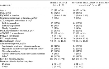
From June 2004 to March 2006, blood was collected for measurement of IGF-1 from 110 patients. Baseline demographics and clinical characteristics of these patients are described in Table 1. The median (IQR) time between ICU admission and study enrollment was 1.9 (1.2–3.3) days. Acute brain dysfunction was common, with 78% delirious and 74% of patients comatose at some point during the 14-day study period. Thirty (25%) patients died during the study period. On the day following IGF-1 measurement, two patients had died and 46 patients were comatose, precluding delirium assessment at that time. Of the 62 patients who were alive and not comatose, and therefore assessed with the CAM-ICU, 40 (64%) were delirious.
Demographics and clinical characteristics of the study population

IQCODE = Short Informant Questionnaire of Cognitive Decline in the Elderly; ADL = activities of daily living; IADL = Instrumental Activities of Daily Living; APACHE II = Acute Physiology and Chronic Health Evaluation II; SOFA = Sequential Organ Failure Assessment; ICU = intensive care unit; IGF-1 = insulin-like growth factor 1.
a Median (interquartile range) unless otherwise noted.
b Clinical and demographic characteristics of patients alive and not comatose on the day after the IGF-1 sample were drawn. These patients were included in the primary analysis to test the association between IGF-1 concentrations and the probability of delirium the day following IGF-1 measurement.
c Patients with an IQCODE score ≥ 4 were considered to have pre-existing cognitive impairment, presumably of mild to moderate severity, because we excluded patients from study enrollment with dementia that prevented them from living independently.
d ADL were defined according to the Katz scale: 0–1 = “independent,” 2–4 = “partial disability,” and 5–6 = “total dependence” (Katz et al., Reference Katz, Ford, Moskowitz, Jackson and Jaffe1963).
e IADL were defined with the Functional Assessment (FAQ), with score ≥ 10 indicating impaired functioning (Pfeffer et al., Reference Pfeffer, Kurosaki, Harrah, Chance and Filos1982).
f Including gastrointestinal bleeding, metabolic disarray, haemoptysis, pulmonary embolism, and status epilepticus.
IGF-1 concentrations were not associated with delirium on the day after IGF-1 measurement, with or without adjustment for sepsis (p = 0.97 and 0.93, respectively) (Table 2, Figure 1). IGF-1 concentrations were also not significantly associated with duration of normal mental state (Table 2) either before or after adjustment for sepsis (p = 0.27 and 0.23, respectively).
Insulin-like growth factor-1 and neurologic outcomesa

a All associations were adjusted for age, severity of illness, and severe sepsis at ICU admission.
b The relationship between insulin-like-growth factor-1 and delirium was nonlinear; thus a single odds ratio cannot estimate this association.
Insulin-like growth factor 1 (IGF-1) and delirium. IGF-1 concentrations were not associated with risk of delirium on the day after IGF-1 measurement (p = 0.97), adjusting for age, severity of illness, and severe sepsis at ICU admission. The solid black line shows the point estimate of the association between IGF-1 and probability of delirium, and the gray ribbon indicates the 95% confidence interval. The rug plot above the x axis shows the distribution of IGF-1 concentrations measured at study baseline.

Discussion
In this pilot investigation, the first to our knowledge to evaluate the relationship between IGF-1 and delirium in the ICU, IGF-1 concentrations early in critical illness were not associated with delirium or duration of normal mental state.
IGF-1 is a hormone produced primarily in the liver when stimulated by growth hormone. Although many of the effects of growth hormone on cell growth are mediated directly through the growth hormone receptor, the hormone also affects body growth and metabolism through stimulation of IGF-1 production. Reinhardt and Bondy (Reference Reinhardt and Bondy1994) demonstrated that liver-derived circulating IGF-1 crosses the blood-brain barrier, after which it can bind IGF-1 receptors believed to be expressed throughout the brain, especially in the superficial and deep cortical layers, olfactory bulb, amygdala, thalamic nuclei and hippocampus. In animal studies, IGF-1 has been shown to regulate stem cell differentiation into neurons (Brooker et al., Reference Brooker, Kalloniatis, Russo, Murphy, Werther and Bartlett2000) and induce neurogenesis of the hippocampus (Aberg et al., Reference Aberg, Aberg, Hedbacker, Oscarsson and Eriksson2000). IGF-1 and its binding proteins have therefore been examined in a variety of brain diseases (e.g. Alzheimer's disease, stroke, and multiple sclerosis) (Trejo et al., Reference Trejo, Carro, Garcia-Galloway and Torres-Aleman2004).
Few studies to date, all conducted outside the ICU and including from 67 to 164 patients, have analyzed IGF-1 as a risk factor for or predictor of delirium (Wilson et al., Reference Wilson, Broadhurst, Diver, Jackson and Mottram2005; Adamis et al., Reference Adamis, Treloar, Martin, Gregson, Hamilton and Macdonald2007; Reference Adamis2009). The lack of an association between IGF-1 and delirium in our investigation is similar to the findings of Lemstra et al. (Reference Lemstra, Kalisvaart, Vreeswijk, van Gool and Eikelenboom2008), who found that preoperative IGF-1 concentrations did not predict the occurrence of postoperative delirium in their study of 68 older hip surgery patients. Other studies, alternatively, have found low IGF-1 to be associated with incident and prevalent delirium in older patients admitted to medical wards (Wilson et al., Reference Wilson, Broadhurst, Diver, Jackson and Mottram2005; Adamis et al., Reference Adamis, Treloar, Martin, Gregson, Hamilton and Macdonald2007; Reference Adamis2009). In contrast to these investigations, we studied a population consisting of critically ill ICU patients. For example, Wilson et al. (Reference Wilson, Broadhurst, Diver, Jackson and Mottram2005) excluded severely ill patients who could not provide consent, a characteristic common during the early days of an ICU stay; Adamis et al. (Reference Adamis, Treloar, Martin, Gregson, Hamilton and Macdonald2007; Reference Adamis2009) excluded intubated patients from their studies. The often multifactorial nature of delirium in the ICU may obscure the role of IGF-1 previously noted, especially given the higher number of predisposing and precipitating risk factors affecting severely ill patients. In light of earlier results, future larger studies including serial measurements of IGF-1 in the ICU are needed before a relationship between this neuroprotective hormone and delirium during critical illness can be ruled out.
Our study has important strengths including prospective, daily neurologic assessments by trained research personnel using a validated delirium assessment tool (i.e. the CAM-ICU) and a validated sedation scale (i.e. the RASS), as well as a study population at high risk for delirium, i.e. critically ill patients, half of whom were older patients.
This study, however, has limitations. We did not measure, for example, other related biomarkers including growth hormone and IGF-binding proteins. High levels of growth hormone may lead to insulin resistance, which has been thought to influence clinical outcomes through alterations of IGF-1 and insulin-like growth factor 1 binding proteins (IGFBP) in critically ill patients (Basi et al., Reference Basi2005). Timmins et al. (Reference Timmins1996) reported low levels of IGFBP-3 in ICU patients. IGFBP-3 is the main IGF-1 binding protein; it carries 90%–95% of the circulating IGF-1 in a ternary complex with acid-labile subunit (Baxter et al., Reference Baxter, Martin and Beniac1989). Low levels of IGFBP-3 are considered to increase the bioavailability of IGF-1 to counteract the effect of insulin resistance and subsequently a hyperglycemic status. IGF-1 actually increases insulin sensitivity, through enhanced peripheral glucose re-uptake and decreased hepatic glucose production (Boulware et al., Reference Boulware, Tamborlane, Rennert, Gesundheit and Sherwin1994). However, high levels of IGF-1 are reported to be responsible for symptomatic hypoglycemia (Zenobi et al., Reference Zenobi, Graf, Ursprung and Froesch1992). The glycemic perturbation might also be related to neurological outcomes. In the current investigation, no data were collected on the glycemic changes during the course of the critical illness.
Studies show that IGF-1 and IGFBP-3 concentrations are low during the course of a critical illness, and these can persist for seven days or more (Timmins et al., Reference Timmins1996; Baxter et al., Reference Baxter, Hawker, To, Stewart and Holman1998). Alternatively, increases in IGF-1 levels during the course of illness have been noted and reported to be a sign of recovery from the critical illness (Timmins et al., Reference Timmins1996; Baxter et al., Reference Baxter, Hawker, To, Stewart and Holman1998); changes in IGF-1 levels over time, therefore, might have a relationship with recovery from delirium. Since we only measured IGF-1 at baseline, we were unable to examine potential associations between changes over time in IGF-1 concentrations and delirium. Because the median ICU length of stay was about a week in our study, we suspect that IGF-1 concentrations were low during the entire course of the ICU stay. Nevertheless, future studies should include serial measurements of IGF-1 and IGFBP-3 to better clarify the role of the growth hormone-IGF-1 axis in the neurological effects of critical illness. Changes in IGF-1 and IGFBP-3 over time might be important determinants of neurological outcomes even if baseline IGF-1 concentrations are not.
Finally, because of the relatively small size of our pilot study, we were limited in the number of covariates we could adjust for in the regression models; the inclusion of additional covariates, such as pre-existing functional and cognitive impairment – both potential risk factors for delirium – would increase the risk of overfitting in the regression models, i.e., increasing the chance of obtaining a significant result due to random error or noise. Future larger studies that adjust for these and other potential confounders as well as include serial measurements of IGF-1 and IGFBP-3 may better clarify the role of the growth hormone-IGF-1 axis in the neurological effects of critical illness.
Conclusions
This pilot study is the first to analyze potential associations between IGF-1 concentrations early in critical illness and delirium in the ICU. No association was found between baseline IGF-1 levels and either delirium or duration of normal mental state in critically ill patients. Future studies, including sequential samples of IGF-1 and its related binding proteins, are needed to further clarify the relationship between the growth hormone-IGF-1 axis and acute neurological outcomes during critical illness.
Conflict of interest
None.
Description of authors' roles
All authors were involved in the study's conception and design, the interpretation of results, revision and final approval of the paper. A. Morandi and T.D. Girard were responsible for the acquisition of data; and J.L. Thompson and A.K. Shintani were responsible for the data analysis.
Acknowledgments
This research was supported by the Saint Thomas Foundation (Nashville, TN) and the National Institutes of Health (RR024975). In addition, Dr. Pandharipande is supported by the VA Clinical Science Research and Development Service (VA Career Development Award), Dr. Ely is supported by the VA Clinical Science Research and Development Service (VA Merit Review Award) and the National Institutes of Health (AG027472), Dr. Girard is supported by the National Institutes of Health (AG034257), and Drs. Ely and Girard are both supported by the Veterans Affairs Tennessee Valley Geriatric Research, Education and Clinical Center (GRECC). Dr Jackson is supported by the National Institute of Health (AG031322).