


Cervical spine injections, including epidural and facet joint injections, are routinely used for the management of neck pain and cervical radiculopathy. Although generally considered safe, each procedure carries distinct but serious risks: epidural injections are more commonly implicated in pneumocephalus via inadvertent dural puncture, whereas facet injections may contribute to direct mechanical cord injury. Pneumocephalus, defined as intracranial air within the ventricular or subarachnoid spaces, is most often seen following trauma or neurosurgery. Reference Schirmer, Heilman and Bhardwaj1 Its occurrence after cervical injections is scarcely documented in the literature, particularly with associated post-procedural stroke-like deficits. We present a case of acute hemiparesis following cervical epidural and facet joint injections with imaging evidence of intracranial pneumocephalus and spinal cord edema, showing rapid improvement with conservative inpatient management. Recognition of this complication is crucial, as its presentation can closely mimic acute cerebrovascular events.
A 56-year-old woman with a history of type 2 diabetes, hypertension and prior myocardial infarction presented to the emergency department with the sudden onset of right-sided weakness. Symptoms occurred immediately following right-sided cervical facet joint injections at C3–4, C4–5 and C5–6 levels and a concurrent cervical epidural injection performed under fluoroscopic guidance at an outpatient pain management clinic for chronic neck pain. She experienced chest pain, somnolence and new right arm and leg weakness with paresthesia. On arrival, her vital signs were within normal limits, and she was alert and oriented with fluent speech, intact comprehension and intact cranial nerves. Initial neurological examination revealed 4/5 strength in the left extremities and variable right-sided weakness, decreasing from 3/5 at the deltoids to 1/5 in finger flexion and extension. The patient also exhibited a wide-based gait, limited by pain and decreased right-sided sensation to light touch and pinprick. Additionally, deep tendon reflexes were absent in the right upper extremity, with a negative Babinski sign.
Initial non-contrast CT scan of the head revealed persistent scattered foci of subarachnoid pneumocephalus, mostly visualized at the bifrontal regions, and no acute intracranial hemorrhage, midline shift or hydrocephalus (Figure 1A). Brain MRI showed no acute infarct or hemorrhage with similar findings of pneumocephalus and mild chronic microvascular ischemic changes (Figure 1B). Additionally, a cervical MRI revealed a long-segment T2-hyperintense cord signal spanning from C3 through the partially visualized T5 level, with no evidence of a cord infarct (Figure 1C). Scattered foci of intrathecal and epidural air were also visualized throughout the spine, with mild nonspecific prevertebral soft tissue edema and moderate spinal canal stenosis at the C6 to C7 level. Comparison with pre-procedural cervical MRI demonstrated pre-existing spinal canal stenosis at C6–C7 without cord signal abnormality, suggesting that the observed T2 hyperintensity represents a new post-procedural finding. Laboratory studies were unremarkable.
(A) Axial non-contrast head CT (bone window) demonstrating scattered foci of intracranial air (arrows) within the bifrontal subarachnoid spaces, consistent with pneumocephalus. (B) Axial susceptibility-weighted imaging (SWI) brain MRI demonstrating corresponding susceptibility artifact (arrows) within the bifrontal subarachnoid spaces, supporting pneumocephalus findings on CT. No evidence of acute infarction or intracranial hemorrhage. (C) Sagittal T2-weighted short tau inversion recovery (STIR) cervical MRI showing a right-sided long-segment intramedullary T2-hyperintense signal abnormality (bottom arrow) extending from C3 through the partially visualized T5 level. Additional scattered foci of intrathecal and epidural air are present within the spinal canal (top arrow).
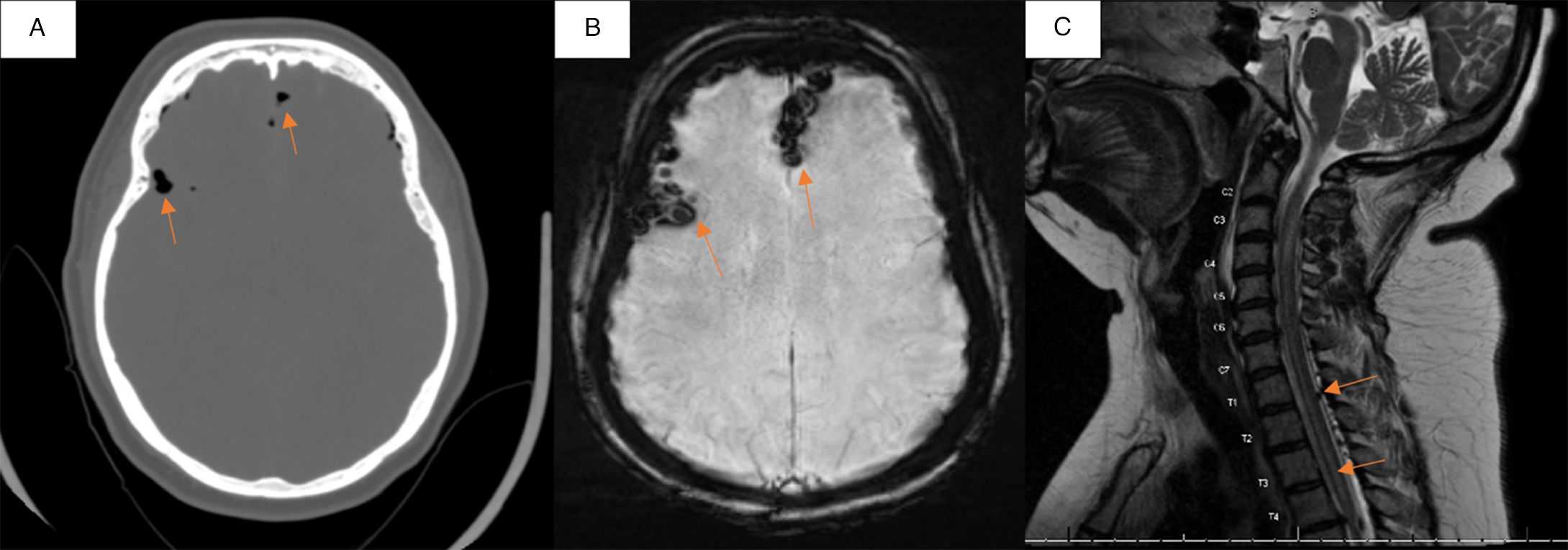
Figure 1. Long description
Panel A: An axial non-contrast head CT scan in bone window setting showing scattered foci of intracranial air within the bifrontal subarachnoid spaces, indicated by arrows. Panel B: An axial susceptibility-weighted imaging (SWI) brain MRI demonstrating corresponding susceptibility artifact within the bifrontal subarachnoid spaces, also indicated by arrows, supporting pneumocephalus findings on CT. No evidence of acute infarction or intracranial hemorrhage is visible. Panel C: A sagittal T2-weighted short tau inversion recovery (STIR) cervical MRI showing a right-sided long-segment intramedullary T2-hyperintense signal abnormality extending from C3 through the partially visualized T5 level, indicated by the bottom arrow. Additional scattered foci of intrathecal and epidural air are present within the spinal canal, indicated by the top arrow.
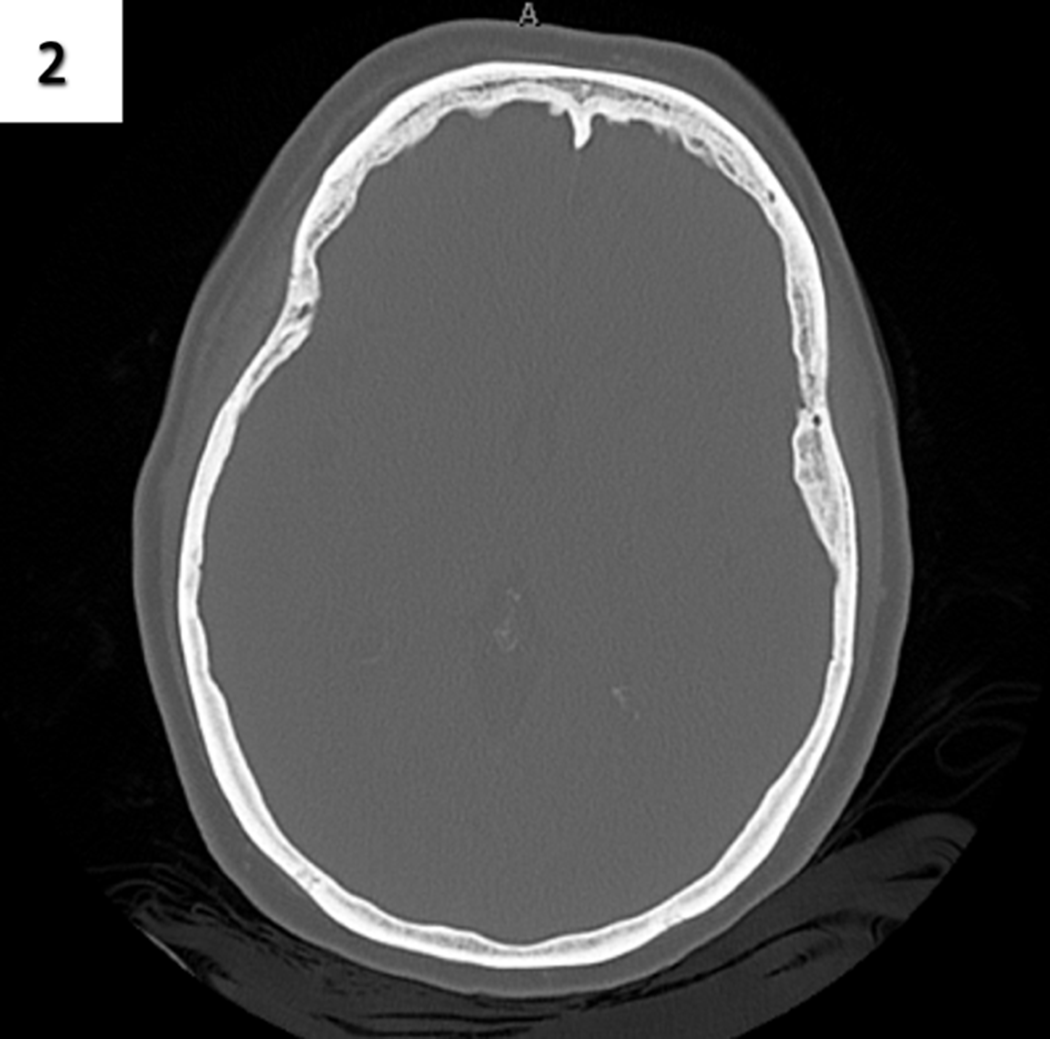
The patient was managed conservatively with high-flow oxygen via a non-rebreather mask, head elevation and intravenous dexamethasone tapered over several days. On postadmission day 4, the patient’s neck and back pain improved to a 7/10 in severity from a baseline of 10/10, and she began ambulating independently. She continued to complain of intermittent shooting pain down her left arm, although it was mildly improving. The patient’s right-sided weakness persisted, particularly in the distal right upper extremity (wrist flexion/extension 2/5, finger flexion/extension 1/5, absent reflexes, decreased sensation). However, she regained near-baseline motor strength in all other extremities. A repeat brain CT on day 5 (Figure 2) showed complete resorption of air and resolution of the pneumocephalus. The patient was discharged to an acute inpatient rehabilitation facility to continue improving her right-sided motor deficits and regaining functional independence.
Follow-up non-contrast head CT (bone window) obtained 5 days after presentation demonstrating complete resolution of the previously identified pneumocephalus.
Pneumocephalus following cervical epidural injection is thought to result from inadvertent dural puncture with air entry during the loss-of-resistance (LOR) technique, allowing air to enter the subarachnoid or subdural compartment and migrate intracranially along pressure gradients. Reference Das, Munakomi and Bajaj2 The air typically collects anteriorly due to positioning and gravity, producing mild symptoms such as headache or dizziness, though larger volumes may cause severe manifestations via mass effect or cord ischemia. Reference Abbasi, Malhotra and Malanga3 The concurrent cervical cord T2 hyperintensity in our case suggests additional mechanisms, including mechanical trauma, inflammation or air-induced mass effect. The facet injections may have contributed to direct cord injury via misdirected needle placement or periarticular trauma, while the epidural injection may have produced local ischemia secondary to transient pressure changes. This multifactorial etiology likely explains the patient’s transient neurologic deficits and gradual clinical improvement.
Management of pneumocephalus typically involves high-concentration oxygen therapy to accelerate the resorption of intracranial air, steroids to address inflammation and supportive measures, as most cases resolve spontaneously. Data comparing hyperbaric oxygen therapy (HBOT) and high-flow normobaric oxygen use in such clinical scenarios remain limited. A 2014 review by Paiva et al. reported faster resolution of pneumocephalus with HBOT but found no difference in neurologic outcomes when compared with 100% FiO2 via a non-rebreather mask. Reference Paiva, de Andrade and Figueiredo4 Additionally, a recent systematic review identified only three relevant studies, all using different oxygen delivery methods, and none directly comparing HBOT with high-flow oxygen for pneumocephalus management. Reference Kim, Ji and Chen5 These findings highlight a gap in the literature, particularly for complex presentations with concurrent cord abnormalities, where management decisions are largely guided by clinical judgment rather than robust evidence. Our patient improved steadily with high-flow oxygen delivered via a non-rebreather mask, the more accessible and cost-effective option.
To our knowledge, reports describing concurrent intracranial pneumocephalus and cervical cord signal abnormality presenting as a stroke mimic are limited, and no clear consensus exists regarding optimal management in such cases. We present a rare case of this entity following cervical epidural and facet injections, manifesting as acute hemiparesis mimicking stroke. While both complications have been individually described, their simultaneous presentation as a stroke mimic remains poorly characterized. This case highlights the importance of maintaining a broad differential when evaluating focal neurologic deficits after spinal procedures and recognizing the potentially multifactorial etiology. Furthermore, the absence of established guidelines for managing complex pneumocephalus with concurrent cord abnormalities underscores a critical gap in the literature. Early recognition and conservative management may lead to favorable outcomes while avoiding unnecessary interventions.
Acknowledgments
The authors thank the patient for granting permission to share her case. The authors declare no conflicts of interest and received no financial support for this work.
Author contributions
JA led the case conception and coordination, had direct involvement in the patient’s clinical care and drafted the initial manuscript. AS contributed to manuscript writing and literature review. RDO provided critical revisions and editorial feedback. MA served as the attending physician overseeing the patient’s care and provided clinical supervision and intellectual input. All authors reviewed and approved the final manuscript.

 Open access

