Introduction
Following fertilization the oocyte completes meiosis division and immediately enters into the mitotic cell-cycle process. Meiotic control machinery functions as an internal pacemarker that propels oocytes toward embryogenesis (Stitzel and Seydoux, Reference Stitzel and Seydoux2007). During the transition from matured oocyte to 1-cell embryo, mRNA transcripts employed during oocyte maturation are selectively and gradually degraded. Although, the new transcriptional activity can be detected as early as the pronuclear stage of the 1-cell stage, the synthesis of embryonic proteins occurs shortly after the first mitotic division, and another wave of translation follows the S-phase in the 2-cell stage embryo (for review Corry et al., Reference Corry, Tanasijevic, Barry, Krueger and Rasmussen2009). Thus all essential processes required for early embryonic development are controlled by maternally inherent checkpoint machinery.
Entry into mitosis is immediately induced and catalyzed by the kinase activity of cyclin-dependent kinase 1 (CDK1) in a complex with cyclin B (also referred to as MPF factor = M-phase promoting factor; Nurse, Reference Nurse1990). The highly conserved complex of CDK1-cyclin B triggers nuclear envelope breakdown, chromosome condensation and mitotic spindle assembly. In addition, it has been shown recently that low activity of CDK1-cyclin B is indispensable for pronuclear union occurring during the first embryonic cell cycle after fertilization (Tachibana et al., Reference Tachibana, Hara, Hattori and Kishimoto2008). These new observations reveal the involvement of CDK1 in pronuclear congression and pronuclear fusion (karyogamy) after fertilization. These data were confirmed recently by Gavet & Pines (Reference Gavet and Pines2010), who revealed the link between CDK1-cyclin B and reorganization of the nucleus and cytoplasm at mitotic entry.
The preimplantation embryo is dependent on signals generated by growth factors produced by the embryo itself or present in the maternal environment. The developing embryo expresses a number of growth factor receptors known to activate the phosphatidylinositol 3-kinase (PI3K) pathway, including insulin and insulin-like growth factor-I receptors (Lighten et al., Reference Lighten, Hardy, Winston and Moore1997).
Protein kinase B (Akt/PKB) was primarily identified as a serine-threonine kinase that functions downstream of PI3K (Bellacosa et al., Reference Bellacosa, Sakaue, Nedachi, Kovacina, Clayberger, Conti and Roth1991), and activation of Akt/PKB is thought to be a critical step in the PI3K signalling pathway. It was shown later that the downstream signalling protein kinase B plays a transduction role in multiple cellular signals targeted on initial activation of CDK1-cyclin B complex (Okumura et al., Reference Okumura, Fukuhara, Yoshida, Hanada, Koyutsumi, Mori, Tachibana and Kishimoto2002). Thus, kinase Akt/PKB acts as a cell relay at cell entry into the mitotic phase. In this context, it was shown on the mouse oocyte that Akt/PKB activation precedes breakdown of the oocyte's nucleus, referred to as germinal vesicle breakdown (GVBD), and that inhibition of the PI3K-Akt/PKB pathway prevents both CDK1 activation and resumption of meiosis (Kalous et al., Reference Kalous, Solc, Baran, Kubelka, Schultz and Motlik2006). Similarly to oocyte maturation events, Akt/PKB signalling is present also after fertilization (Riley et al., Reference Riley, Carayannopoulos, Wyman, Chi, Ratajczak and Moley2005) in the mouse preimplantation embryo, in which the activity of Akt/PKB is critical for glucose metabolism and embryo survival (Riley et al., Reference Riley, Carayannopoulos, Wyman, Chi and Moley2006). In addition, it was shown that Akt/PKB causes the activation of MPF and promotes development of the 1-cell stage mouse embryo by inducing Akt-dependent phosphorylation of CDC25B phosphatase (Feng et al., Reference Feng, Yu, Liu, Zhang, Zong, Su, Zhang, Yu, Sun and Yu2007).
The molecular processes of G2/M transition have been extensively studied on somatic cells but some key aspects of the molecular mechanisms involved in the first embryonic cell cycle are less known. It has been shown that kinase Akt/PKB downregulates the expression of pro-apoptotic proteins (Kops & Burgering, Reference Kops and Burgering1999; Kops et al., Reference Kops, deRuiter, deVries-Smith, Powell, Bos and Burgering1999; Brunet et al., Reference Brunet, Park, Tran, Hu, Hemmings and Greenberg2001) and thus blocks phosphorylation and activation of the key components in the apoptotic cascade (Kim et al., Reference Kim, Khursigara, Sun, Franke and Chao2001), by which promotes cell survival. Moreover, activated Akt/PKB kinase can phosphorylate a wide range of downstream targets, many of which are required for normal cell survival. In this respect, this knowledge provides important guidance in selecting chemotherapeutic agents for Akt/PKB deregulation in cancer treatment (Shi et al., Reference Shi, Liu, Han, Guan, Shoemaker, Oleksijew, Woods, Fisher, Klinghofer, Lasko, McGonidal, Li, Rosenberg, Giranda and Luo2005). This multiplicity of downstream effectors provides an idea of the wide range of mechanisms through which ligand activation of PI3K may induce cellular response in the embryo (for a review see O'Neill, Reference O'Neill2008a). Many studies on somatic cancer cells (Hemstrom et al., Reference Hemstrom, Sandstrom and Zhivotovsky2006; Chao et al., Reference Chao, Zao, Xiao-Yi, Li-Jun and Tao2010; Mikami et al., Reference Mikami, Zhang, Hirata, Okamoto, Koizumi, Shimizu, Jablons and He2010) as well as embryonic cells (Li et al., Reference Li, Chandrakanthan, Day and O'Neill2007) have shown that inhibition of the PI3K pathway leads to the induction of apoptosis. In the previous studies, PI3 kinase inhibitor (LY-294002) was applied, but the impact of selective inhibition of other kinases employed in the has not been studied in cleaving mammalian embryos. In this study we show that protein kinase Akt/PKB plays an important role during entry of the 1-cell mouse embryo into the first mitosis, and functions as a relay which is responsible for mediating an anti-apoptotic signal after fertilization.
Materials and methods
Embryo collection and culture conditions
Six- to 8-week-old female mice of the C57BL/6J strain (Velaz, Prague, Czech Republic) were super-ovulated with 5 IU of pregnant mare's serum gonadotropin (eCG; Folligon, Intervet International, Boxmeer, Holland) followed by 5 IU human chorionic gonadotropin (hCG; Pregnyl, Organon, Oss, Holland) 46 h later, and were mated with males of the BALB/c strain (Velaz). All animal experiments were reviewed and approved by the Ethical Committee for animal experimentation at the Institute of Animal Physiology, approved by the State Veterinary and Food Administration of the Slovak Republic, and were performed in accordance with Slovak legislation based on Directive 86/609/EEC on the protection of animals used for experimental and other scientific purposes. One-cell embryos were collected by flushing the oviducts 17 h post hCG administration. The cumulus cells were removed with 0.25% hyaluronidase and immediately cultured in mouse embryo potassium simplex optimization medium (KSOM; Millipore) under 5% CO2 in air at 37.5°C until fixation. The collected early 1-cell embryos were divided randomly into control and inhibited batches. Akt/PKB activity was inhibited in a medium supplemented with specific SH-6 inhibitor at final concentration 50 μM (Enzo Life Sciences). In order to inhibit CDK1, the culture medium was supplemented with roscovitine at final concentration 25 μM (Enzo Life Sciences). Freshly isolated embryos (17 h post hCG administration) as well as embryos cultured in vitro for 12 or 24 h were briefly washed in phosphate-buffered saline (PBS) and immediately fixed in 4% (w/v) paraformaldehyde dissolved in PBS for 45 min at room temperature (RT). Fixed embryos were used for immunocytochemistry staining or cell death assay. In the case of vital staining with propidium iodide, the embryos were fixed in 4% (w/v) paraformaldehyde after the first staining with propidium iodide (10 μg/ml, Sigma-Aldrich).
Immunocytochemistry
Fixed embryos were washed in PBS and free aldehydic groups were blocked with 50 mM ammonium chloride in PBS at RT for 30 min. After washing in PBS containing 0.25% bovine serum albumin (PBS/BSA), the embryos were permeabilized with 0.5% (w/v) Triton-X100 in PBS for 15 min at RT and pre-incubated with inactivated 2% normal goat serum in PBS/BSA for 60 min at RT. After brief washing in PBS/BSA the embryos were separately incubated with anti-Ser473Akt/PKB (overnight at 4°C) followed by incubation with primary antibody anti-pericentrin (3 h at RT) diluted in PBS/BSA. After exhaustive washing (in PBS/BSA) affinity-purified secondary antibodies coupled with fluorescent dye fluorescein isothiocyanate (FITC) or Texas Red were applied for 60 min. In the double-staining experiments, both primary antibodies were employed separately followed by detection with fluorescent immunoconjugates applied simultaneously. Chromatin was stained with standard 4′,6-diamidino-2-phenylindole (DAPI; Sigma-Aldrich) staining procedure. Ser473-phosphorylated protein kinase Akt/PKB was detected using rabbit polyclonal antibody pAkt (Ser473)-R (Santa Cruz Biotechnology, Inc.) diluted 1:100. The protein marker of pericentriolar matrix – pericentrin was detected using mouse monoclonal antibody anti-Pericentrin (BD Biosciences Pharmingen) at dilution 1:40–50.
Cell death assay
Immediately after 12 h culture with or without the inhibitor, mouse embryos were divided randomly in two groups, subjected to two different staining protocols (Fabian et al., Reference Fabian, Juhás, Ilkova and Koppel2007) and evaluated under a fluorescence microscope (BX50 Olympus, Japan) for the presence of specific features of apoptotic or necrotic cell death.
Terminal deoxynucleotidyl transferase dUTP nick end labelling (TUNEL)/propidium iodide/Hoechst 33342 fluorescence staining
Vital embryos were first stained with propidium iodide (PI; 10 μg/ml; Sigma–Aldrich; 5 min at 37°C), which stains DNA of dead cells only, and then fixed with 4% paraformaldehyde in PBS for 60 min at RT. Fixed embryos were washed, permeabilized with 0.5% (w/v) Triton X-100 for 60 min and then incubated in 10 μl of terminal deoxynucleotidyl transferase and 90 μl of fluorescein-conjugated dUTP (TUNEL, In Situ Cell Death Detection Kit; Roche, Penzberg, Germany) for 60 min at 37°C stored in a dark box. After the TUNEL reaction, nuclear chromatin in all embryos was stained with Hoechst 33342 at concentration 20 μg/ml (Sigma–Aldrich) for 5 min at RT. In positive staining controls, several embryos were pre-incubated in deoxyribonuclease I (DNase I; 100 U/ml; Invitrogen) for 30 min at 37°C before the TUNEL reaction.
TUNEL/caspase-3/Hoechst 33342 fluorescence staining
Fixed embryos were washed and permeabilized in 1% (w/v) Triton X-100 for 2 h at RT and subjected to the TUNEL reaction (see above). TUNEL-reacted embryos were then blocked for 4 h in PBS containing 5% normal goat serum and 0.1% Triton X-100. Washed embryos were incubated overnight with a primary polyclonal rabbit antibody against active caspase-3 (cleaved caspase-3 (Asp175), Cell Signaling Technology) followed by incubation in Texas Red-conjugated secondary antibody for 90 min. Finally, embryos were extensively washed in 0.3% Triton X-100 in PBS and stained with Hoechst 33342 stain (20 μg/ml). Immunocytochemical negative control staining was performed by pre-absorption of the primary antibody with specific cleaved caspase-3 (Asp175) blocking peptide (Cell Signaling Technology).
Statistical analysis
Standard chi-squared tests were used to detect differences in developmental abilities (stage-distribution) of embryos between control and experimental groups. P-values < 0.05 were considered to be significant.
Results
Embryo development
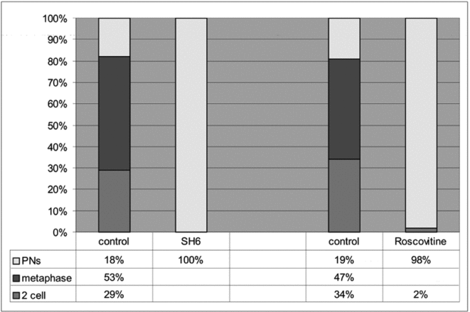
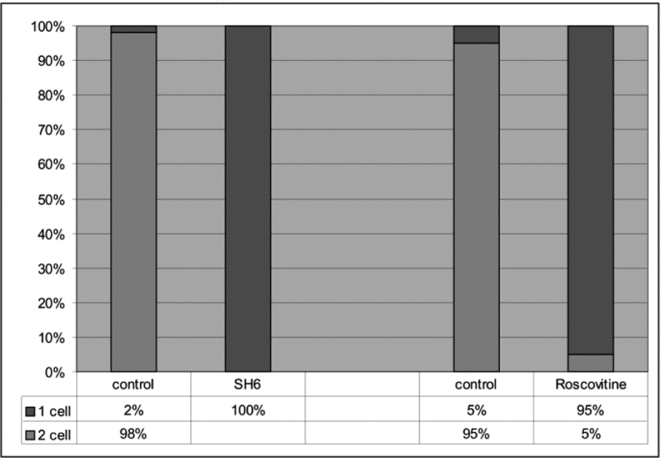
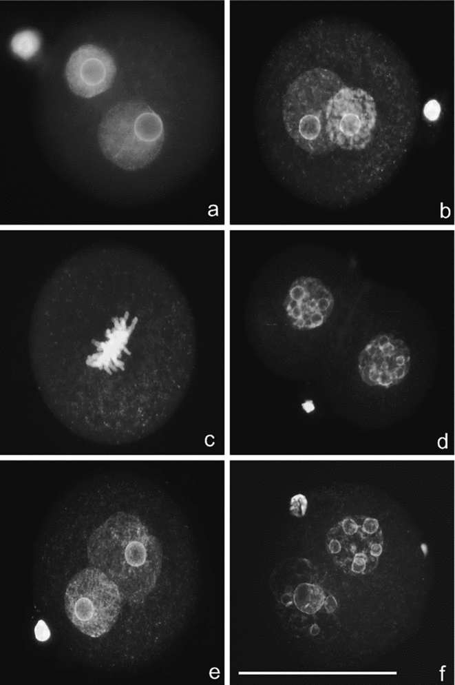
One-cell mouse embryo development following culture either with SH-6 (inhibitor Akt/PKB) or roscovitine (inhibitor CDK1) was monitored by stereomicroscopic imaging of live cells during 1-cell embryo stage and evaluated also on fixed embryos after DAPI staining procedure. As shown in Figs. 1 and 2, the development of 1-cell embryos was arrested when the embryos were cultured in SH6-containing medium or in roscovitine-containing medium. These embryos were not able to enter into the mitotic cleavage. After 12-h culture (Fig. 1), the percentage of control embryos that arrived at metaphase was 53 or 47%, respectively. The incidence of 2-cell embryos in the control groups was 29 or 34%, respectively. In the overall population of the control embryos, the percentage of arrested embryos in the 1-cell stage (before entry into mitosis) was 18 or 19%, respectively. In the control embryos, DAPI staining showed standard appearance of pronuclear bodies (Fig. 3a,b), metaphase arranged chromosomes (Fig. 3c) and normal nuclei of 2-cell embryos (Fig. 3d). In inhibited embryos (Fig. 1), all (100%) embryos cultured in the SH6-containing medium and 98% of embryos cultured in the roscovitine-containing medium were developmentally arrested before the first embryonal cleavage. Only 2% of the embryos incubated with roscovitine were cleaved into the 2-cell stage. DAPI staining revealed that the embryo development was stopped during the pronuclear stage. In roscovitine-inhibited embryos, the pronuclei displayed a round shape without disturbances of the pronuclear envelope. Chromatin was arranged homogeneously throughout the pronuclear bodies (Fig. 3e). In SH6-inhibited embryos, the pronuclei usually displayed changes in chromatin structure and pronuclear shape when compared with control embryos or embryos cultured in the roscovitine-containing medium. In the SH6-inhibited embryos, chromatin texture was usually more condensed, the roundish shape of pronuclei was disturbed and pronucleolar disintegration was observed (Fig. 3f). After 24-h culture (Fig. 2), the percentage of control embryos, which arrived at the 2-cell stage, was 98 or 95%, respectively. In inhibited embryos, all (100%) embryos treated with SH-6 and 95% of embryos treated with roscovitine were stopped in the 1-cell stage. Only 5% of embryos cultured in roscovitine-containing medium achieved the 2-cell stage.
Mouse embryos (control and treated with inhibitor SH6 or roscovitine) scored for the first cleavage after 12-h culture in vitro. PNs, pronuclei.

Mouse embryos (control and treated with inhibitor SH6 or roscovitine) scored for the first cleavage after 24-h culture in vitro.
Mouse 1-cell embryos stained with 4′,6-diamidino-2-phenylindole (DAPI). Control 1-cell embryos (a–c) standard appearance of pronuclear bodies (a,b), metaphase arranged chromosomes (c); and control normal nuclei of 2-cell embryo (d). Embryo treated with inhibitor roscovitine (e) or SH6 (f). Bar = 50 μm.
Ser473-Akt/PKB localization
Immediately after isolation of 1-cell embryos at 17 h post hCG administration, both male and female pronuclei were visible in the periphery of the 1-cell embryo (Fig. 4a). The pronuclei displayed decondensed chromatin and they were localized in opposite peripheral regions. Akt/PKB kinase phosphorylated on serine 473 (Ser473-Akt/PKB) was not detected by immunocytochemistry at this developmental stage (Fig. 4e). After 12-h cultivation in control medium, the pronuclei were localized in the central region of the cell (Fig. 4b). Ser473-Akt/PKB was detected in both male and female pronuclei of the control embryos (Fig. 4f – arrows). In the experimental group with embryos cultured in medium supplemented with inhibitor SH6 for 12 h, Ser473-Akt/PKB was not detected immunocytochemically either in male or female pronuclei (Fig. 4g). In the experimental group with embryos cultured in medium supplemented with inhibitor roscovitine for 12 h, Ser473-Akt/PKB was positively detected in both male and female pronuclei (Fig 4h – arrows). Pericentrin (centrosomal protein marker) was positively immunostained in all experimental groups. In the control embryos, fixed and immunostained at 17 h post hCG administration, pericentrin-positive foci were arranged loosely throughout the cytoplasm (Fig. 4i – arrow). After 12-h cultivation, the pericentrin-positive foci were localized closely around both male and female pronuclei in both control (Fig. 2j) and also inhibited embryos with SH6 (Fig. 4k) or roscovitine (Fig. 4l). Immunosignal of Ser473-Akt/PKB was not detected in the pericentrin-positive foci in either control or inhibited embryos.
4′,6-Diamidino-2-phenylindole (DAPI) staining (blue) and double immunostaining of Ser473-Akt/PKB (green) and pericentrin (red) in control freshly isolated (17 h post hCG) embryos (a, e, i) or control embryos cultured for 12-h in pure medium (b, f, j) and also embryos cultured for 12-h in medium supplemented with inhibitor SH6 (c, g, k) or inhibitor roscovitine (d, h, l). Bar = 50 μm. Arrows (c,f,h) = pronuclei, (i) = pericentrin-positive focus.
Cell death assay
The specificities of TUNEL and active caspase-3 immunostaining were confirmed by positive and negative control staining. Pre-incubation of 1-cell embryos from additional cultures with DNase I led to clear and uniform positive TUNEL in all of their nuclei (Fig. 5b – arrows). Moreover, the apparent TUNEL reaction was regularly observed in polar bodies localized in the perivitelline space (Fig. 5b – arrowhead). The pre-adsorption of the primary antibody with corresponding immunizing peptide led to significant reduction of caspase-3 immunostaining signal (Fig. 5f).
Cell death assay on control 1-cell mouse embryos. Positive staining control embryo pre-incubated with DNase I (a–c) stained with Hoechst 33342 (a) arrows; terminal deoxynucleotidyl transferase dUTP nick end labelling (TUNEL) assay (b) arrows; and propidium iodide (c). Positive TUNEL in polar body (b) arrowhead. Negative control staining (d–f) on embryos stained with Hoechst 33342 (d), TUNEL (e) and immunostaining of active caspase-3 (f). Control embryos with SH6 treatment (g–i) stained with Hoechst 33342 (g), TUNEL (h) and propidium iodide (i). Control embryos with roscovitine treatment (j–l) stained with Hoechst 33342 (j), TUNEL (k) and immunostaining of active caspase-3 (l). Bar = 50 μm.
In the control groups for roscovitine (n = 76) and for SH6 (n = 83), no signs of cell death were observed. All control 1-cell (Fig. 5g, 5j) and 2-cell embryos (not shown) showed normal nuclear morphology and absence of TUNEL (Fig. 5h, k) or PI staining (Fig. 5i) in the nucleoplasm. Only 12 1-cell embryos (from the total number of immunostained embryos, n = 41) showed weak signal for active caspase-3 in the nucleoplasm (slightly distinct from background staining) (Fig. 5l).
In the SH6 group (n = 83), frequent incidence of cell death was observed. The majority of embryos showed various features of apoptosis or secondary necrosis (n = 57). Although Hoechst 33342 stain revealed usually non-apoptotic nuclear morphology in them (Fig. 6a, d), some 1-cell embryos showed massive nuclear fragmentation (n = 2). A relatively high number of embryos (n = 23) showed the presence of TUNEL in their pronuclei or in their cytoplasm (Fig. 6b, e – arrows). In some cases, degraded DNA also showed weak PI staining (Fig. 6c – arrows). Finally, 20 1-cell embryos (from the total number of immunostained embryos, n = 42) showed strong staining signal for active caspase-3 in their nucleoplasm (Fig. 4f – arrow) or cytoplasm. Furthermore, in another 12 embryos, weak immunostaining signal was observed. Eight of the total SH6-treated embryos showed typical features of necrosis, represented by cytoplasmic shrinkage, pycnotic nuclear morphology and strong TUNEL (in both nucleoplasm and cytoplasm) accompanied (if stained) with nuclear PI-staining (not shown).
Cell death assay on 1-cell mouse embryos treated with inhibitor SH6 (a–f) and with roscovitine (g–l). Hoechst 33342 staining (a, d, g, j), terminal deoxynucleotidyl transferase dUTP nick end labelling (TUNEL) assay (b, e, h, k) and propidium iodide staining (c, i) or immunostaining of active caspase-3 (f, l). Arrows – positive TUNEL (b, e), positive propidium iodide staining (c), and positive immunostaining of active caspase-3 (f). Bar = 50 μm.
Conversely, in the roscovitine group (n = 79), the incidence of cell death features was low. All treated embryos showed normal nuclear morphology (Fig. 6g, j), absence of TUNEL (Fig. 6h, k) and absence of PI staining (Fig. 6i). Seven embryos (from the total number of immunostained embryos, n = 41) showed the presence of active caspase-3 in their nucleoplasm (Fig. 6l), and another 17 embryos showed weak staining signal in their pronuclear area.
Discussion
In this study, we report that selective inhibition of Akt/PKB (with SH6) and CDK1 (with roscovitine) during the 1-cell stage of the mouse embryo arrested the embryo development before entry into the first mitosis. In the Akt/PKB-inhibited 1-cell embryos, significant occurrence of apoptosis was observed when compared with the CDK1-inhibited 1-cell embryos, in which apoptotic cell death features were almost completely absent. Thus, Akt/PKB seems to be the primary target responsible for mediating anti-apoptotic signals at the 1-cell mouse embryo stage. In the experiments, the working concentration of inhibitor SH-6 was 50 μM. Tomek & Smiljakovic (Reference Tomek and Smiljakovic2005) showed that SH-6 concentration lower than 20 μM had no effect, whereas a SH-6 concentration higher than 75 μM had a cytotoxic effect. In the case of CDK inhibition, Mermillod et al. (Reference Mermillod, Tomanek, Marchal and Maier2000) showed a that lower concentration (12.5 μM) of roscovitine arrested oocytes during metaphase I. Conversely, at higher doses of roscovitine (from 25 μM) the oocytes do not resume meiosis but they are blocked at the germinal vesicle stage. In this case, the oocytes were able to maintain an intact nuclear (germinal vesicle) membrane (Saskova et al., Reference Saskova, Solc, Baran, Kubelka, Schultz and Motlik2008).
Akt/PKB is known as a downstream factor of phosphoinositide 3-kinase (PI3K) and an upstream regulator of cyclin-dependent kinase 1 (CDK1). It has been shown that Akt/PKB is involved in mouse oocyte meiosis (Kalous et al., Reference Kalous, Solc, Baran, Kubelka, Schultz and Motlik2006). Our results suggest a key role of Akt/PKB at the entry of fertilized mouse oocytes into the first mitotic division. The PI3K – Akt/PKB pathway is a well known link that mediates cell survival signals in which Akt/PKB kinase plays an important effector role in the machinery. Activated Akt/PKB phosphorylates a wide range of downstream targets, many of which are required for normal cell survival (Downward, Reference Downward2004). Conversely, the cleaving early embryo shows apparent autonomy from external survival factors. This phenomenon is apparent during the culture of embryos in vitro, when they develop in media free of hormones or growth factors (Li et al., Reference Li, Chandrakanthan, Day and O'Neill2007). Embryonic developmental autonomy might be explained by the activity of trophic ligands released by the embryo itself (O'Neill, Reference O'Neill2008b).
It has been shown in mouse oocytes that phosphorylation of Akt/PKB on serine473 is located on the nuclear membrane, and that active Ser473Akt/PKB acts on the centrosomes shortly before GVBD. In addition to phosphorylation of Akt/PKB on serine473, Akt/PKB kinase is phosphorylated on threonine308 (Kalous et al., Reference Kalous, Solc, Baran, Kubelka, Schultz and Motlik2006). Thr308Akt/PKB was detected (by immunofluorescence) on the oocyte centrosomes alone. On the basis of the data, Kalous et al. (Reference Kalous, Solc, Baran, Kubelka, Schultz and Motlik2006) assumed that Thr308Akt/PKB is not phosphorylated on the nuclear membrane but rather on the centrosome, or Thr308Akt/PKB is phosphorylated on the nuclear membrane similarly to Ser473Akt/PKB, but then very rapidly translocated to the centrosome. Interestingly, the immunostaining experiments presented in this study, did not reveal presence of Ser473Akt/PKB in the centrosomes (detected by immunostaining to pericentrin) arranged around both male and female pronuclei. In this context, both Ser473Akt/PKB and Thr308Akt/PKB phosphorylated forms were detected in the centrosomes of control embryos as late as the pronucleus envelope breakdown, during the prometaphase and metaphase stages of the first mitosis (not shown). This finding implies that phosphorylated Akt/PKB kinase is translocated from pronuclei to centrosomes during or after the pronucleus envelope breakdown, at the time of formation of the mitotic spindle. In the experimental group of embryos in which Akt/PKB or CDK1 was inhibited by SH-6 or roscovitine, the process of pronucleus envelope breakdown and spindle assembly was stopped. Those embryos inhibited by inhibitor SH6 underwent apoptotic cell death in contrast with the embryos inhibited by roscovitine.
Cell death assays showed that control embryos were vital and revealed no signs of cell death. Occasional presence of weak signal for active caspase-3 in the nucleoplasm of several control 1-cell embryos might reflect the presence of some basal activity of this enzyme, which was previously documented in intact oocytes and zygotes (Papandile et al., Reference Papandile, Tyas, O'Malley and Warner2004). As hypothesized by other authors, this activity could be unrelated to cell death (Martinez et al., Reference Martinez, Rienzi, Iacobelli, Ubaldi, Mendoza, Greco and Tesarik2002). Nevertheless, the unspecific affinity of primary antibody to unknown protein in pronuclear area cannot be excluded either. In this context, the presence of active caspase-3 only (without any other apoptotic features) in a low number of roscovitine-treated embryos does not give exact proof that these embryos were undergoing apoptotic cell death. However, the possibility of their so doing should not be excluded, as the process could be simply delayed due to growth arrest, and other signs of apoptosis could be manifested later (Fabian et al., Reference Fabian, Bukovska, Juhas and Koppel2009). On the other hand, SH6-treated embryos showed relatively frequent occurrence of cell death. In the majority of these 1-cell embryos, typical features of apoptosis were recorded–nuclear fragmentation, TUNEL positivity and the presence of active caspase-3 in both the cytoplasm and the nucleoplasm. Occurrence of untypical cytoplasmic localization of TUNEL might appear slightly confusing; however, cytoplasmic TUNEL has also been observed in other embryonic, mesenchymal or epithelial cells. It probably represents leakage of DNA fragments out of the nucleus of the apoptotic cells during advanced phases of the apoptotic process (Wijsman et al., Reference Wijsman, Jonker, Keijzer, van de Velde, Cornelisse and van Dierendonck1993; Wheeldon et al., Reference Wheeldon, Williams, Soames, James and Roberts1995; Rojo & Gonzalez, Reference Rojo and Gonzalez1998; Perez et al., Reference Perez, Tao and Tilly1999), and it might be connected with the onset of secondary necrosis, which is the typical terminal stage of apoptosis in embryonic cells (Fabian et al., Reference Fabian, Koppel and Maddox-Hyttel2005). A small portion of SH6-treated 1-cell embryos apparently reached this autolytic stage even after 12 h, since they presented typical signs of necrosis (cell shrinkage, pycnosis and membrane damage).
Evidently, the PI3K pathway may be implicated in the suppression of apoptosis mediated with growth factor signalling, as presented in the functional importance in maintaining glucose metabolism and survival of the early preimplantation embryo. In addition, Akt/PKB plays an important role in the mammalian cell cycle as an mitosis-phase initiator (G2/M transition) and serves as a cell-cycle relay. In our study, similarly to SH6 treatment, roscovitine inhibition also effectively blocked initiation of the first mitosis and resulted in undulation of pronuclear membranes, similar to that observed by Lonergan et al. (Reference Lonergan, Faerge, Maddox-Hyttel, Boland and Fair2003) on bovine maturating oocytes. Conversely, the induced process of apoptosis was not observed in roscovitine-treated embryos in comparison with those treated with inhibitor SH6. In this respect, it was established earlier that roscovitine, a potent inhibitor of M-phase factor, was been able to maintain cattle oocytes at the GV stage without affecting their developmental potential (Mermillod et al., Reference Mermillod, Tomanek, Marchal and Maier2000). Reversibility of the inhibitory effect on porcine oocytes (Marchal et al., Reference Marchal, Tomanek, Terqui and Mermillod2001) suggests the idea that roscovitine is not responsible for initiation of the apoptosis machinery.
In earlier studies it was shown repeatedly that PI3 kinase inhibitor LY294002 primarily induced apoptotic cell death (e.g. Riley et al., Reference Riley, Carayannopoulos, Wyman, Chi and Moley2006), but it is very likely that this inhibitor influences a wider range of kinases related to the PI3K cascade (Vlahos et al., Reference Vlahos, Matter, Hui and Brown1994). In addition, the LY294002 inhibitory effects block phosphorylation of Akt/PKB (Hosino et al., Reference Hosino, Yokoo, Yoshida, Sasada, Matsumoto and Satao2004) in dependence on the concentration used (O'Neill, Reference O'Neill2008a). Side effects of inhibitor LY294002 must therefore be taken into consideration (Schied & Woodget, Reference Schied and Woodget2003). Although Akt/PKB can also phosphorylate also a wide range of downstream targets (CDC25B, MPF, GSK3) responsible for cell survival, Akt/PKB itself acts as a downstream effector of the PI3K-Akt/PKB cascade that regulates fundamental cellular functions such as transcription, translation, growth and survival. Selective specific inhibition of Akt/PKB by SH6 inhibitor (Kozikowski et al., Reference Kozikowski, Sun, Brognard and Dennis2003) and of CDK1 by roscovitine inhibitor (Meijer et al., Reference Meijer, Borgne, Mulner, Chong, Blow, Delcros and Moulinoux1997) suggested that Akt/PKB is the pivotal kinase mediating the anti-apoptotic inductors targeted on the PI3K-Akt/PKB-CDK1 cascade. Thus, our results demonstrate the key importance of Akt/PKB in preimplantation embryo survival. The SH6 inhibitor has been shown to prevent Akt/PKB activation on cycling somatic cells (Kozikowski et al., Reference Kozikowski, Sun, Brognard and Dennis2003; Chrysis et al., Reference Chrysis, Zaman, Chagin, Takigawa and Savendahl2005), but it has not been tested in the first mitosis of the cleaving embryo. It was presented earlier on somatic cells that, dominant-negative mutants of Akt/PKB enhance cytotoxicity through chemotherapeutic agents (Clark et al., Reference Clark, West, Streicher and Dennis2002). In this context, specific exogeneous inhibition of Akt/PKB activity may be an effective approach to overcoming the resistance of cancer cells to chemotherapy (Osaki et al., Reference Osaki, Oshimura and Ito2004). These data promote the idea of combining chemotherapeutic agents with Akt/PKB specific inhibitors in cancer treatment (Shi et al., Reference Shi, Liu, Han, Guan, Shoemaker, Oleksijew, Woods, Fisher, Klinghofer, Lasko, McGonidal, Li, Rosenberg, Giranda and Luo2005).
In conclusion, our data suggest an important role of Akt/PKB during resumption of the first mitosis in the 1-cell mouse embryo, in which Akt/PKB functions as a relay. Thus, Akt/PKB seems to be the primary agent responsible for mediating anti-apoptotic signals in the 1-cell mouse embryo.
Acknowledgements
This study was supported by grant APVV-0237–10 from the Slovak Grant Agency and grant VEGA 2/0041/09.