Introduction
The host response to biomaterials forms the foundation for the integration of natural and synthetic materials with the human body.Reference Ziats, Miller and Anderson1, Reference Anderson, Rodriguez and Chang2 An understanding of cell-biomaterials interactions within this framework provides clinically relevant insights for the design of new biomedical innovations ranging from medical implants to nanoparticle therapies.Reference Voskerician, Shive, Shawgo, Recum, Anderson, Cima and Langer3 The depth and breadth of knowledge in this domain can be expanded by adopting new tools and innovative methods. Improved technological capabilities can be used to propose increasingly sophisticated biological questions and test more complex hypotheses. Natural and synthetic materials along with applied chemistry and advanced microfabrication techniques play a central role in advancing our collective knowledge base of biomaterials-tissue interactions and biocompatibility across many length scales.Reference Chen, Jones, Xu, Low, Anderson and Leong4, Reference Anderson and McNally5 The inherent biological cues that are presented to cells at interfaces can be precisely controlled by defining the physical properties, including surface chemistry, mechanical properties, and microstructure.Reference Chen, Mrksich, Huang, Whitesides and Ingber6–Reference Charest, Eliason, García and King9 Modulating these parameters in the context of biomaterials design can provide additional insight into mechanisms associated with cell adhesion, migration, spreading, and differentiation. Consider the now well-established strategies of selective protein adsorption using soft lithographyReference Xia and Whitesides10, Reference Lee, Blumenkranz, Fishman and Bent11 and controlled substrate stiffness using engineered polymer networks.Reference Duffy, McDonald, Schueller and Whitesides12These elegant approaches have produced remarkable fundamental discoveries.Reference Chen, Mrksich, Huang, Whitesides and Ingber6, Reference Engler, Sen, Sweeney and Discher13
Static materials with well-defined chemical, physical, and mechanical properties have contributed significantly to the identification of mechanisms that underpin cell-biomaterials interactions. Assessing cell responses to biomaterials or interfaces with novel compositions, structures, or functionality typically involves the characterization of dynamic reactions in a static biomaterial environment.Reference Bettinger, Orrick, Misra, Langer and Borenstein14–Reference Abrams, Goodman, Nealey, Franco and Murphy17 Interrogating cell populations through fluorescence microscopy, immunohistological characterization, mRNA quantification, and cytokine profiling provides information that describes the composite of a cell’s observable characteristics (phenotype) and insight into ongoing active biological responses at discrete time points. While static materials can provide some element of insight into these investigations, they are fundamentally limited with respect to measuring transient responses. Living cells are inherently dynamic and continuously adapt to their environment in the relentless pursuit of homeostasis. Many of the kinetic aspects of cytoskeleton responses are difficult to interpret when using static surfaces. Such processes may include adhesion, spreading, and migration.Reference Engler, Sen, Sweeney and Discher13Furthermore, seeding cells on engineered surfaces that present time-invariant bioactive signals (static cues) leads to a convolution of nascent adhesion and spreading with other types of cellular responses that may be of interest.
The properties that define the extracellular matrix (ECM) microenvironment, either natural or synthetic, can be roughly classified into the following broad categories: chemical and biochemical (surface energy, protein adsorption, integrin binding),Reference García and Boettiger18, Reference Petrie, Capadona, Reyes and García19 mechanical (viscoelasticity, stiffness),Reference Lo, Wang, Dembo and Wang20 and topographic (geometry of ECM structures).Reference Harrison21–Reference Bettinger, Langer and Borenstein24 The bioactive signals in the native ECM can be reproduced in synthetic environments through a variety of techniques, including protein-patterning,Reference Bernard, Renault, Michel, Bosshard and Delamarche25 materials engineering,Reference Dalby, Riehle, Johnstone, Affrossman and Curtis15 and microfabrication.Reference Bettinger, Weinberg, Kulig, Vacanti, Wang, Borenstein and Langer26–Reference Engelmayr, Cheng, Bettinger, Borenstein, Langer and Freed28 For example, the ECM is composed of proteinaceous fibers with a characteristic size, geometry, and orientation. These properties collectively define the topography of the ECM, which can be mimicked using synthetic substrates.Reference Abrams, Goodman, Nealey, Franco and Murphy17 Controlling the topography of a biomaterial, either natural or synthetic, is a powerful way to modulate a broad spectrum of cell functions,Reference Bettinger, Langer and Borenstein24 including morphology,Reference Yim, Reano, Pang, Yee, Chen and Leong7, Reference Choi, Hagvall, Wu, Dunn, Beygui and Kim29 proliferation,Reference Gerecht, Bettinger, Zhang, Borenstein, Vunjak-Novakovic and Langer30 migration,Reference Bettinger, Zhang, Gerecht, Borenstein and Langer31, Reference Diehl, Foley, Nealey and Murphy32 and differentiation.Reference Yim, Pang and Leong8, Reference Dalby, Gadegaard, Tare, Andar, Riehle, Herzyk, Wilkinson and Oreffo33 Engineered cell-topography interactions using synthetic materials have been explored in numerous contexts, including controlled differentiationReference Yim, Pang and Leong8, Reference Dalby, Gadegaard, Tare, Andar, Riehle, Herzyk, Wilkinson and Oreffo33, Reference Foley, Grunwald, Nealey and Murphy34 and regenerative medicine.Reference Foley, Grunwald, Nealey and Murphy34, Reference Patel, Kurpinski, Quigley, Gao, Hsiao, Poo and Li35 Hence, topography is an important aspect of an ECM that can be engineered to study cell-biomaterial interactions using model in vitro environments.
There have been many technological advances in real-time characterization techniques, including sophisticated microscopy tools and imaging agents. However, parallel advances in the real-time control of materials are also required. Coordinated progress in the design and synthesis of smart materials with downstream characterization techniques can lead to a deeper understanding of the kinetics of cell-biomaterials interactions. For example, a variety of cytoskeleton remodeling processes can be quantified, including microtubule organization, filopodia formation, and other structural changes.Reference Gardel, Schneider, Aratyn-Schaus and Waterman36Precise measurements of these processes may serve as biomarkers that yield insight into the identification and treatment of disease. In addition to measuring the characteristic time scales of cytoskeleton remodeling events, dynamic systems can be used as controlled environments to study complex processes such as stem cell differentiation or tissue development.
This article focuses on the design of next-generation polymeric materials and functional interfaces for applications in elucidating the fundamentals that drive cell-biomaterials interactions. Specific focus is granted to the governing philosophy that precise spatiotemporal control of cue presentation to cells using smart materials is beneficial. Recent advances in programmable microenvironments that provide new capabilities in testing advanced hypotheses in biology will be discussed. The article first outlines the benefits of using dynamic systems to study biology and then provides recent examples from the literature in the general areas of programmable interfaces and dynamic polymeric networks. Emerging trends and future challenges are also highlighted.
Programmable surface chemistry
Design considerations in switchable surfaces
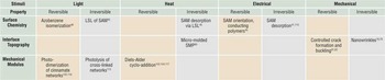
There are two essential components of a dynamic bioactive surface: (1) a stimulus that can be arbitrarily applied to the surface and (2) a material system that is designed to transduce the stimulus into a signal. The mechanism of stimulus transduction is ideally reversible and very precise. There is an abundance of materials that satisfy the latter criterion, while there are relatively few stimuli that are both mild and benign and therefore appropriate in studying dynamic cell-materials interactions. Most programmable stimuli for these applications rely on the selective application of heat, light, mechanical strain, or electrical signals to the surface. Perhaps the simplest demonstration of a dynamic surface is reversible stretching, which has been extensively studied primarily in the context of measuring the impact of cell mechanics on downstream function.Reference Geiger, Taylor, Glucksberg and Dean37–Reference Jungbauer, Gao, Spatz and Kemkemer39 Systems that employ increased complexity by utilizing alternative stimuli-responsive surfaces will be discussed in this section. Table I highlights representative examples of responsive materials that are able to alter physical properties of the surface (left column) via an external stimulus.
Summary of dynamic materials and their associated stimuli.

This non-exhaustive list represents a brief survey of the relevant materials that are discussed in this work. LSL, laser-scanning lithography; SAM, self-assembled monolayers; SMP, shape-memory polymer.
Electronic control of self-assembled monolayers
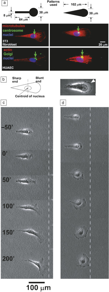
Dynamic control of surface chemistry represents a general tool that is widely applicable, including for self-cleaning surfaces, the reduction of biofilm formation, and selective cellular adhesion. There are many possible modes to control the local surface chemistry, which in turn can be used to modulate the adsorption of proteins and other functional groups to promote or hinder cell adhesion. Early examples of this approach have used applied voltages to alter the configuration of self-assembled monolayers (SAM) on gold surfaces.Reference Lahann, Mitragotri, Tran, Kaido, Sundaram, Choi, Hoffer, Somorjai and Langer40 Under small positive voltages, the terminal anionic carboxylic acid groups of low-density, flexible alkane thiols are temporary localized to the surface in a reversible manner. This transient configuration leads to the presentation of hydrophobic domains located in the mid-chain of the SAM to the free surface. When the voltage is removed, the carboxylic acid groups become unpinned from the surface and relax, thereby recovering the anionic surface. This process is rapid, reversible, and nearly instantaneous. Cell adhesion can be controlled by manipulating surface chemistry through the application of external voltages. In one demonstration, SAM attached to gold surfaces can be patterned into arbitrary micron-scale geometries through soft lithography and irreversible removal through the application of a reductive potential. This technique can be used to study the migration of single cells that are pre-patterned into select shapes.Reference Jiang, Bruzewicz, Wong, Piel and Whitesides41 The guiding hypothesis for this work is that cells temporarily physically constrained into a shape that represents an actively migrating cell will assume a preference for biased migration once the barriers to spontaneous migration (the SAM in this case) are removed. This hypothesis has been proven correct: cells that are pre-positioned with asymmetric geometries showed enhanced migration in terms of both directionality and velocity compared to cells that are pre-patterned into isotropic shapes. Figure 1 summarizes the results of these experiments. The asymmetric shapes used in these studies are shown in Figure 1a where the dark area represents the cell adherent area (no SAM). Confining cells within these geometries leads to segregation of certain cellular components toward the blunt end of the cell (Figure 1b). Figure 1c–dshows time lapse images of two different cell types as the physical barrier is released at time t = 0’. This demonstration is a salient example of a material that can be utilized to test an intriguing hypothesis that would otherwise be impossible using traditional cell culture methods.
Controlled electrical desorption of self-assembled monolayers to assess polarized cell migration. (a) Two representative asymmetric patterns used in biased migration experiments are shown. The dark area represents the cell adhesive domain and the approximate shape of the cell prior to release of the migration barrier (the self-assembled monolayer). The Golgi and the centrosome are located closer to the half of a cell with the blunt end. The various components of the cell are identified as follows: actin (red), the Golgi (green), the nucleus (blue), microtubules (red), and the centrosome (green), respectively. HUAEC, human umbilical artery endothelial cells. (b) Asymmetric pattern and corresponding bright field image of a fibroblast are shown. (c) Time-lapse images (in minutes) show the motility of a polarized fibroblast after the removal of the migration barrier. (d) Kidney cells derived from monkeys (COS-7 cells) show a similar behavior to fibroblasts. This system depicts the ability to observe real-time changes in a cytoskeletal structure. Reprinted with permission from Reference Jiang, Bruzewicz, Wong, Piel and WhitesidesReference 41. ©2005, National Academy of Sciences.

It should also be noted that the direct modification of surface energy can be achieved by simply altering surface charge through electrical stimulation.Reference Gumus, Califano, Wan, Huynh, Reinhart-King and Malliaras42 This approach has been utilized in a number of advanced materials for studying cell-biomaterials interactions, including conducting polymers. Electronically conductive cell-adhesive substrates composed of naturally occurring compounds, such as melanins, may also serve as candidate materials for this strategy.Reference Bettinger, Bruggeman, Misra, Borenstein and Langer43The advantages of this method include the reversibility of surface properties, robust and predictable responses, facile fabrication, and micron-scale resolution. This topic has been reviewed extensively elsewhere.Reference Muskovich and Bettinger44
Optical control of surface ligands
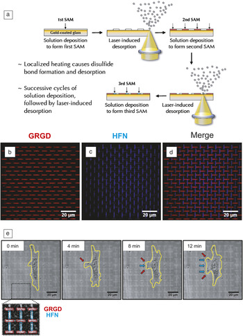
Spatially selective removal of SAM can also be engineered through the application of optical stimulus. Laser-scanning lithography (LSL) is a convenient tool to deliver such a stimulus with resolutions that are relevant to cell-biomaterial interactions. One such application of this technique utilizes gold-thiol SAM that are modified with cell adhesive ligands such as Arg-Gly-Asp residues.Reference Slater, Miller, Yu and West45 The principle of operation is to selectively pattern successive SAM compositions through cycles of blanket absorption and subsequent localized desorption (Figure 2). The process begins by depositing the first SAM layer on a gold-coated glass. A laser is raster scanned on the surface to induce selective desorption through localized heating. A second SAM layer can then be deposited onto available substrate domains. This process can be repeated in succession to create complete patterns of complex surface chemistries. The desorption process is sufficiently rapid enough to enable spatially targeted removal of ligands from cell-seeded substrates. The development of this tool is predicated on the desired ability to study the response of single cells to the selective removal of substrate cues, including chemical domains that are known to promote cell adhesion. It may be possible to selectively detach certain parts of a cell in a controlled manner. For example, an area of a cell that expresses a certain protein can be removed, and the downstream alterations in morphology or regulatory behavior can then be quantified with high-resolution live-cell imaging. Although the process of selective ligand removal is irreversible, the ability to manipulate cytoskeleton components with subcellular resolution is attractive from a technological standpoint. There are other prominent demonstrations in which the surface energy and presentation of ligands can be controlled reversibly through optical methods by employing photo-switchable chemistries based on azobenzene derivatives.Reference Lim, Han, Kwak, Jin and Cho46
Dynamic desorption of self-assembled monolayers (SAMs) using laser-scanning lithography. (a) Gold-coated glass coverslips are functionalized with alkanethiols. Select regions are thermally desorbed using laser-scanning lithography (LSL). Bare regions are exposed to additional alkanethiols. This process is continued to allow for patterning of multiple SAMs. (b–d) Multifaceted surfaces can be created to present coordinated ligands, including adhesion-promoting peptides (GRGD) and fibronectin (HFN). The multifaceted surface displays independent arrays of GRGD (red horizontal patterns) and HFN (blue vertical patterns). These dynamic interfaces can be used to study lamella formation and migration in endothelial cells. (e) Endothelial cells are initially elongated in a direction parallel to the long axis of the HFN patterns. The cells then produce membrane extensions along GRGD patterns and form lamella near fibronectin. Scale bars represent 20 microns. Reprinted with permission from Reference Slater, Miller, Yu and WestReference 45. ©2011, Wiley.

Dynamic topography
Substrate topography with features that span the micron- and submicron length scales serves as a potent cue in directing cell function.Reference Dalby, Riehle, Sutherland, Agheli and Curtis47, Reference Dalby, Biggs, Gadegaard, Kalna, Wilkinson and Curtis48 Reactions to topography are highly conserved across many phenotypes, including neurons, endothelial cells, smooth muscle cells, and stem cells.Reference Yim, Reano, Pang, Yee, Chen and Leong7, Reference Bettinger, Orrick, Misra, Langer and Borenstein14, Reference Bettinger, Langer and Borenstein24, Reference Bettinger, Kulig, Vacanti, Langer and Borenstein27, Reference Gerecht, Bettinger, Zhang, Borenstein, Vunjak-Novakovic and Langer30, Reference Bettinger, Zhang, Gerecht, Borenstein and Langer31, Reference Peng, Eltgroth, LaTempa, Grimes and Desai49 Natural topography in ECM proteins influences contact guidance responses and directs in vivo migration.Reference Campbell and Marcum50–Reference Wolf, Muller, Borgmann, Brocker and Friedl53 Native structures can be reproduced in vitro using synthetic substrates with micron-scale features.Reference Weiss54, Reference Dunn and Heath55 Microfabrication techniques have been employed to study the role of topographical cues on cell function.Reference Flemming, Murphy, Abrams, Goodman and Nealey23, Reference Andersson, Olsson, Lidberg and Sutherland56 Synthetic topography uniformly induces altered cellular morphologies as well as subsequent downstream signaling that affect proliferation,Reference Choi, Hagvall, Wu, Dunn, Beygui and Kim29, Reference Rebollar, Frischauf, Olbrich, Peterbauer, Hering, Preiner, Hinterdorfer, Romanin and Heitz57, Reference Lim and Donahue58 migration,Reference Abrams, Goodman, Nealey, Franco and Murphy17, Reference Bettinger, Langer and Borenstein24, Reference Diehl, Foley, Nealey and Murphy32, Reference Patel, Kurpinski, Quigley, Gao, Hsiao, Poo and Li35, Reference Lenhert, Meier, Meyer, Chi and Wiesmann59–Reference Cavalcanti-Adam, Volberg, Micoulet, Kessler, Geiger and Spatz63 differentiation,Reference Yim, Pang and Leong8, Reference Dalby, Gadegaard, Tare, Andar, Riehle, Herzyk, Wilkinson and Oreffo33, Reference Foley, Grunwald, Nealey and Murphy34, Reference Martínez, Engel, Planell and Samitier62 and ECM protein production.Reference Chou, Firth, Uitto and Brunette64–Reference Rosenberg66 These responses are likely to govern decision-making processes regarding interactions with micro- and nanoparticles such as opsonization and phagocytosis, mechanisms by which cells can uptake micron-scale particles.Reference Champion, Katare and Mitragotri67 One theory suggests that filopodia, small actin-rich fragments that extend from the cytoskeleton, engage in an active substrate exploring function.Reference Albrecht-Buehler68 Although the impact of topographical microenvironment on gross morphology has been studied extensively,Reference Carrel and Burrows69, Reference Weiss and Garber70 the mechanisms that govern cell-topography interactions are only starting to be unraveled.Reference Dalby, Hart and Yarwood71Thus, topography is a cue that warrants parallel investigation along with other substrate properties such as substrate stiffness and surface chemistry.Reference Johansson, Carlberg, Danielsen, Montelius and Kanje72
Previous studies have investigated populations of fixed cells that are seeded on engineered topographic substrates with well-defined features,Reference Yim, Pang and Leong8, Reference Bettinger, Orrick, Misra, Langer and Borenstein14, Reference Dalby, Riehle, Johnstone, Affrossman and Curtis15, Reference Abrams, Goodman, Nealey, Franco and Murphy17, Reference Oakley and Brunette73 including bilayer films with controlled buckling.Reference Jiang, Takayama, Q, Ostuni, Wu, Bowden, LeDuc, Ingber and Whitesides74 The insight in these studies is limited in part by the static nature of the substrate. Scant information on the kinetic responses of these interactions can be extracted using static surfaces.Reference Oakley and Brunette16 There are several prominent examples of substrates with unique structures that can be used as sensing elements. For example, substrates can be decorated with micron- or nanometer-scale vertical pillars that extend from the surface. Adherent cells exert forces on these structures, which distort the shape of the pillars.Reference Tan, Tien, Pirone, Gray, Bhadriraju and Chen75, Reference Li, Song, Mantini, Lu, Fang, Falconi, Chen and Wang76 The degree of tip deflection is directly related to the force generated by the cell and can be accurately measured through microscopy (light, fluorescent, or scanning electron).Reference Tan, Tien, Pirone, Gray, Bhadriraju and Chen75, Reference Li, Song, Mantini, Lu, Fang, Falconi, Chen and Wang76 This technique can measure the forces generated by a cell that is migrating on a surface with subcellular-scale precision.Reference du Roure, Saez, Buguin, Austin, Chavrier, Siberzan and Ladoux77There is considerable interest in using structures on surfaces as a characterization tool to measure responses. However, there is also merit in designing dynamic surfaces that can serve as a stimulus as well. Dynamic substrate topography enables the quantification of temporal responses of the cytoskeleton, including reorganization kinetics. Surfaces that can actively switch between defined surface topographies may serve as a powerful tool in cell biology with the potential for broad impact in a variety of disciplines. This system could be used to quantify the kinetics associated with microtubule organization, filopodia formation, or other structural changes. Dynamic topography is an attractive tool to study cell-biomaterial interactions for several reasons: (1) interfaces can be designed using a variety of smart and stimuli-responsive materials; (2) the fabrication of these surfaces is compatible with many standardized techniques such as replica molding and photolithography; (3) topographic states are binary, dynamic, and potentially reversible; (4) there are many available approaches to fabricate structures across a broad spectrum of length scales; and (5) the presence or absence of topography on a surface can be precisely controlled in time by employing the strategies described herein.
Nanowrinkles
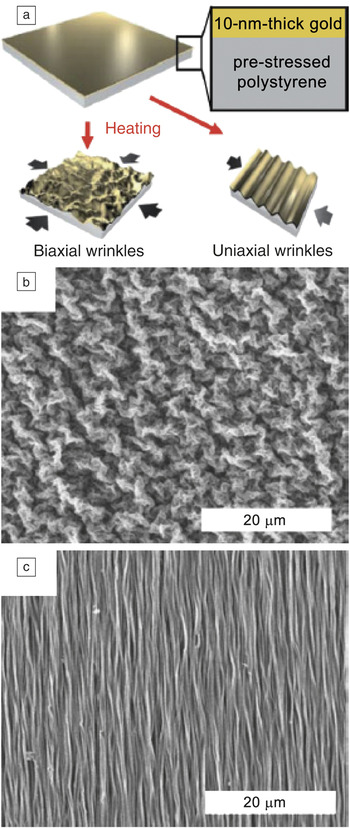
Next-generation programmable surfaces will benefit from the transduction of benign external stimuli into arbitrary presentation of topographical features at the material interface in a reversible manner. There are many candidate materials that can harness this principle to ultimately overcome the limitations of static surface systems. Thermal loads have been used as stimuli to generate tunable nanowrinkles.Reference Fu, Grimes, Long, Ferri, Rich, Ghosh, Ghosh, Lee, Gopinathan and Khine78 Specifically, temporary flat substrates are prepared by depositing thin gold membranes on bulk polystyrene films. The composites are then heated, which shrinks the polystyrene films and subsequently introduces strain-induced nanowrinkle formation in the gold membrane (Figure 3). Biaxial compression produces disordered submicron-scale features through isotropic wrinkling (Figure 3b). Uniaxial compression produces nanograting features with short-range order and wavelengths between 200 nm and 1 μm (Figure 3c). The advantages of this approach include the ability to functionalize gold with a variety of thiol-containing compounds and the relatively straightforward fabrication process. An alternative technique utilizes bilayer substrates that consist of a shape-memory polymer (SMP) with a thin oxide film that is formed using oxygen plasma. The composite is uniaxially compressed through thermal activation by heating the films to 150°C. This technique has been adopted to enhance the assembly of tissue. More specifically, substrates with nanoscale features can organize cells that are precursors to forming functional cardiac tissue. Using topography to organize these cells produces cardiac tissue that functions more like the native heart.Reference Chen, Lieu, Freschauf, Lew, Sharma, Wang, Nguyen, Karakikes, Hajjar, Gopinathan, Botvinick, Fowlkes, Li and Khine79 However, the formation of dynamic topography using these materials requires high temperatures that are incompatible with in situ cell culture and ultimately restrict the ability to signal viable cell populations in real time.
(a) Fabrication of uniaxial and biaxial nanowrinkles for use as a cell culture platform. (b) Biaxial and (c) uniaxial scanning electron microscopy images of shrunk polystyrene sheets with a layer of gold (10 nm). Controlled wrinkling patterns can be used as a cell culture platform for real-time microscopy and cytoskeletal dynamic studies. Reprinted with permission from Reference Fu, Grimes, Long, Ferri, Rich, Ghosh, Ghosh, Lee, Gopinathan and KhineReference 78. ©2009, Wiley.

Shape-memory polymers
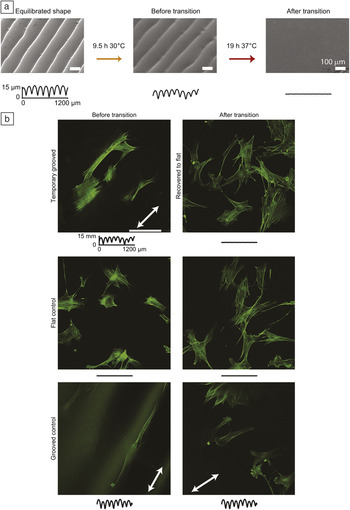
Thermal stimuli can be directly integrated with cell-seeded surfaces by adopting phase transitions at or near physiological temperatures. In work by Henderson et al., a thermally activated SMP network is utilized as a material that can present dynamic substrate topography.Reference Davis, Burke, Mather and Henderson80 External heating serves as an irreversible stimulus, which is transduced through a polyurethane-based SMP. The SMP network (Norland Optical Adhesive-63) is initially cured on microfabricated negative molds by photo-cross-linking via thiolene chemistry. Fibroblasts are cultured on surfaces with temporary micron-scale grating features. Thermal activation leads to the recovery of a flat substrate. Abolishing the grating features by heating the material from 30 to 37°C leads to a gradual decay of cell alignment and a recovery of isotropic cell spreading. Hence, there is a gradual restoration to a morphology that is representative of cells cultured on flat substrates (Figure 4). The next generation of materials for this application should be reversible with minimal hysteresis and exhibit narrower temperature ranges for thermal transitions.
(a) Thermal effect of cell culture substrate transition from a grooved surface to a flat surface using an SMP network. After cells were cultured for 9.5 h at 30°C, the substrate was triggered by placing a sample in a 37°C incubator. The traces below the image represent a cross-section of the contours of the culture surface. (b) The change in substrate topography controls cell behavior. Confocal images of cells stained with phalloidin on a temporary grooved structure have microstructures in alignment with the groove direction (white arrow). After the surface transitions, the cells rearrange, are randomly oriented, and show stress fiber formation. Scale bar in (b) is 100 μm. Reprinted with permission from Reference Davis, Burke, Mather and HendersonReference 80. ©2011, Elsevier.

Reversible topography
The central ideal of reversible topography is to apply aspects of materials engineering to fabricate synthetic substrates with topographic features that can signal cells in a dynamic manner. Prospective studies in this area will ultimately lead to a deeper understanding of the role of cytoskeleton function in response to the selective presentation of topographic cues. These systems can be used to elucidate cytoskeleton reorganization kinetics in a variety of cells, including phenotypes that approximate progenitor or disease states. The design and fabrication of reversible topography for studying cell-biomaterials interactions requires a cross-cutting interdisciplinary effort that integrates materials science and engineering, polymer fabrication, solid mechanics, and cell biology.
Mechanical strain is an effective stimulus that can be leveraged for use in reversible topography. Takayama et al. described the reversible mechanical compression of oxidized polydimethylsiloxane (PDMS) bi-layer substrates for use in measuring dynamic cell-substrate interactions.Reference Lam, Clem and Takayama81 In this study, uniaxial compression of a rigid film on an elastomeric substrate at strains of 11–15% produces grating features with wavelengths of approximately 7 μm and amplitudes of 670 nm. Alternating the presentation and withdrawal of substrate topography signals induces aligned and random orientation of C2C12 murine myoblasts, respectively. This process can be repeated up to nine times without any observable hysteresis. There are several technical challenges in this approach, including the limited range of mechanical strains that can induce feature formation without macroscopic deformation of the substrate. Mechanical strains over 3.5% can lead to a convolution of mechanical strain in conjunction with the presentation of topographic cues.Reference Wang, Ip, Boissy and Grood38 By comparison, a typical cell stretching experiment would strain a substrate by approximately 10%.Reference Wang, Goldschmidt-Clermont, Wille and Yin82 Reversible topography has also been utilized as a method to control the presentation of adhesion ligands.Reference Zhu, Mills, Peters, Bahng, Liu, Shim, Naruse, Csete, Thouless and Takayama83Briefly, submicron-scale cracks can be induced by uniaxially straining bilayers composed of elastomeric PDMS substrates and thin silica membranes. First, the unstrained substrate is coated using cell repulsive detergent (Pluronic F108). Next, the substrate is strained, thereby forming micro-cracks that can be chemically modified with collagen. The resulting surface consists of a field of cell repulsive pluronic with cell adhesive collagen that is confined within the micro-cracks. The adhesion domains are masked under zero strain and exposed during the application of positive strain induced by uniaxial tension. The time course and density of collagen domains on the substrate can therefore be controlled by simply adjusting the amount of strain. This approach has been used to validate the dynamic responses of muscle precursor cells to pulsed collagen exposure. These cells align and elongate in response to the exposed collagen domains that are present under 10% strain. The spreading and alignment can be reversibly controlled by cycling between strains of zero and ten percent.
Reconfigurable polymer networks
The intrinsic mechanical properties of a cell can play a prevalent role in determining cell fate.Reference Engler, Sen, Sweeney and Discher13, Reference McBeath, Pirone, Nelson, Bhadriraju and Chen84 Pathologies in cellular mechanics can lead to debilitating disease states.Reference Suresh, Spatz, Mills, Micoulet, Dao, Lim, Beil and Seufferlein85 Conversely, the intrinsic mechanical properties of cytoskeleton components may serve as a valuable biomarker for diagnostics.Reference Suresh86 For example, the cytoskeleton of metastatic cancer cells is 70% softer than corresponding non-metastatic cancer cells.Reference Cross, Jin, Rao and Gimzewski87 Cell mechanics play a critical role in hematologic diseases as well.Reference Rosenbluth, Lam and Fletcher88 Hence, the role of cellular biomechanics in maintaining homeostasis cannot be diminished. There are several established theories of mechanotransduction that have been supported by subsequent biophysical studies. The tensegrity model proposed by Ingber posits that pre-stressed microfilaments and microtubules, structural components of the cytoskeleton, produce tension in the underlying network that ultimately yields a stable structure which, in part, serves as a physical signal transduction pathway.Reference Ingber89, Reference Ingber90 Tensegrity, or tensional integrity, is a concept in which the mechanical stability of a structure, in this case the cytoskeleton, is structurally stable through the appropriate combination of components that are either under tension or compression and assembled in a coordinated manner. A complementary theory posed by Charras and Horton suggested that tensegrity is maintained by intermediate filaments, cytoskeleton protein structures that have a characteristic size that is between microfilaments and microtubules,Reference Charras and Horton91 which is supported by relevant observations.Reference Hotani and Miyamoto92, Reference MacKintosh, Kas and Janmey93 Another theory posed by Forgacs suggested that critical percolation of actin filaments is essential for mechanotransduction.Reference Forgacs94, Reference Shafrir and Forgacs95 These theories have been collectively tested in the context of constrained cell shape,Reference Chen, Mrksich, Huang, Whitesides and Ingber6, Reference Parker, Brock, Brangwynne, Mannix, Wang, Ostuni, Geisse, Adams, Whitesides and Ingber96 altered substrate stiffness,Reference Discher, Janmey and Wang97, Reference Yeung, Georges, Flanagan, Marg, Ortiz, Funaki, Zahir, Ming, Weaver and Janmey98or deformed substrates.Reference Cheng, Steward and LeDuc99
Cell mechanics is an important design criterion in the broader effort to transform two-dimensional cell culture models into more biologically relevant three-dimensional constructs. This overarching strategy has influenced the design of many bioactive materials, including porous polymer networks and hydrogels. A parallel strategy may also be devised for the design of dynamic three-dimensional networks that are able to control the spatiotemporal presentation of cell signaling proteins and peptides. Recent efforts have been predicated on repurposing reversible coupling chemistries that can be applied to control molecular topology and the extent of cross-linking in the context of controlling the elasticity of synthetic ECM. Similar approaches can also be used to control the presentation of bound ligands for controlled adhesion and integrin activation. This section intentionally omits the vast literature on ionic cross-linking of networks and phase change materials, including stimulus-responsive hydrogels, which have been extensively reviewed elsewhere.Reference Gupta, Vermani and Garg100, Reference Peppas and Leobandung101Instead, a dedicated focus is granted toward modular covalent coupling in the context of precisely controlling the mechanical properties of the network and the ability to present chemical cues with spatiotemporal specificity.
Reversible cross-linking
Controlling the cross-linking density in polymeric networks is a convenient method to modulate the bulk mechanical properties of synthetic ECM analogues. Furthermore, networks consisting of hydrogels or elastomers are able to achieve elastic moduli that are similar to a wide range of naturally occurring tissues.Reference Bettinger102The ideal design criteria in engineering reversible polymer networks are as follows: (1) Hyperbranched network precursors are functionalized with homobifunctional or complementary bifunctional cross-linking moieties; (2) the chosen cross-linking chemistries are reversible; and (3) forward and reverse cross-linking reactions are both inducible through the application of mild stimuli.
There is a wide range of reversible coupling chemistries that are suitable for modulating the bulk properties of polymer networks. Diels–Alder cycloaddition reactions provide an efficient [2+4] cycloaddition mechanism. Diels–Alder coupling using furan-based dienes and maleimide-based dienophiles has been used as a strategy for the room temperature cross-linking of hydrogels.Reference Kosif, Park, Sanyal and Sanyal103, Reference Nimmo, Owen and Shoichet104One potential limitation in applying reversible Diels–Alder chemistry is the large temperature window that is required for promoting forward and reverse coupling reactions. The kinetics of forward Diels–Alder cycloaddition is somewhat slow at room temperature, while retrocycloaddition reactions require temperatures of 70°C and higher, depending upon the composition of the substituents and the solvent environment.
Cinnamates, a class of aromatic molecules based on cinnamic acid, represent an alternative chemistry that is capable of reversible coupling via photoexcitation.Reference Shi, Matsusaki, Kaneko and Akashi105 Cinnamate derivatives undergo reversible photodimerization upon UV irradiation.Reference Pinazzi and Fernandez106 Dimerization forms a cyclobutane ring by [2+2] photocycloaddition at wavelengths greater than 260 nm, while photocleavage of the cyclobutane ring is possible by irradiation at a wavelength less than 260 nm. Hyperbranched pre-polymers derivatized with pendant cinnamate can produce reversibly photo-cross-linkable networks.Reference Shi, Matsusaki, Kaneko and Akashi105 There are several advantages of synthesizing networks using precursors that feature cinnamate functionalization. First, cinnamates are a class of naturally occurring aromatic compounds that are found in some fruits. Second, photodimerization and cleavage can be controlled in two dimensions through simple masking techniques. Third, coupling occurs through homodimerization, which can simplify the synthesis of network precursors. Despite these advantages, the high intensities and low wavelengths used in reversible photodimerization typically preclude the incorporation of viable cell populations. There are numerous other reconfigurable cross-linking chemistries, including those based on isocyanates/imidazole coupling and free radical polymerization of thiolene compounds. This class of cross-linking chemistry is broadly categorized as covalent adaptive networks (CAN), which are described in more detail elsewhere.Reference Kloxin, Scott, Adzima and Bowman107Despite the potential capabilities in utilizing CAN as programmable polymer networks and interfaces, there are several practical processing limitations that can preclude the widespread application of these materials for studying dynamic cell-biomaterial interactions in real time.
Dynamic 3D environments
The advantages of reconfigurability in covalent networks must be balanced with the benefits of in situ manipulation of cell-seeded networks. Hydrogels offer an attractive platform to create covalent networks that are capable of presenting dynamic three-dimensional cellular microenvironments. Specifically, light-activated networks offer an opportunity to control the spatiotemporal presentation of cues that cells can recognize in real time. The core material property that has been utilized in a number of demonstrations is the ability to modify polymer side chains and backbones using photolysis. Under this scheme, the single- (λ = 365 nm) or two-photon (λ = 740 nm) absorption of select aromatic species can initiate rearrangement reactions that uncage molecules or modify the properties of polymer networks. For example, photo-induced rearrangement in polymer backbones can be used for the instantaneous decomposition of synthetic polymers in response to benign levels of IR irradiation.Reference Fomina, McFearin, Sermsakdi, Morachis and Almutairi108In this study, pendant photosensitizers based on 4-bromo-7-hydroxycoumarin protecting groups are incorporated within linear polyester precursors. Upon excitation at λ = 740 nm, a cascade of cyclization and rearrangement reactions proceeds, which leads to systemic degradation of the network. This rapid degradation reaction yields relatively non-toxic low-molecular weight by-products, including adipic acid.
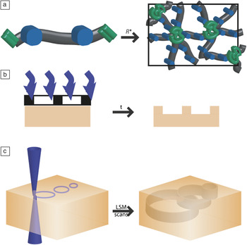
Photodeprotection chemistries generally describe the ability to cleave a portion of a molecule in a selective manner using light. Photodeprotection can serve as an efficient method to selectively alter the compositions of side chains in polymeric materials. Functional groups containing pendant 2-nitrobenzyl groups via ester linkages can be cleaved to produce the nascently deprotected carboxylic acid and the newly formed 2-nitrobenzaldehyde by-product. Variations on this chemistry have also been used for three-dimensional patterning of recognition molecules in hydrogels via single- or two-photon patterning of static networks.Reference Luo and Shoichet109, Reference Wylie and Shoichet110 This synthetic strategy can also be used to create dynamic hydrogel biomaterial networks. Anseth et al. demonstrated the formation of photolytically cleavable cell-seeded hydrogel networks based on poly(ethylene glycol) (PEG) functionalized with 2-nitrobenzyl groups.Reference Kloxin, Kasko, Salinas and Anseth111 Briefly, linear PEG precursors are functionalized with acrylate-containing 2-nitrobenzyl groups via ester linkages. These photolabile PEG precursors form cross-linked hydrogel networks. The storage modulus of the networks, which is directly proportional to the cross-link density, can be reduced irreversibly by over 90% after exposure to benign UV irradiation of λ = 365 nm at 10 mW/cm2 for 10 minutes. Therefore, the mechanical properties of the network can be dynamically tuned in space and time through controlled irradiation. A parallel synthetic approach can be used to create networks in which cell adhesion domains can be selectively removed using photolysis. Human mesenchymal stem cells cultured in PEG hydrogels with photocleavable cell-binding domains engage in accelerated chrondrogenesis upon selective removal of this integrin binding motif. Similar chemistries can be utilized for real time control of elasticity in hydrogel substrates as well.Reference Kloxin, Tibbitt and Anseth112, Reference Kloxin, Benton and Anseth113 Practical aspects of these techniques are summarized in Figure 5. Figure 5a is a schematic of the formation of a photodegradable polymer network through free radical photopolymerization. The green groups represent dienes that can undergo free radical polymerization to create the cross-linked network. The blue groups represent photodegradable segments that can be cleaved to selectively remove parts of a network. One strategy for selective photocleavage of cross-linked networks uses masking combined with single-photon excitation to create two-dimensional structures in a manner that is similar to photolithography (Figure 5b). Another strategy uses two-photon absorption in which laser scanning microscopy can be used to create arbitrary three-dimensional features by selective photocleavage (Figure 5c).
Methods for photodegradable hydrogel synthesis and patterning. (a) Photodegradable hydrogels formed by redox-initiated, free-radical chain polymerization, which is indicated by R*. The resultant network is comprised of polyacrylate kinetic chains (green) connected by PEG-based cross-links (black) with photocleavable moieties (blue) in the backbone. (b) Using a chrome mask, the surface of the photocleavable hydrogel can be patterned with features of depths that depend upon exposure time. Features can be generated within minutes of irradiation time. (c) Use of a laser-scanning microscope (LSM) with a 405 nm laser can also be used to create three-dimensional patterns by precisely moving the laser in the x–y plane using a scanner. The z-dimension is controlled with a higher minimum feature resolution by either controlling the focal plane of the laser or the location of the stage. Reprinted with permission from Reference Kloxin, Tibbitt and AnsethReference 112. ©2010, Nature Publishing Group.

Light-activated hydrogel networks are advantageous because direct manipulation of cell-biomaterial interactions is achievable, although irreversible. Nonetheless, there is a broad set of biological questions that can be answered using material systems that are able to administer step changes in biomaterial response. The emerging challenge is to design materials, systems, and experiments that are able to eventually provide insight into disease progression such that complementary treatments may eventually be discovered.
Conclusions and outlook
The continued interest in dynamic cell-biomaterials interactions using smart materials will lead to discoveries that could resolve key aspects of cytoskeleton function in a variety of model systems such as neurons, cardiomyocytes, and stem cells. In addition to fundamental scientific discovery, this approach may be utilized in application-based studies such as in vitro disease models or assays for drug development. Despite the proliferation of materials and the rapidly growing set of fabrication capabilities, the field of dynamic materials for elucidating cell responses remains in its infancy. The utility of these systems continues to expand with help from parallel advances in materials design strategies. Next-generation dynamic materials will gradually incorporate cues beyond traditional first-order engineering properties such as surface energy, feature topography, or elastic modulus. For example, a dedicated focus may be the control of dynamic cell-cell interactions. Preliminary examples exist, but they will benefit from continued refinement.Reference Hui and Bhatia114Prospective dynamic systems will require new materials with increased sophistication in polymer synthesis and macromolecular engineering approaches.
The prospect for increasing complexity in these systems must be balanced with the practical utility of such inventions. There may be reduced scientific merit and unrealized downstream clinical benefit by arbitrarily increasing the complexity of dynamic cell culture materials without regard to the ease of widespread adoption. One example of a technology that has successfully navigated this tradeoff is soft lithography. Microfluidics and micropatterning in biomaterials have been widely adopted as tools by both biomedical engineers and biologists alike. This prominent success story can provide a roadmap for the adoption of future technologies that are predicated on dynamic materials. Traction of early-stage demonstrations can be accelerated through two types of research environments: (1) strategic collaborations between materials engineers and biomedical researchers who are early adopters of innovative techniques; and (2) laboratories that are able to comprehensively integrate the design, fabrication, and validation of dynamic materials in model biological systems under one roof. Taken together, the prospects for dynamic biomaterials remain bright and have the potential to enable high impact discoveries in numerous fields ranging from the design of medical implants to engineering immunotherapies.
Acknowledgments
Funding was provided by the following organizations: the Berkman Foundation; the American Chemical Society Petroleum Research Fund (ACS PRF #51980-DNI7); the Proctor & Gamble Education Grant Program; and the Carnegie Mellon University School of Engineering.

Stephen Kustra received his master of science degree in biomedical engineering at Carnegie Mellon University and his bachelor of science degree in biomedical engineering from the University of Connecticut. His current research interests include biomaterials-based medical devices and smart interfaces for understanding fundamentals of cell-biomaterials interactions. Kustra can be reached by email at skustra@andrew.cmu.edu.

Christopher J. Bettinger is an assistant professor at Carnegie Mellon University (CMU) in the Departments of Materials Science and Biomedical Engineering. He directs the laboratory for Biomaterials-Based Microsystems and Electronics at CMU, which is broadly interested in the design of novel materials and interfaces that promote the integration of medical devices with the human body. Bettinger has received many honors, is a co-inventor on several patents, and was a finalist in the MIT $100K Entrepreneurship Competition. Bettinger received an SB degree in chemical engineering, an M.Eng. degree in biomedical engineering, and a PhD degree in materials science and engineering as a Charles Stark Draper Fellow, all from the Massachusetts Institute of Technology. He completed his post-doctoral fellowship at Stanford University in the Department of Chemical Engineering as an NIH Ruth Kirschstein Fellow. Bettinger can be reached by email at cbetting@andrew.cmu.edu.