Idiopathic dilated cardiomyopathy in children is a well-recognized cause of congestive heart failure in the absence of congenital cardiac disease. It is responsible for high rates of medical care and mortality, ranging from 16% at 10 years to 49% and 80%, at 5 years.1–5
Cardiac transplantation is the treatment of choice in patients who fail to respond to medical treatment. The cumulative survival rates vary from 75 to 80% at 1 year, and from 60 to 75% at five years.6–8 Several studies have been carried out to establish unfavourable prognostic parameters, and hence provide an early indication for cardiac transplantation.2–4, 9–12 No consensus on the matter, however, has been reached so far.
In the light of this lack of consensus on the predictors for death in the setting of infantile idiopathic dilated cardiomyopathy, we carried out a retrospective review study of all children presenting to our hospital over a period of 22 years, analyzing the evolution of clinical factors, and chest X-ray, electrocardiographic, and echocardiographic data. The suspected risk factors were then submitted to statistical analysis.
Methods
We trawled the database of the National Institute of Cardiology Laranjeiras, Rio de Janeiro, Brazil, and identified 142 children seen consecutively as suitable for retrospective analysis. The children had been diagnosed with idiopathic dilated cardiomyopathy from November 1982 to September 2004. The criterions for inclusion were the presence of congestive heart failure or the appearance of cardiomegaly in a routine chest X-ray, confirmed by enlargement and hypo kinesis of the left ventricle observed in the echocardiogram. We also included asymptomatic children in functional class I of the categorization of the New York Heart Association. In the first three patients, diagnosis had been made on the basis of angiographic studies. Echocardiographic studies were started from June, 1984, and used from this time to confirm the diagnosis. Patients with congenital cardiac disease, anomalous origin of the coronary arteries, excluded by angiographic study if necessary, Kawasaki's disease, arrhythmogenic cardiomyopathy, ischaemic injury due to neonatal asphyxia or cardiorespiratory arrest, those exposed to cardiotoxic agents, such as anthracycline, and those with inborn errors of metabolism, primary arrhythmias, rheumatic heart disease, neuromuscular disease, systemic arterial hypertension, septicaemia, infection with human immunodeficiency virus, Chagas' disease, and diphtheria were all excluded.
We analysed age at presentation, gender, ethnic origin, history of a preceding viral disease within 3 months before diagnosis, signs and symptoms at presentation, and functional class within the categorization of the New York Heart Association. Functional class I was defined as no limitations in normal activities for age, class II as comfortable at rest, but with normal activities for age resulting in symptoms of cardiac failure, class III as comfortable at rest, but with mild activities for age resulting in symptoms of cardiac failure, and class IV as symptoms of cardiac failure at rest. In addition, 2 of the authors reviewed 806 radiographic films from all 142 patients, giving an average of 5.67 examinations per patients, 479 tracings of the surface 12 lead electrocardiogram from 131 patients, averaging 3.66 recordings per patient, and 490 echocardiograms from 141 patients, averaging 3.47 recordings per patient. The inter-observer and intra-observer rates of reliability were 94.7%, with kappa of 0.938, for the chest radiographs, 90.4% with kappa of 0.907 for the electrocardiograms, and 92.3%, with kappa of 0.918, for the echocardiograms.
The 12 lead electrocardiograms all included 3 to 5 complexes by lead and lead DII taken over 15 to 30 seconds. If any arrhythmia was found on the surface electrocardiogram, or if the patient had symptoms of tachycardia or bradycardia, thoracic pain, or syncope, a 24-hour electrocardiographic Holter recording was performed.
Mitral regurgitation was assessed by pulsed and colour Doppler according to the era of examination, using the golden standard to each era, by the guidelines from the American Society of Echocardiography.13 Left ventricle ejection fraction was measured by Simpson's method, at examination or by reviewing the tapes.
The medical treatment employed was optimized for the clinical and functional class of the patients, and varied during the period of study. It was the conventional regime employed at the time, and included digitalis in 109 patients (76.8%), furosemide or hydrochlorothiazide in 132 patients (93.0%) and spironolactone in 87 patients (61.3%), inhibitors of angiotensin converting enzyme in 90 patients (63.4%), hydralazine in 37 patients (26.1%), and acetylsalicylic acid in 35 patients (24.6%) for prevention of thromboembolic events. The use of inhibitors of angiotensin converting enzyme started in 1986, and after then became a routine treatment. Intravenous inotropic drugs were administered at least once in the 45 patients (31.7%) in functional class IV. The analysis of the influence of treatment on outcome was not the primary purpose of this study. Nevertheless, since use of inhibitors of angiotensin converting enzyme is now considered a corner stone of modern treatment for cardiac failure, we analyzed its impact on outcome.
The assumption of myocarditis was based on clinical findings such as fever and chest pain, laboratory findings such as abnormalities in creatine phosphokinase, troponin and myoglobin, and electrocardiographic criterions such as low voltage of the QRS complexes and abnormalities of conduction. Over the last years, myocarditis was confirmed by endomyocardial biopsy and immunohistochemistry, albeit that only five such investigations were made over the period, thus limiting any analysis in terms of survival.
The statistical analysis was carried out using the Epi Info 6.04 from Centers for Disease Control & Prevention software and the Statistica 6 from Statsoft Inc software. Dichotomous data was evaluated by Chi-Square test, with confidence intervals of 95% being calculated. Descriptive data was expressed as means and standard deviation, and analyzed by Student's t-test. The Receiver Operating Curve was built in order to test the best cut-off of continuous variables. Continuous dependent variables of time were evaluated by analysis of variance for repeated measures. Survival was analyzed by Kaplan-Meier method. Cox's analysis was used to predict the association of the clinical signs and abnormalities with death. Variables that may have predicted a bad prognosis in the univariate analysis (p equal to 0.1) were divided into groups and analyzed using Cox's regression model with casewise deletion, when any parameters were absent, to determine independent predictors of death. Any value was considered significant when alpha was less than 0.05. The study complies with the Declaration of Helsinki, and was approved by our institutional review committee, with all the subjects or their families or guardians giving informed consent.
Results
The incidence was approximately 6.4 new cases per year, or 0.39 per 100,000. Mean age at presentation was 2.48 years, with a median of 0.83 year, and a range from 1 month to 15.7 years, with follow-up of 4.0 years, ranging from 1 day to 15.96 years. At the end of the study, 64 (45.1%) patients were symptom-free, 42 (29.6%) had developed chronic congestive heart failure, and 36 (25.3%) had died. Survival rates, with confidence intervals of 95%, and the numbers of patients at risk, are presented in Figure 1.

Figure 1. Cumulative proportional survival and confidence interval of 95% of the 142 children and adolescents with idiopathic dilated cardiomyopathy.
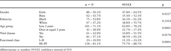
The results and confidence limits for the presumed risk factors are demonstrated in Tables 1–3. In this cohort, most of patients (71.1%) were younger than 2 years old, with a mean of 0.67 years plus or minus 0.47, and a majority was female (56.3%), but no difference was observed in ethnic origin (Table 1).
Table 1. Epidemiological characteristic of the cohort, confidence interval of 95% and significance.

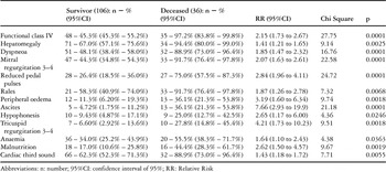
Table 2. Clinical markers of death.

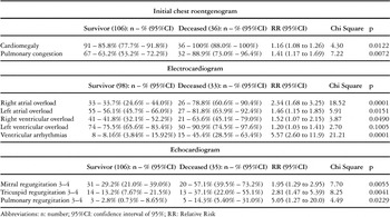
Table 3. The markers of death in complementary exams.

A history of a preceding viral disease within 3 months before diagnosis was reported by 61 (42.9%) patients, with respiratory viral disease reported in 54 patients (88.5%), and gastrointestinal viral infection in 7 patients (11.5% – p equal to 0.0001). Myocarditis was presumed in 60 (42.2% – confidence interval 34.1% to 50.8%) patients on the basis of clinical, laboratory and electrocardiogram criterions as described above. Presence or absence of myocarditis, however, did not confer any difference with regard to death (p equal to 0.1472). There was no difference in survival between those presenting at age younger or older than 2 years (p equal to 0.751), nor for the genders (p equal to 0.477), for any ethnic group (p equal to 0.083), nor the presence of previous viral disease (p equal to 0.226) or the assumption of myocarditis (p equal to 0.241).
At presentation, 11 (7.7%) patients were in functional class I, 13 (9.2%) in functional class II, 35 (24.6%) in functional class III, and 83 (58.5%) in functional class IV. The majority (83.1%) was in functional class III and IV (p equal to 0.0001). All 36 deaths occurred in patients in these functional classes (p equal to 0.0040). Presentation in functional class IV was a strong marker of death (p equal to 0.0001), with relative risk of 2.15, and confidence intervals from 1.73 to 2.67 (chi square 27.75).
The clinical markers of death at presentation in univariate analysis are presented in Table 2. Multivariate analysis showed that the presence of dyspnea (p equal to 0.0096), and reduced pedal pulses (p equal to 0.0413), were independent clinical markers of death. Mitral regurgitation of grade 3 or 4 (p equal to 0.0570), and auscultation of rales (p equal to 0.0573), came close to achieving significance.
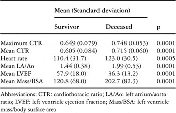
In the initial chest X-ray, the majority of children had cardiomegaly (89.4% – p equal to 0.0122), with relative risk of 1.16 (confidence intervals from 1.08 to 1.26), and pulmonary congestion (69.7% – p equal to 0.0072), with relative risk of 1.41, and confidence intervals from 1.17 to 1.69. These features proved to be markers for death (Table 3). The analysis of the cardiothoracic ratios is summarized in Table 4. A maximal cardiothoracic ratio was also a strong marker of death (p equal to 0.0001). Analysis of variance demonstrated a division by groups, starting at one month of follow-up (p equal to 0.0001 – Fig. 2). Interestingly, no difference was detected in terms of the cardiothoracic ratio between the groups previously exposed to viral disease (p equal to 0.6114), but the ratio was lower in those with myocarditis (p equal to 0.0192).
Table 4. Univariate analysis of complementary exams.


Figure 2. Cardiothoracic ratio and confidence interval of 95%, by time of follow-up (months), between groups (alive versus dead).
Holter recordings were performed in 35 patients (24.6%), and confirmed the presence of arrhythmias in 31 patients (88.6%, confidence interval 72.3% to 96.3%). Premature ventricular complexes, seen in 9 patients, non-sustained ventricular tachycardia in 10 patients, and sustained ventricular tachycardia that degenerated in non-reversible ventricular fibrillation in 4 patients, were all observed during follow-up. Supraventricular complexes at greater than 30 per hour were seen in 2 patients, supraventricular tachycardia in 5 patients, and disturbances of conduction in 1 patient.
Electrocardiographic predictors of mortality are shown in Table 3. Right atrial overload (p equal to 0.0118) and ventricular arrhythmias (p equal to 0.0148) were confirmed as independent predictors of death by multivariate analysis. Ventricular arrhythmias were also deleterious to survival (Fig. 3). The analysis of heart rate is summarized in Table 4. Analysis of variance demonstrated a higher heart rate in those dying (p equal to 0.0275 – Fig. 4).

Figure 3. Survival analysis by Kaplan-Meier method and log-rank test by groups of ventricular arrhythmias with confidence interval of 95%.
Figure 4. Heart rate on electrocardiogram and confidence interval of 95%, by time of follow-up (months), between groups (alive versus dead).
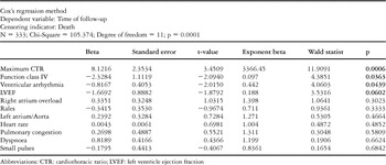
In univariate analysis of the echocardiograms, mitral, tricuspid and pulmonary regurgitations of grades 3 or 4 all emerged as markers of death (Table 3). Multivariate analysis confirmed that mitral regurgitation at grade 3 or 4 (p equal to 0.0017) was an independent echocardiographic marker of death, with a relative risk of 1.95, and intervals of 1.29 to 2.95 (chi square 7.70). Tricuspid regurgitation of grade 3 or 4 was weak associated with death (p equal to 0.0885). The continuous variables are summarized in Table 4. Analysis of variance demonstrated two different groups, for left ventricular ejection fraction (p equal to 0.0266) and the left atrial aortic ratio (p equal to 0.0001), starting at three months of follow-up (Figs. 5, 6), but did not show any effect of left ventricular mass indexed to body surface area (p equal to 0.1997). The multivariate analysis confirmed left ventricular ejection fraction (p equal to 0.0001), and the left atrial aortic ratio (p equal to 0.0073), as independent predictors of death. Cox's regression method was performed using the potentially prognostic factors identified in the previous analyses of clinical and laboratory data. To avoid bias, multivariate analysis was performed on the basis of grouping by the severity of mitral regurgitation. The dependent variable was time of follow-up. Examinations that did not have one of the parameters were discarded from the analysis. The analysis was carried out in two steps, with the second step carried out only using those parameters producing p values of less than 0.10 in the first analysis (Table 5).

Figure 5. Left ventricle ejection fraction and confidence interval of 95%, by time of follow-up (months), between groups (alive versus dead).

Figure 6. Left atrium/aorta ratio and confidence interval of 95%, by time of follow-up (months), between groups (alive versus dead).
Table 5. Table 5. Multivariate analysis through Cox's regression method of prognostic factors of death considering mitral regurgitation severity (covariate) – Phase 1.

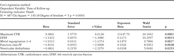
The independent predictors of death proved to be ventricular arrhythmias (p equal to 0.0253), maximal cardiothoracic ratio in the chest radiograph (p equal to 0.0001), presentation in functional class IV (p equal to 0.0028), and left ventricular ejection fraction (p equal to 0.0013 – Table 6). Mitral regurgitation of grade 3 or 4 identified in the echocardiogram had proved to be an independent marker of death in previously analysis (p equal to 0.0017).
Table 6. Multivariate analysis through Cox's regression method of prognostic factors of death – Phase 2.

With regard to therapeutic regimes, only use of inhibitors of angiotensin converting enzyme reached near significance in multivariate analysis when considering all functional classes (p equal to 0.0856), or when selecting those patients in functional classes III and IV (p equal to 0.0656). When contrasting the independent predictors of death described above, the use of inhibitors of angiotensin converting enzyme did not produce any interaction (p equal to 0.1138).
Our next step was to find the cut-offs for the maximal cardiothoracic ratio and left ventricular ejection fraction. We used receiver operating curve to determine the best values (Fig. 7). The cut-off for maximal cardiothoracic ratio reached was 0.69, this value having 91.7% sensitivity, 65.1% specificity, and a fitted area within the receiver operating curve of 0.848. The cut-off for left ventricular ejection fraction was 0.32, with 73.3% sensitivity, 68.6% specificity, and fitted area within the receiver operating curve of 0.752.

Figure 7. Receiver operating curve of maximum cardiothoracic ratio reached (a) and left ventricular ejection fraction (b).
Discussion
Our Institute is a tertiary referral centre that concentrates the follow-up of all children with idiopathic dilated cardiomyopathy in Rio de Janeiro, Brazil. The last census, in 2000, estimated a population for our catchment area of 14.4 million people, with 3.6 million of these being under the age of 15 years. The incidence of dilated cardiomyopathy, therefore, was 6.4 new cases per year, or 0.39 per 100,000 children. This incidence of new cases per year is similar to that found in a Finnish study,3 but was lower proportionately than the figures cited in the Finnish and Australian studies.3, 14 These differences may be due to our rural population, that has more difficulties in gaining access to medical facilities. As far as we are aware, our cohort is the largest thus far collected, and has been followed over the longest period. Previous reports have studied cohorts varying from 1515 to 81 children,16 albeit that Lipshultz et al.17 reported findings in 196 children with human immunodeficiency virus infection and left ventricular dysfunction due to viral myocarditis.
In our study, most of patients had presented symptoms before 2 years of age, in keeping with a previous study.18 This may reflect greater susceptibility for viral diseases in our cohort. Such susceptibility has also been suggested by Griffin et al.,9 and Burch et al.,10 albeit that it has not emerged from other reports.11, 19 Mean age at presentation of our patients was 2.48 years, this being in agreement with some previous series,2, 4, 5, 15, 20–22 but not with others.3, 10
We confirmed previous reports showing gender to have no influence on survival,1–5, 9, 11, 14, 15, 23–25 and similarly with ethnicity and age at presentation.14, 16, 25–26 Arola et al.3 found greater mortality in patients with endocardial fibroelastosis who were less than 1 year old and in male teenagers, while for others, the mortality was higher in patients over 2 years old.9, 10, 25
A history of previous viral disease was present in just over two-fifths of our patients, with previous studies showing a range from 4.8% to 54.2%.4, 10 Taliercio et al.4 described lower mortality in such patients, but this observation is neither by us nor by other series.3
At presentation, most of our patients were already seriously ill, and all deaths occurred in this group, again in keeping with the experience of others.18, 27 Silva et al.11 reported that the majority of their patients were in functional class I and II, but they did not mention the influence of functional class on mortality. Nogueira et al.25 like us, found that death was related to initial severity of illness, and they also identified presentation in functional class IV as an important marker of death.
Perhaps surprisingly, few have demonstrated a relationship between clinical presentation and death. Friedman et al.2 reported that the persistence of congestive heart failure in follow-up was a predictor of death, despite the clinical therapeutics. Taliercio et al.4 did not find any correlations, but reported that severe mitral regurgitation as discovered at cardiac catheterization was a bad prognostic marker. Right ventricular failure at presentation has also been described as a predictor of death,3 although Nogueira et al.25 focussed attention on low systemic perfusion. Clinical data at presentation that are supposed to be predictors of death in univariate analysis reflect the severity of initial cardiac failure, which implies a worse prognosis. Multivariate analysis of our data reveals dyspneoa and reduced pedal pulses as the highest markers of death, with mitral regurgitation of grade 3 or 4, and auscultation of rales, as approaching significance. These symptoms are simple to detect at the bedside, and suggest severe cardiac failure, and hence an increased risk of death. This indicates the need for closer follow-up, and optimization of therapeutic regimes, aiming at improving the catastrophic natural history.
The majority of children had cardiomegaly, which is a characteristic of idiopathic dilated cardiomyopathy, along with pulmonary congestion on the initial chest X-ray. Pulmonary congestion is due to diastolic left ventricular dysfunction, made worse by significant mitral regurgitation, and could predict death. Cardiomegaly has previously been reported in over three-quarters of some series of patients,11, 28 depending on the severity of the studied population, but as far as we know it has not previously been related to death. We found that pulmonary congestion had been present in nine-tenths of our patients dying, and it clearly represents a marker of worse prognosis.
The maximal cardiothoracic ratio reached was lower in those surviving that in those who died, as was the case for the mean cardiothoracic ratio. Our calculated cut-off value was 0.69, above which our patients had a higher risk of death. Measurement of cardiothoracic ratio is very easy, and it was the most powerful method of predicting death in our cohort, as with others.27 Others have found cardiothoracic ratios at presentation of between 0.60 and 0.66.9, 15 Griffin et al.9 did not find any differences regarding death at presentation, but subsequently found ratios of 0.57 in those who survived, in contrast to 0.69 for those who died. Akagi et al.,5 however, reported ratios of 0.57 for survivors, and 0.65 for those dying, at presentation. We found that the cardiothoracic ratio was lower in our patients with myocarditis. It could be argued that, in this subgroup, the compensatory mechanisms had not been overcome by the relative shorter time of disease.
In terms of electrocardiographic findings, our multivariate analysis identified right atrial overload as a marker of death. This finding was found in one-twentieth of the cases seen by Ciszewski et al.,22 and in one-quarter of the patients studied by Friedman et al.,2 although neither group described its influence on mortality. Such right atrial overload reflects the severity of tricuspid regurgitation, itself due to pulmonary arterial hypertension secondary to increased left atrial pressure in consequence of left ventricular failure and severe mitral regurgitation. Our analysis showed that ventricular arrhythmias indicated a worse prognosis after both univariate and multivariate calculations. In this respect, Nogueira et al.25 also found a higher frequency of malignant arrhythmias in those who died, while Griffin et al.9 found an association between supraventricular arrhythmias and complex ventricular arrhythmias with a bad prognosis, although Müller et al.20 failed to find such a correlation.
Mean heart rate was higher in those who died (Fig. 4). As far as we could establish, there are no previous accounts of the significance of heart rate in follow-up, nor its correlation with the outcome in children. Perhaps our finding supports the benefits of beta blockade in the treatment of idiopathic dilated cardiomyopathy in childhood.
Echocardiographic findings supported our previous comments with regard to the severity of mitral regurgitation.29, 30 In this respect, the mean left atrial aortic ratio was greater than 38.2% in those dying. This ratio was also increased in those who died as from 6 months of follow-up, a finding also reported previously.29 Cabrera et al.24 described a mean ratio of 1.50, but did not mention if it had any significance for follow-up. Mean left ventricular ejection fraction was greater in those who survived, and the analysis of variance showed an increase in survivors from 3 months of follow-up. Using Cox's analysis of the echocardiographic parameters, we determined that an increased ratio of the left atrium to the aorta, and a decreased left ventricular ejection fraction, were independent predictors of death. The literature also shows series in which decreased left ventricular ejection fraction, and similar features, were predictors of death.1, 5, 19, 25, 26, 31
In conclusion, recognising the lack of current consensus on predictors of mortality in children with idiopathic dilated cardiomyopathy, we propose that clinical and complementary laboratory data, which are easy to be obtained, and which can be performed everywhere from a tertiary referral center to a communitarian center with few resources, can function as prognostic markers. These markers may be helpful in establishing those children at a high risk of death, and these patients should then be considered for early cardiac transplantation, particularly if they fail to respond to conventional clinical treatment. This might, of course, mean that some patients could be subject to unnecessary procedures, or might even be referred for transplantation too early. In this era of scarce donors, it is probably better to be more discriminating when it comes to referrals, although we suppose that it is better to refer too early rather than too late. Therein continues to lie the dilemma.