Introduction
Head and neck cancer incidence and mortality rates continue to rise in England as set out in a recent UK government report published in October 2024.1 Over half of cases are diagnosed at an advanced stage, resulting in more challenging clinical complexity and worse prognosis. It therefore follows that prompt clinical assessment of suspected head and neck malignancy is vital to optimise patient management and outcomes. One method of this presumed rapid assessment is through the two-week-wait (2WW) suspected cancer pathway for specialist assessment in the UK. For head and neck cancer, the referral criteria include a persistent (more than three weeks) and unexplained neck lump.2 Following cancer suspicion, the National Health Service (NHS) aims for 85 per cent of patients to begin their first treatment within 62 days of initial referral.3 Despite this, a performance of 68.4 per cent was reported in the year 2024/25, significantly below the operational standard.
A potential solution to increasing the rapid assessment and diagnosis of suspected head and neck cancer patients is through a One-Stop Neck Lump Clinic (OSNLC). The integration of such clinics has expanded throughout the UK over the past two decades but is still absent in many units. National Institute for Health and Care Excellence (NICE) guidance titled Improving Outcomes in Head and Neck Cancers was published in 2004, recommending patients with persisting neck lumps should be seen in a specialist clinic, akin to breast diagnosis clinics.4 Whilst initially it was suggested that there was a lack of objective evidence to support these recommendations,4 the concept has since become broadly accepted as the gold-standard for neck lump assessment, improving efficiency of resource utilisation and stream-lining diagnosis.Reference Hilmi, Yeo, O’Neill, McPhaden and MacKenzie5 Despite this, long-term data reporting is limited, with the majority of studies reporting over a period of less than 24 months.Reference Hariri, Jawad, Otero, Lechner, Morley and Beale6–Reference Howlett, Harper, Quante, Berresford, Morley and Grant10 In addition, the number of patients evaluated is relatively small in most published studies on OSNLCs.Reference Hariri, Jawad, Otero, Lechner, Morley and Beale6–Reference Al Hamarneh, Liew and Shortridge13
Patients referred with a neck lump on to the 2WW pathway are seen in a dedicated OSNLC at our single-centre as per NICE recommendations.2 In this study, we report our OSNLC data over a five-year period, amounting to the longest duration of data reporting on this topic available in current literature. This report therefore aims to add further objective evidence of the utility of the OSNLC in achieving prompt diagnosis and stream-lining management decisions for patients with suspected head and neck malignancy, in a climate of increasing caseload, complexity and pressure to meet cancer referral targets.
Materials & methods
Study design
A prospective cohort study analysing all consecutive patients seen at a single-centre (Worcester Royal Hospital) OSNLC over a five-year period (April 2019 to March 2024) was conducted. No ethical approval was required for this study as confirmed by the NHS Research Ethics Committee tool provided by the Medical Research Council.
OSNLC pathway
The weekly OSNLC at our centre is led by an ENT head and neck surgeon. Patients are mainly referred via the 2WW pathway through primary care. They can also be directly referred from other specialties or facilitated by the ENT team where a referral is deemed appropriate for the OSNLC. Patients are initially assessed with a focussed history and examination (including flexible naso-endoscopy where appropriate) in a dedicated ENT clinic area. If deemed appropriate, patients are then sent to the radiology department for a same day ultrasound scan of the neck with or without fine-needle aspiration (USS +/- FNA) to be performed by a dedicated head and neck sonographer. The sonographer works in conjunction with an on-site biomedical scientist who performs rapid on-site evaluation (ROSE). Cytological specimens are microscopically analysed to immediately verify adequacy based on Royal College of Radiologists guidelines. The biomedical scientist can provide preliminary reports and suggest further appropriate testing for different samples. If necessary, the aspiration can therefore be repeated in the same sitting. Subsequently, the ENT surgeon receives a provisional ultrasound report from the sonographer and calls the patient back to discuss onward management. Patients awaiting FNA results will typically be followed up within 14 days by which time the full histopathological report is available and subsequent actions can be taken as appropriate based on results. Where malignancy is highly suspected, other appropriate actions are taken such as requesting cross sectional imaging, listing for biopsies and referral to the head and neck multidisciplinary team (MDT) meeting.
Data collection and analysis
The patient details of all cases assessed by USS +/- FNA were recorded at the time of assessment. Details including patient hospital number, date of assessment, a brief description of provisional scan findings and if FNA was performed were recorded prospectively in a Microsoft Excel spreadsheet. For the purpose of this study, all scan and FNA reports were reviewed and recorded retrospectively and the radiological or histopathological diagnoses recorded. Diagnoses were categorised into benign or abnormal pathology. Abnormal diagnosis included all cases of confirmed malignancy as well as Thy3a and Thy3f thyroid nodules. Thy3 thyroid nodules were placed in the abnormal category on the basis of potential for malignancy and necessary subsequent assessment to fully exclude this. Inadequate or non-diagnostic FNA samples were also recorded as a third category. Following data extraction, the results for each year (April – March; 2019–2020, 2020–2021, 2021–2022, 2022–2023, 2023–2024) was synthesised and analysed to determine total case numbers and diagnosis categorisations per individual year.
Results
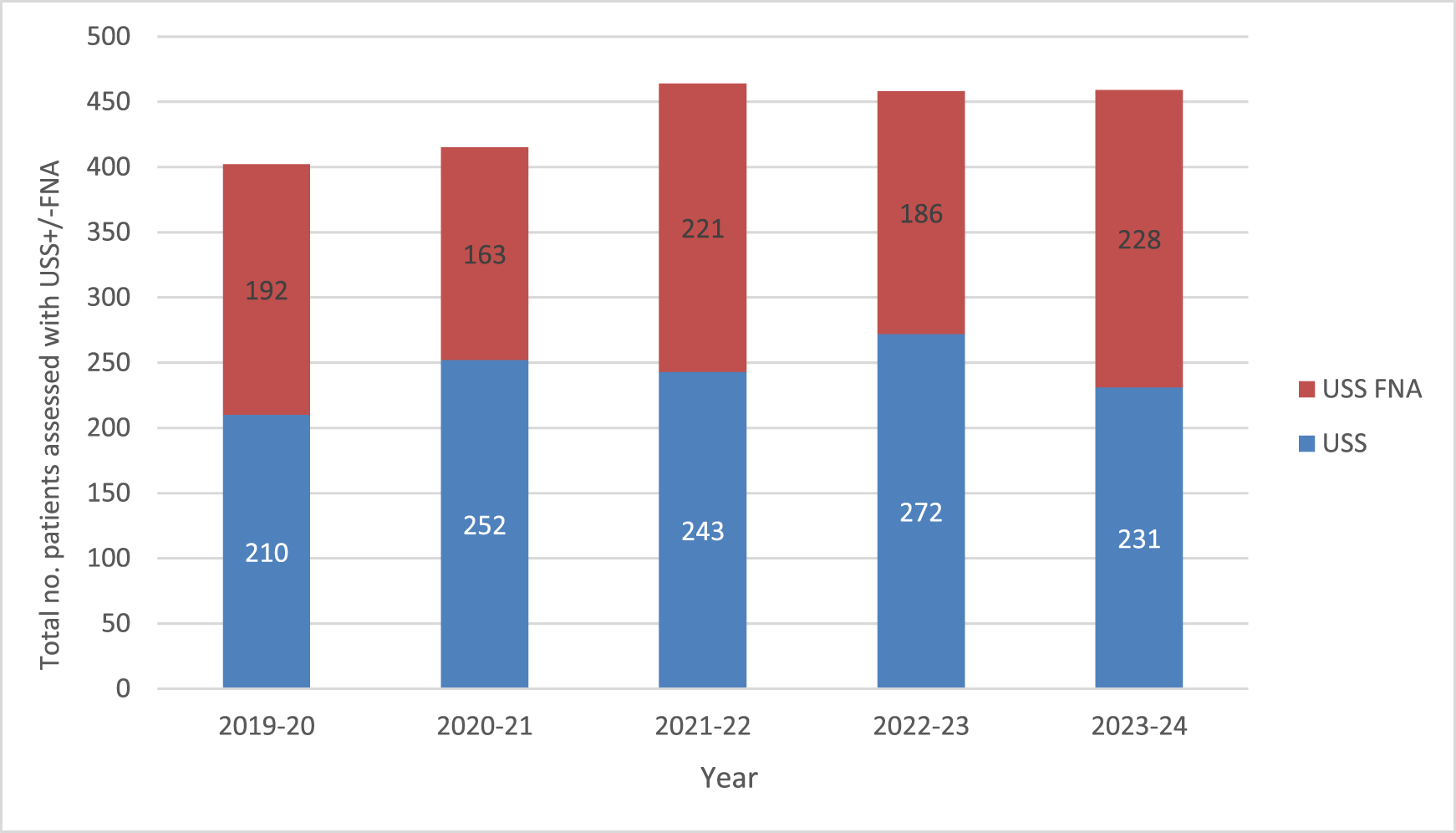
In total, 2198 patients were assessed with USS +/- FNA in the OSNLC over the five-year period. The number of patients seen in each individual year is summarised inFigure 1, with an average of 439.6 per year. Figure 1 also demonstrates the number of patients who underwent USS only compared to those who underwent USS FNA in each year. Overall, USS FNA assessment was performed in just under half of cases (n = 990; 45.0 per cent).
Total number of cases assessed each year showing numbers of patients assessed with USS only and USS FNAs.

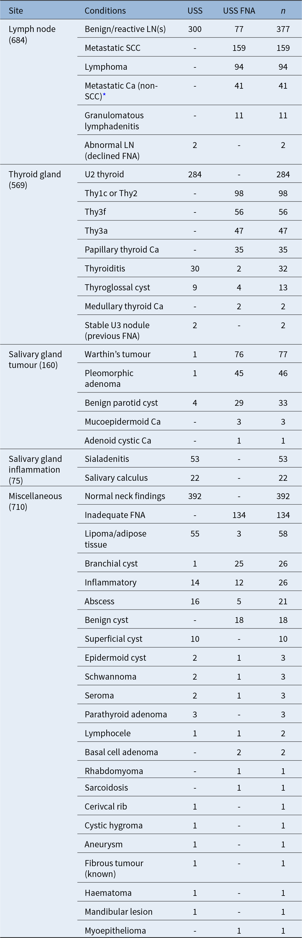
Of patients assessed with USS only (n = 1208), the most common finding was a normal neck, with no pathology demonstrated (n = 392; 17.8 per cent of total), followed by benign/reactive lymph node(s) (n = 300; 24.8 per cent) and U2 thyroid nodules (n = 284; 12.9 per cent). Of patients undergoing USS FNA (n = 990), the commonest benign pathology was a benign parotid tumour (pleomorphic adenoma or Warthin’s) (n = 121; 5.5 per cent), with benign or cystic thyroid nodules (Thy2 or Thy1c) (n = 98; 4.5 per cent) following. Overall, benign diagnoses were observed in 1235 cases (56.2 per cent of total USS +/- FNA). Abnormal findings (malignancy and Thy3a/Thy3f nodules) comprised 437 cases, 19.9 per cent of total cases. Confirmed malignancy (not including Thy3a and Thy3f nodules) was detected in 334 cases (15.2 per cent). The commonest diagnoses were metastatic squamous cell carcinoma (SCC) and lymphoma (n = 159, 7.2 per cent and n = 94, 4.3 per cent, respectively).
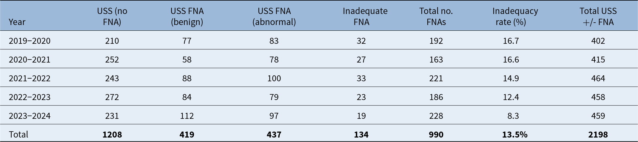
Of the total 990 USS FNAs, 134 were insufficient/ inadequate, yielding an overall inadequacy rate of 13.5 per cent over the five years analysed (seeTable 1). The inadequacy rate decreased each year with a rate of 8.3 per cent in the final year of 2023–2024 analysed. A diagnosis (benign or abnormal) was achieved in 2064 cases (93.9 per cent). A summary of all conditions diagnosed, both through USS alone and USS FNA, over the five years are summarised inTable 2.
Total number of cases per year based on USS or USS FNA (benign vs abnormal vs inadequate)

Summary of all 2198 conditions encountered in five-year period (2019–2024) diagnosed through USS alone vs USS FNA

* Metastatic adenocarcinoma (n = 11), non-small cell Ca (n = 14), metastatic melanoma (n = 7), small cell Ca (n = 1), spindle cell Ca (n = 1), spindle cell sarcoma (n = 1), myoepithelial Ca (n = 1), metastatic clear cell Ca (n = 1), metastatic urotehlial Ca (n = 1), necrotic atypical cells (uncertain aetiology) (n = 2), metastatic basaloid Ca (n = 1)
Discussion
In a post-pandemic era affected by challenging waiting list backlogs and urgency to meet cancer care targets, the need for increasing service efficiency and maximising resource utilisation has never been more pertinent. The OSNLC is one such initiative towards achieving this goal, advocated by NICE guidelines over two decades ago.3 Compared to a general ENT clinic, patients undergo specialist ENT assessment and USS +/- FNA in a single appointment, avoiding the need for multiple hospital visits, reducing the need for repeat scanning and frequency of inadequate sampling, all of which delay achieving definitive diagnosis. Delays in achieving definitive diagnosis and initiating treatment are associated with worse prognosis and locoregional progression.Reference Murphy, Galloway, Handorf, Egleston, Wang and Mehra14, Reference Shaikh, Handorf, Murphy, Mehra, Ridge and Galloway15 Identification of malignancy at an earlier stage serves to streamline treatment pathways and importantly, has been shown to improve overall prognosis.Reference Tumati, Hoang, Sumer, Truelson, Myers and Khan16
The OSNLC in Worcestershire Royal Hospital has been successfully implemented since 2018. Within the five years analysed, 2198 patients were assessed with USS +/- FNA in total, with a diagnosis achieved in 93.9 per cent. One of the key functions of the OSNLC is not only to detect patients with malignancy early, but also to reassure those that do not. The majority of patients had no concerning findings or benign pathology on USS with 392 patients having a “normal neck” on USS and a further 377 found to have benign/reactive lymph nodes with no pathological findings. In total, over half of patients (n = 1208; 55.0 per cent) were assessed with USS alone and found to have benign pathology or normal findings so could be suitably reassured that head and neck malignancy was excluded in their first appointment. A further 419 patients (19.1 per cent) had benign pathology confirmed by FNA, which was conveyed in their follow up appointment, typically two weeks after the initial OSNLC appointment. The anxiety experienced by patients around cancer-related imaging is well described.Reference Hussain, Chau, Turner and Paterson17, Reference Derry-Vick, Heathcote, Glesby, Stribling, Luebke and Epstein18 Thus, being able to reassure patients in a single visit or shortly thereafter is a significant benefit of the OSNLC.
Abnormal FNA results were reported in just under one fifth of cases (n = 437; 19.9 per cent). The majority of the abnormal cases were confirmed malignancy (n = 334; 15.2 per cent) with the remaining (n = 103) comprising Thy3a and Thy3f nodules. Given the potential for malignancy in Thy3 nodules, these were included in the “abnormal” category, whilst the authors recognise a significant proportion will likely be benign. The commonest malignancies detected were metastatic SCC (n = 159) and lymphoma (n = 94). A comparable study published in 2011 reported data on 2183 patients over a similar timeframe.Reference Hilmi, Yeo, O’Neill, McPhaden and MacKenzie5 The current study identified a greater number of metastatic SCC cases than Hilmi etal. (n = 76), reflecting the rising incidence of head and neck SCC in the UK population over the past decade.1 The number of lymphoma cases identified is remarkably similar with the Glasgow study (n = 91).Reference Hilmi, Yeo, O’Neill, McPhaden and MacKenzie5
Our study has shown persistently low sample inadequacy rates year on year. The rate has halved from 16.7 per cent in the initial 2019–2020 year to 8.3 per cent in the 2023–2024 year. Whilst there are no official target adequacy rates for non-thyroid head and neck sampling, the Royal College of Radiologists quote a non-diagnostic yield between 10 and 30 per cent and a target of 70 per cent diagnosis for thyroid nodule FNAs.19 Of 990 patients who underwent USS FNA in the current study, diagnosis was achieved in 856 cases (86.5 per cent) on initial assessment. A meta-analysis assessing FNA of thyroid nodules, including 17 studies with 24,649 nodules in total, reported an inadequacy rate of 24 per cent.Reference Issa, McCarthy, Hussein, Albuck, Emad and Shama20 This reduced to 8 per cent with the use of rapid onsite evaluation (ROSE). This figure only pertains to thyroid nodule assessment specifically rather than wider neck lumps but is almost identical to the 2023–2024 rate in our institution. For direct comparison, a smaller study evaluated inadequacy rates from a neck lump clinic in which the FNA samples were taken by the head and neck surgeons via palpation, without ultrasound guidance.Reference Reddy, Bennett, Bassett, Cunliffe, Fryer and Reece8 The study quoted inadequacy rates of 29.9 per cent and 40.4 per cent with and without on-site technician assessment respectively.Reference Reddy, Bennett, Bassett, Cunliffe, Fryer and Reece8 Even with on-site assessment, the quoted inadequacy rate of 29.9 per cent is significantly higher compared to our unit. Our data demonstrate an effective and high yield service with year-by-year improvement in diagnostic rates.
Additionally, other studies reporting OSNLC data involve smaller patient numbers (<500) and shorter durations of data collection, typically less than 24 months.Reference Hariri, Jawad, Otero, Lechner, Morley and Beale6–Reference Howlett, Harper, Quante, Berresford, Morley and Grant10 Whilst the OSNLC is anecdotally accepted as the gold standard for neck lump assessment, long term data reporting is deficient. Only the aforementioned study by Hilmi etal., which was published in 2011, has reported a similar case number of 2183.Reference Hilmi, Yeo, O’Neill, McPhaden and MacKenzie5 More recent studies published within the last decade report comparatively low case numbers.Reference Hariri, Jawad, Otero, Lechner, Morley and Beale6, Reference Sood, Supriya, Thopil, Surendra and Patel7, Reference Campbell, Huston and Collard11, Reference Sah, Sah and Khan12 The most recent study relating to an alternative approach to the one stop neck lump clinic, using a pre-clinic ultrasound pathway during the coronavirus pandemic, reported on a total of 99 patients.Reference Hariri, Jawad, Otero, Lechner, Morley and Beale6 Another relatively recent one-stop head and neck clinic study compared patients seen in 2004 to those seen in 2017, with 277 and 205 patients, respectively (482 total).Reference Campbell, Huston and Collard11 Two further OSNLC studies published within the past decade involve 146 and 144 patients respectively, with the latter focussing on parotid lesions only.Reference Sood, Supriya, Thopil, Surendra and Patel7, Reference Sah, Sah and Khan12 Pre-2015 studies similarly report data on fewer patients over a shorter time-frame.Reference Reddy, Bennett, Bassett, Cunliffe, Fryer and Reece8–Reference Howlett, Harper, Quante, Berresford, Morley and Grant10, Reference Al Hamarneh, Liew and Shortridge13 Comparatively, the current study provides an up-to-date report of OSNLC data, involving the longest period of data collection and largest case number currently reported in literature.
Clinical implications
The implementation of a OSNLC provides clear benefits in streamlining diagnosis of patients with head and neck malignancy and reassuring patients with benign pathology or normal findings in a single visit. The OSNLC is effective in providing definitive diagnosis with a significantly low cohort of patients requiring repeat cytopathological evaluation. Further, avoidance of multiple clinic appointments and repeat scanning relieves pressure on an already pressured system, improving overall resource utilisation. In cases where malignancy is detected, treatment pathways are promptly initiated to improve prognosis and reduce disease burden. Where resources and personnel are available, similar models of neck lump assessment should continue to be introduced nationally.
• National Institute for Health and Care Excellence (NICE) recommends patients with persisting neck lumps should be seen in a specialist lump clinic, involving ear, nose and throat surgeons, cytologists and radiologists.
• Dedicated neck lump clinics are perceived to improve efficiency and speed of diagnosis, but few studies report long-term and large patient data-sets in support of their implementation.
• This study reporting five-years of data from a single-centre One-Stop Neck Lump Clinic (OSNLC) demonstrates high diagnostic yield and yearly reduction in rates of non-diagnostic fine-needle aspiration (FNA) sampling.
• The OSNLC model improves resource efficiency and streamlines diagnosis and treatment of patients presenting with head and neck malignancy.
Conclusion
The OSNLC represents a significant advancement in the head and neck cancer diagnostic pathway, offering a streamlined, patient-centred model of care. By providing same-day clinical assessment, imaging, and, where appropriate, tissue sampling, the clinic expedites diagnosis and facilitates prompt initiation of treatment. The presence of a multidisciplinary team, including ENT consultants, radiographers and allied health professionals, ensures that patients receive expert input and have their concerns addressed comprehensively during a single visit. This collaborative approach not only improves clinical accuracy and continuity of care but also enhances patient understanding and satisfaction.
Critically, the model reduces the emotional burden associated with diagnostic delays by minimising waiting times, repeat appointments and unnecessary investigations. Faster diagnosis directly enables faster treatment, which is essential for optimising outcomes in head and neck oncology. In addition to its clinical benefits, the OSNLC improves service efficiency and reduces pressure on general outpatient services. It provides an effective, efficient and compassionate model of care that should be more broadly implemented. Adopted models should be encouraged in other specialties involving cancer diagnosis.
Contribution statement
All listed authors made significant contributions to the content, production and review of this article and have approved it prior to submission.
Data sharing
The data that support the findings of this study are available on request from the corresponding author. The data are not publicly available due to privacy or ethical restrictions.
Funding
No funding was required.
Competing interests
The authors declare that there is no conflict of interest.
Ethical approval
The authors confirm that no ethical approval was required for this study.