Case report
A 21-year-old female patient, diagnosed with tetralogy of Fallot, pulmonary atresia, and major aortopulmonary collateral artery at the age of 1 month, was followed up in our department. She was followed up without any surgical intervention based on the decision of the family, and fortunately evolved normally without major complications during her childhood. She had moderate cyanosis with 80% oxygen saturation level in room air, but was asymptomatic without limitation of physical activity (NYHA class I). She did not have any chromosomal abnormalities, including the 22q11.2 deletion. At the age of 19, she presented with frequent cough and wheezing, and occasional haemoptysis.
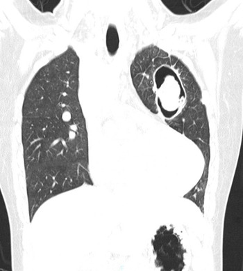
Computed tomography imaging of the chest revealed a 90 mm long cavitary lesion with a 38 mm nodule in the upper left lobe of the lung (Fig 1). Pulmonary aspergillosis was diagnosed by chest computed tomography imaging and detection of antibodies against Aspergillus in patient’s blood. Pharmacological management using voriconazole (200–300 mg/day for 6 months) was not effective. Consequently, thoracoscopic resection of the left upper lobe of the lung was planned.
Non-contrast computed tomography of the chest showing a 90 mm long cavitary lesion with a solid mass, 38 mm in diameter, inside the upper left lung lobe.

Contrast-enhanced computed tomography and cardiac catheterisation were performed. Based on these observations, the right major aortopulmonary collateral artery to the right lung S4–10 region originated directly from the Valsalva sinus, and the other two MAPCAs originated from the aortic arch to the right lung S1–3 region, and from the descending aorta to the left anterior lung, respectively. The tracheal valve was tricuspid without any stenosis or regurgitation. Thus, our working diagnosis was truncus arteriosus communis (Van Praagh type A3) and major aortopulmonary collateral artery without intrapericardial central pulmonary arteries. With the orifice stenosis of all the major aortopulmonary collateral arteries, mean pulmonary arterial pressure was as low as 9 mmHg and 5 mmHg at the right (S1–3 region) and the left pulmonary artery, respectively. The right and left ventricular size and function were observed to be appropriate for biventricular repair. Consequently, we decided to perform biventricular repair after thoracoscopic resection.
Prior to thoracoscopic resection, coil embolisation of many systemic-to-pulmonary collateral vessels in the cavitary lesion was performed to avoid excessive bleeding, and the left upper lobe of the lung was resected. Two weeks after successful thoracoscopic lobectomy, Rastelli operation with unifocalisation of all the major aortopulmonary collateral arteries was performed. Major aortopulmonary collateral arteries were divided from the aorta and were unifocalised using an expanded polytetrafluoroethylene graft. A new outflow from the right ventricle to the bilateral pulmonary artery was established using the expanded polytetrafluoroethylene conduit with bulging sinuses and fan-shaped valves. The ventricular septal defect was closed with an expanded polytetrafluoroethylene patch. Weaning from cardiopulmonary bypass was safe and successful. On the third postoperative day, a small right lung abscess was detected and was successfully treated with antibiotics. Owing to an unsuccessful extubations due to repeated pulmonary hemorrhage, coil embolisation of the small collateral artery including the bronchial artery was performed. The patient was successfully extubated on the 27th postoperative day and was discharged on the 43rd postoperative day.
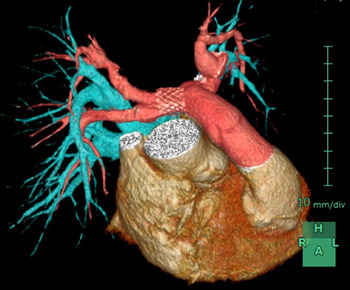
Two months after discharge, the proximal right pulmonary artery stenosis progressed, and stent implantation was performed via cardiac catheterisation. After the stent implantation, the size of the proximal right pulmonary artery increased from 5.3 mm to 12.0 mm and the pressure gradient decreased from 80 mmHg to 30 mmHg (Fig 2).
3D-computed tomography after the Rastelli operation and stent placement in the right pulmonary artery.

At present, the patient is under follow-up at the outpatient clinic and performs well without any late postoperative complications.
Discussion
Pulmonary aspergillosis is classified into four categories: simple pulmonary aspergilloma, chronic progressive lung aspergillosis, invasive aspergillosis, and allergic bronchopulmonary aspergillosis. Simple pulmonary aspergilloma is diagnosed in patients with cavitary lesions and is associated with fungus balls in a single cavity. The disease is also known to occur in compromised patients in some cases, even in the absence of basic pulmonary diseases. It is considered that the mechanism of pathogenesis of the disease involves the infiltrating growth of the fungi in the lung, which consequently leads to the formation of a cavity through the necrosis and destruction of lung tissue. Reference Segretain1,Reference Binder, Faling and Pugatch2 Some cases of aspergillosis associated with tetralogy of Fallot have been reported in the literature Reference Campbell and Clayton3,Reference Paulk, Schlant and Albert4 and these reports presumed that Aspergillus grows saprophytically in the locally immunocompromised lung tissue, owing to pulmonary hypoperfusion. It is possible that in the case presented in the current report, in addition to the unoperated truncus arteriosus communis and major aortopulmonary collateral artery, the patient had chronic local pulmonary hypoperfusion, which resulted in simple pulmonary aspergilloma.
In principle, the first treatment of choice for the management of simple pulmonary aspergilloma is surgical resection. Reference Walsh, Anaissie and Denning5 However, in high-risk surgical patients, such as patients with low lung function, pharmacological management with voriconazole or itraconazole is the preferred treatment modality. In adult patients with unoperated congenital heart diseases, the presence of pulmonary hypertension, including Eisenmenger’s syndrome, is a serious risk factor for surgery. Reference Warnes, Williams and Bashore6 It is recommended that surgery be avoided in such patients. A majority of the unoperated truncus arteriosus communis patients develop Eisenmenger’s syndrome. Reference Perloff, Perloff and Marelli7 It was suspected that the case presented in the current report was also a complication of the same. However, owing to the failure of the pharmacological mode of management, the cardiac function was evaluated in order to reconsider the possibility of a surgical intervention. The results indicated the absence of pulmonary hypertension. Consequently, it was possible to perform lobectomy and intracardiac repair. Even in scenarios involving unoperated cases of cyanotic congenital heart disease, haemodynamics should be evaluated and an adaptation of the surgical modality of treatment should be considered carefully.
In cases of congenital heart diseases associated with cyanosis that last for a long time, the development of bronchial arteries and collateral vessels as a biological response to hypoxia is often a problem, in addition to systemic multiple organ failure (erythropoiesis, coagulopathy, renal dysfunction etc.), secondary to chronic hypoxemia. Increased venous return to the pulmonary artery and rupture of fragile neovascular vessels cause bleeding during and after the surgical procedure. Hence, coil embolisation of collateral blood flow, prior to the surgical procedure, is recommended in patients with intractable pulmonary diseases. In this case, coil embolisation was performed before surgery, owing to the several collateral vessels that developed around the lung cavitary lesion. The total volume of intraoperative blood loss during lung resection was observed to be small, and the aforementioned pretreatment involving coil embolisation might have contributed to the prevention of excessive bleeding during the surgical procedure.
Conclusion
If patients with unoperated cyanotic congenital heart disease have complicated, intractable pulmonary cavitary lesions, surgical treatment, and intracardiac repair should be considered in the absence of pulmonary hypertension.
In view of the fact that intraoperative bleeding can be a major risk factor, it is necessary to take measures to minimise that risk.
Acknowledgements
We would like to thank Dr. Kichizo Kaga, Department of Cardiovascular and Thoracic Surgery, Hokkaido University Hospital for surgical management of pulmonary aspergillosis.
Financial support
This research received no specific grant from any funding agency, commercial, or not-for-profit sectors.
Conflicts of interest
None.