Introduction
Immense efforts are on to find the exact beginning of the pathway of life on earth 3.5 million years ago. It is assumed that life occurred on earth surface by the self-reproduction of organic compounds in the presence of water and some inorganic minerals such as metal oxides, clay, metal cyanides, etc. Inorganic minerals provide the surface for the adsorption and chemical reactions to carry out the prebiotic process. Hence, it was proposed that these minerals played a vital role in the origins of life (Cleaves et al., Reference Cleaves, Scott, Hill, Leszczynski, Sahai and Hazen2012). Research is in progress to study the interaction and catalytic studies of the nucleic acids with different inorganic compounds. Cains-Smith proposed the role of clay in the origin of life. They suggested that clay minerals could store and replicate structural defects, dislocations and ionic substitutions, and act as ‘genetic candidates’ (Cains-Smith, Reference Cains-Smith1982). Choy et al. discussed the role of clay minerals and layered double hydroxides in various biological applications (Choy et al., Reference Choy, Choi, Oh and Park2007). Intercalation of DNA into CTAB-modified clay minerals for gene protection concluded that CTAB-clay/DNA hybrids are potential materials for the storage of genetic information (Hou et al., Reference Hou, Wu, Huang, Ruan, Liu and Zhu2014). Brack (Reference Brack2013) and Ponnamperuma et al. (Reference Patel, Patel, Chauhan, Chawla and Rawal1982) also worked on the role of clay in the origin of life. The interaction of ribose nucleotides with zinc and copper ferrocyanides has been studied and its role in the origin of life has been discussed (Kamaluddin et al., Reference Kamaluddin and Sharma1994a, Reference Kamaluddin, Nath and Sharmab). Anand et al. worked on the role of double metal ferrocyanides in chemical evolution by studying the interactive studies between RNA components with metal (II) hexacyanocobaltate . Shankar et al. (Reference Shanker, Bhushan, Bhattacharjee and Kamaluddin2012) have reviewed the procedure of synthesis, characterization and role of nanometal hexacyanoferrates. Studies have been carried out on the role of metal oxides in chemical evolution. Oligomerization of glycine and alanine with iron oxide, and its implication in the origins of life have been studied (Shanker et al., Reference Shanker, Bhushan, Bhattacharjee and Kamaluddin2012). The interaction of valine and glycine with aluminium, iron and zinc oxides was studied and hence the role of metal oxides in the origin of life has been supported (Tewari Reference Tewari2017). Arora and Kamaluddin (Reference Arora and Kamaluddin2009) studied the interaction of ribonucleotides with acidic, neutral and basic alumina. Purine nucleotides adsorption was more in acidic conditions (acidic alumina). Adsorption studies of ribose nucleotides with zinc oxide and their role in chemical evolution have been studied (Arora and Kamaluddin, Reference Arora and Kamaluddin2007). Maximum adsorption was observed at neutral pH. The role of hematite–water system in the origins of life on Mars has been reported (Arora et al., Reference Arora, Tomar, Arya and Venkateswararao2006); hematite is one of the most abundant oxides on planets like Mars. Rutile–nucleotide interaction and its role in chemical evolution have been reported (Cleaves et al., Reference Cleaves, Jonsson, Jonsson, Sverjensky and Hazen2010). Feuillie et al. studied the adsorption of ribonucleotides on α-alumina as a function of pH, ionic strength and surface loading (Feuillie et al., Reference Feuillie, Sverjensky and Hazen2015). The role of manganese oxides in chemical evolution has also been studied. Different forms of manganese oxides were used as a catalyst for the polymerization of bio monomers (Bhushan et al., Reference Bhushan, Shanker and Kamaluddin2011). These entities were found to be an efficient catalyst for the polymerization reaction. The interaction of ribonucleic acid constituents has been studied with different spinel ferrite nanoparticles (Iqbal et al., Reference Iqbal, Sharma and Kamaluddin2016). Maximum adsorption takes place in acidic medium. Nickel ferrite (NiFe2O4) showed a higher per cent of binding while manganese ferrite (MnFe2O4) exhibited the lowest. Zirconium is a common element in the earth crust. Moon rocks have more zirconium than earth's rocks. Sun also contains a tiny amount of zirconium. Some cooler stars, which glow with a red light, contain a larger amount of zirconium. Zirconium being a reactive metal forms bonds with other elements to form minerals ( Fig. 1). Zirconium oxide has wide applications in cosmetics, artificial jewellery, abrasive, ceramics, etc. Li et al. have worked on copper oxide-doped zirconium oxide composite microfibres. Their further applications as non-enzymatic MP sensor have been studied (Li et al., Reference Li, Wang, Wang, Gong and Qi2019). Atalay et al. synthesized zirconium oxide nanoparticles of size 20 nm and studied the genotoxic and apoptotic effects of the synthesized zirconium oxide on L929 mouse fibroblast cell line (Atalay et al., Reference Atalay, Celik and Ayaz2018). Zirconium oxide films have been synthesized by the magnetron sputtering method and their various properties have been studied (Patel et al., Reference Ponnamperuma, Shimoyama and Friebele2016). The 3D-characterization of deuterium distributions in zirconium oxide scale has been carried out by using high-resolution SIMS (Kexuel et al., Reference Kexuel, Thomas, Liu, Hulme, Garner, Sergio and Grovenor2019). Ali and coworkers used zirconium oxide as a catalyst for biofuel production by cracking of waste cooking oil (Ali et al., Reference Ali, Reshad, Machhindra, Bhalerao and Goud2018). Zirconium oxide/PAN nanofibres have been used as an adsorbent for the removal of fluoride from groundwater by Wang et al. (Reference Wang, Pan, Zhang, Qi, Sun, Gu, Wang and Li2019). Branched polyethyleneimine has been used for the modification of zirconium oxide nanofibres. Nanocomposites of zirconium oxide nanoparticles and poly (3,4-ethylenedioxythiophene) have been synthesized (Arvand et al., Reference Arvand, Elyan and Ardaki2019). These nanocomposites have been applied for electrocatalytic oxidation and detection of progesterone. Ni/ZrO2 catalyst has been synthesized and applied for the hydrodeoxygenation reaction of guaiacol (Christian et al., Reference Christian, Pichler, Gu, Joshi and Schuth2018). Nanocrystalline zirconium oxide powder has been synthesized and their structural and optical properties have been studied (Waghmar et al., Reference Waghmar, Pawar, Pathan and Ubale2017). As metal oxides are under study for their role in the origins of life for providing their surfaces for selective adsorption, this work is focused on the interaction of ribose nucleotides namely, 5′-AMP, 5′-GMP, 5′-CMP and 5′-UMP with zirconium oxide. It is well known that the RNA molecule is a polymer of ribonucleotides viz., 5′-AMP, 5′-GMP, 5′-CMP and 5′-UMP, linked through phosphodiester bonds. It is, therefore, a matter of growing interest to understand how the nucleotide molecules were concentrated from their dilute aqueous solutions and subsequently polymerized into RNA and DNA molecules. Since ribose nucleotides have negatively charged phosphate groups, lone pair of electrons on nitrogen atoms and π-electron clouds on aromatic rings, therefore, may interact through these negatively charged moieties with the positively charged surface of zirconium oxide. Present work shows zirconium oxide to be a good adsorbent for ribonucleotides. Therefore, it strongly supports the role of metal oxides in concentrating biomolecules during the course of chemical evolution. No work has been reported till date for the role of zirconium oxide in chemical evolution and origins of life and its interaction with ribose nucleotides namely, 5′-AMP, 5′-GMP, 5′-CMP and 5′-UMP.
The oldest rock in the world which has been magnified 5000 times is nearly 4 billion years old and contains tiny zirconia crystals.

Experimental
Material
Zirconium oxychloride (ZrOCl2), 5′-AMP, 5′-CMP, 5′-GMP and 5′-UMP were purchased from Sigma Aldrich. Analytical grade reagents were used throughout the experiments (https://www.nanopartikel.info/en/nanoinfo/materials/zirconium-dioxide/material-information.)
Preparation of zirconia nanoparticles
Zirconium oxychloride (ZrOCl2) solution (750 ml of 0.05 M solution) was taken in a 2 l beaker. Aqueous ammonia (0.05 M) was added to this solution dropwise. The solution was mechanically stirred throughout the experiment. The pH of the solution was controlled with the help of pH meter. Addition of liquor ammonia was stopped when no more precipitates were observed. The pH of the solution was 10.5 at the completion of the experiment at 60°C. The obtained precipitates were filtered and washed to remove the excess of ammonia. The precipitates were kept in oven at 60oC. The dried precipitates were calcined at 500°C in a muffle furnace for about 6 h. After calcination, crystalline oxide was ground with the help of pestle and mortar, sieved through 100-mesh size sieve, and characterized.
Characterization of zirconia nanoparticles
Bruker, D-8 advanced X-ray diffractometer was used for X-Ray Diffraction (XRD) analysis. Transmission electron microscopy (TEM) measurements of the sample were carried out on FEI Tecnai G2 20 S-Twin TEM. Scanning electron microscopy (SEM) studies were performed with (LEO 435VP) model. The magnetic measurements were carried out by using Model PAR 155. The surface area of the synthesized zirconia was found out using micrometrics ASAP 2010 (UK).
Adsorption studies
Different concentrations of the nucleotides (5 × 10⁻⁵M–300 × 10−5 M) were chosen for adsorption studies. A total of 25 mg of the zirconia was taken along with 5 ml of the nucleotide. A total of 25 mg of the zirconia was added to 5 ml of the nucleotide solution having varying concentrations. This mixture was shaken vigorously for 2 min and kept at room temperature for approximately 24 h. It was centrifuged for 30 min at 4000 rpm using REMI, centrifuge R4C. The optical density of the filtrate was observed at the characteristic λmax of the nucleotide. This O.D. was compared with the standard and the post-adsorption concentration of the nucleotide was calculated using the standard calibration curve. Nucleotide–zirconia adduct was washed with water, dried and was subjected to spectral studies.
Spectral studies:
UV–Visible studies were performed on Shimadzu UV-1601 spectrophotometer. The characteristic λmax values for 5′-AMP, 5′-GMP, 5′-CMP and 5′-UMP are 259, 254, 278 and 262 nm, respectively. IR studies of the adsorbent, adsorbate and adsorption adduct were recorded using Perkin Elmer FTIR spectrophotometer (Model Perkin Elmer-1600 Series).
Results and discussion
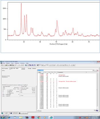
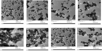
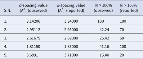
X-ray diffraction studies (Fig. 2) showed that the synthesized metal oxide is pure ZrO2 and exists in monoclinic form (Table 1). Magnetic susceptibility measurements showed that synthesized zirconium oxide is diamagnetic in nature. There are no unpaired electrons in ZrO2.. Observed magnetic moment, μobs, of the synthesized oxide was observed to be 0.34B.M. TEM study was performed to find out the particle size of synthesized zirconium oxide and these images are presented in Fig. 3. TEM studies showed that the synthesized ZrO2 particles were in the range from 14 to 27 nm.
XRD spectrum of zirconia nanoparticles.

TEM images of synthesized zirconia nanoparticles.

X-ray diffraction data for zirconia nanoparticles

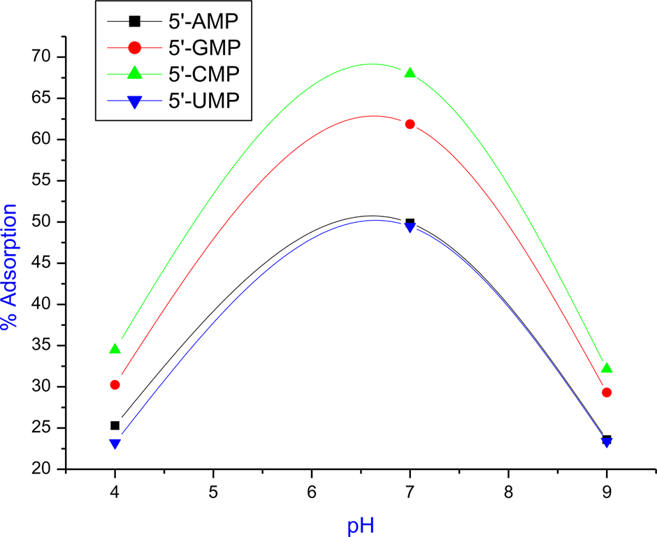
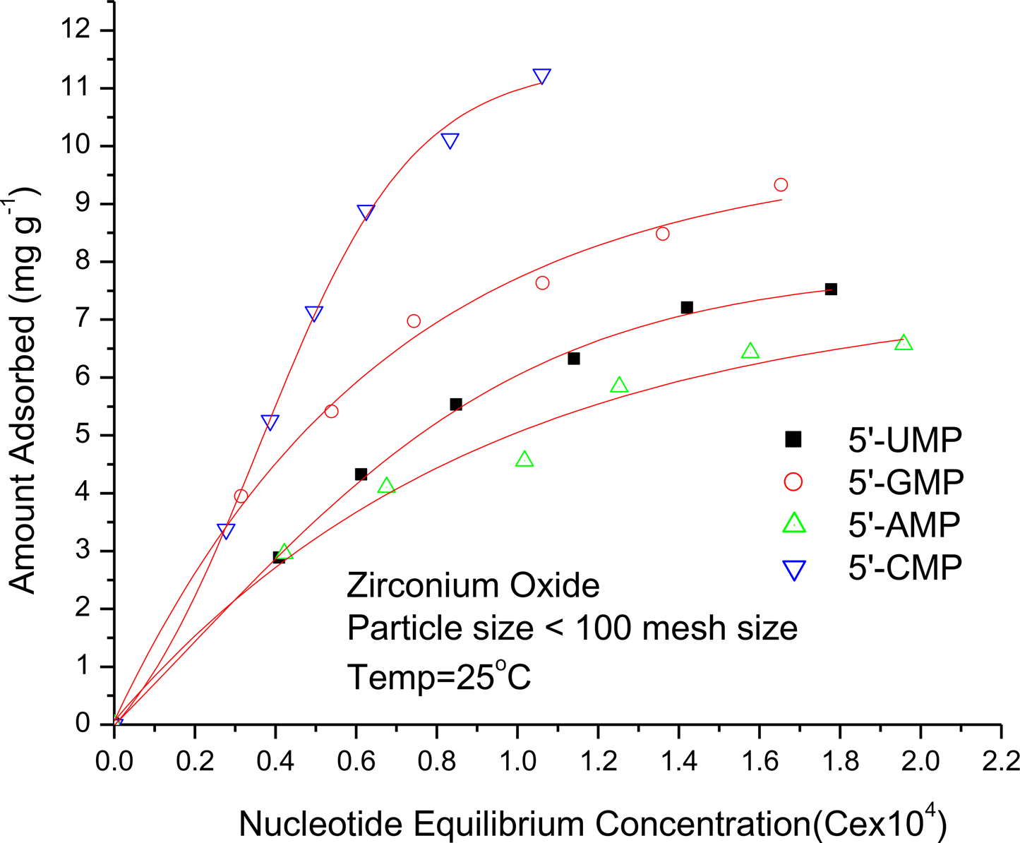
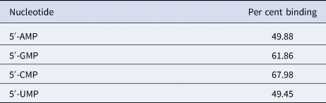
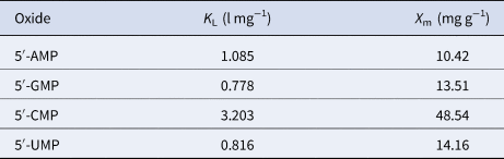
The preliminary studies on the interaction of 5′-AMP, 5′-GMP, 5′-CMP and 5′-UMP with zirconium oxide were carried out at pH 7.0. Point of zero charge (PZC) for zirconium oxide varies from 4 to 11 (Kosmulski, Reference Kosmulski2001). The surface of zirconium oxide is positive below PZC while it is negative above PZC. P Ka2 values for 5′-AMP, 5′-GMP, 5′-CMP and 5′-UMP are 6.1, 6.1, 6.3 and 6.3, respectively (Fig. 4). Dianion nucleotides were formed by the dissociation of second proton of phosphate group. This implies that at neutral pH, all the four nucleotides will exist in their dianionic form. Therefore, at pH above P Ka2, nucleotides should show higher interaction between negatively charged nucleotide and positive charged surface of zirconium oxide. Nucleotides get protonated in acidic medium, so they have some more positive charge. Therefore, at lower pH (in acidic pH), interaction between positively charged surface of zirconium oxide and nucleotides is not much. At higher pH, the surface of zirconium oxide becomes negative in nature, and therefore, there is less interaction between negative moieties of nucleotides and zirconium oxide. At neutral pH, nucleotides are anionic in nature and zirconium oxide surface remains positive in nature, so higher adsorption is found at neutral pH. The equilibrium concentration of nucleotide (Ce) versus the amount of nucleotide adsorbed (Xe) plot was sigmoid in nature (Fig. 5). The initial portion of the isotherms showed a linear relationship between the amount adsorbed and the equilibrium concentration of the ribonucleotides. As the concentration increases, saturation phenomenon appears showing no further adsorption takes place. From the data of the curve at saturation, the per cent binding was calculated and listed in Table 2. The adsorption data were fitted in Langmuir adsorption isotherm. A typical graph of Ce versus Ce/Xe is a straight line (Fig. 6). K L and X m values were determined and are shown in Table 3. In general, zirconium oxide was found to have an extremely high affinity for ribonucleotides as the extent of the surface covered (θ = K LCe/(1 + K LCe)) was determined to be about 100% for all the four nucleotides. The type and number of surface groups on ZrO2 particles determine the extent of nucleotide adsorption. The expected form of zirconium oxide could be, −ZrO2H+, −ZrOH, −ZrO2− and their concentration depends on the pH of the solution. PZC for zirconium oxide varies from 4.0 to 11.0. So decreasing the pH value, concentrations of ZrO2H+ group and −ZrOH group increase markedly, while that of ZrO2− group decreases. At pH, below PZC, the surface of zirconium oxide is positively charged (ZrO2H+), and therefore, adsorption takes place due to attraction between negatively charged groups of nucleotides and positively charged surface of zirconium oxide. In addition, negatively charged oxygen atom of the ribose nucleotide molecule may establish hydrogen bonding with the ZrOH groups on zirconium oxide surfaces, resulting in the adsorption of ribose nucleotides onto the zirconium oxide surface. In alkaline medium, the surface of zirconium oxide becomes negatively charged, due to the formation of ZrO2− group on its surface. There will be repulsion between ZrO2− and the negatively charged oxygen atoms of nucleotides. So the adsorption is very low (~1%). The interaction of nucleotides could also take place due to the interaction of negatively charged oxygen atoms in phosphate group, π-electrons of aromatic ring and lone pair of N and O atoms with positive zirconium oxide. Purines (5′-AMP, 5′-GMP) having one extra ring and lone pair of electrons (N–7) are adsorbed more on zirconium oxide as compared to pyrimidines (5′-CMP and 5′-UMP).
Effect of pH on the adsorption of nucleotides.

Adsorption isotherms of nucleotides on zirconium oxide.

Langmuir isotherm plots of nucleotides on zirconium oxide.

Per cent binding of nucleotides on zirconia nanoparticles

Langmuir constants for adsorption of nucleotides on zirconium oxides

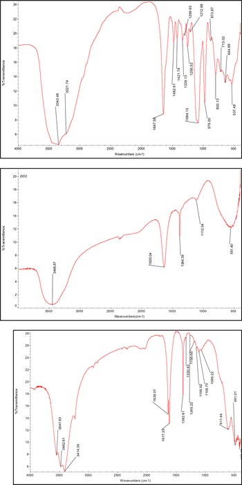
Infrared studies were carried out to find out the point of interaction between ribonucleotides and zirconium oxide. The infrared spectrum of 5′-AMP, zirconium oxide and zirconium oxide–nucleotide adduct are presented in Fig. 7(a)–(c), respectively. The point of interaction is shown by the shift in wavelength of the ribonucleotides after interaction with zirconium oxide. Shift in characteristic frequencies of ribonucleotides in nucleotide–zirconium oxide adduct has been reported in Table 4. It seems that the binding sites of nucleotides are N-1 and N-7 for 5′-AMP, N-1 and N-7 for 5′-GMP, N-3 for 5′-CMP and N-3 for 5′-UMP. The adsorption is presumably related to the involvement of N-1, N-3 and N-7 of the base residues, lone pair of nitrogen atom as well as to the dissociation of two available protons of the phosphate group. A shift towards lower wavelength of N–H bending from 1647 to 1617 cm−1 shows the involvement of –NH2 group in the interaction of ribonucleotide with zirconium oxide. A strong band at 1084 cm−1 in 5′-AMP corresponding to C–O–P vibration has been shifted to 1108 cm−1 indicating an interaction between amino and phosphate group of ribonucleotides with zirconium oxide. No significant change in the typical infrared frequencies of zirconium oxide suggests that the ribonucleotide molecules do not enter into the lattice of zirconium oxide.
IR spectra of AMP, zirconium oxide and AMP–zirconium oxide adduct.

Typical infrared spectral frequencies (cm−1) of ribonucleotides before and after absorption on zirconia nanoparticles

a Bracket values indicate typical infrared spectral frequencies after adsorption.
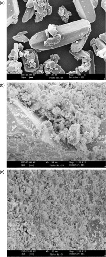
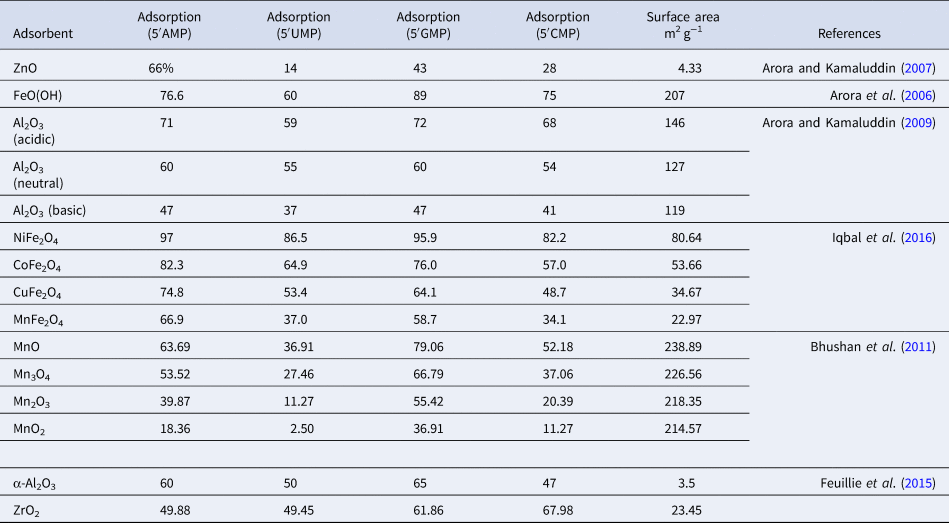
SEM images of the AMP before and after adsorption on zirconium oxide nanoparticles have been presented in Fig. 8. It is clear from SEM images that the adsorption of AMP takes place on zirconium oxide nanoparticles. Surface area of the synthesized zirconium oxide was found out experimentally as 23.45 m2 g−1, using BET. Zirconium oxide shows good adsorbing property towards nucleotides in comparison to other minerals, e.g. montmorillonite (750 m2 g−1) and aluminium oxide (146 m2 g−1) having more surface area.
SEM photographs of AMP, zirconium oxide and AMP–zirconium oxide adduct.

Conclusion
This work supports the role of zirconium oxide in concentrating the biomolecules during chemical evolution. Better adsorption of zirconium oxide as compared to other minerals shows its importance in chemical evolution (Table 5). Zirconium oxide was in nanosize range and with high surface area. The results of the present work for the adsorption of ribose nucleotides on zirconium oxide support that metal oxides present on primitive earth surface might have protected biomolecules from degradation and helped for further polymerization reactions.
Percentage adsorption of nucleotides on different metal oxides

Acknowledgements
The authors thank Dr Kamaluddin, Professor, Department of Chemistry, Indian Institute of Technology, Roorkee for his valuable suggestion to carry out this research.
Author contributions
The authors contributed equally to analysing data and reaching conclusions, and in writing the paper.
Financial support
None.
Conflict of interest
None.