Introduction
Haemangiopericytomas are rare, vascular neoplasms that are derived from pericytes, which surround all capillaries, and which account for only 1 per cent of all vascular tumours.Reference Kanazawa, Nishino, Miyata, Kuriki, Abe and Ichimura1 Between 7.5 and 25 per cent of all haemangiopericytomas are found in the head and neck region, with a marked tendency to occur in the nasal cavity and paranasal sinuses.Reference Kanazawa, Nishino, Miyata, Kuriki, Abe and Ichimura1, Reference Thiringer, Costantino and Houston2
Due to the variation in both histological and clinical features of sinonasal-type haemangiopericytomas, compared with their soft tissue counterparts, it is now widely accepted that these tumours represent an independent clinical entity, referred to as a glomangiopericytoma.Reference Worden, Getz, Luo and Hwang3–Reference Gengler and Guillou6
The effective management of these lesions depends on complete extirpation, with clear margins during initial surgery in order to reduce the potential for recurrence.Reference Snyderman, Carrau, Kassam, Zanation, Predevello and Gardner7 This has traditionally been achieved through either craniofacial resection, mid-facial degloving or lateral rhinotomy approaches, all of which are associated with significant morbidity and major complications.Reference Schlosser, Woodworth, Gillespie and Day8, Reference Leong, Citardi and Batra9
Recent advances in endoscopic surgery have led to the development of techniques that now permit the endoscopic resection of neoplastic lesions not only in the nasal cavity but also the anterior skull base.
In this report, we describe the histopathological features of glomangiopericytoma, and also the complete endoscopic resection of an extensive glomangiopericytoma with associated involvement of the anterior skull base.
Case report
A 32-year-old woman presented to our service with a six-month history of worsening nasal obstruction, anosmia, severe frontal headaches and right-sided proptosis.
Endoscopic examination of the nasal cavity revealed a right-sided, vascular, sinonasal mass abutting the superior turbinate, with associated deviation of the nasal septum to the contralateral side.
An urgent computed tomography (CT) scan of the paranasal sinuses was performed, and the patient was scheduled for endoscopic examination and biopsy under general anaesthesia.
The CT scan showed a large soft tissue mass occupying the right nasal cavity, with involvement of the sphenoid sinus as well as the anterior skull base.
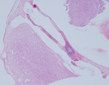
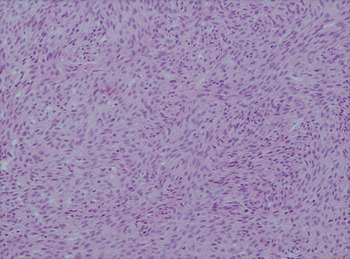
Histopathological examination of the biopsy specimen (Figures 1 and 2) revealed a submucosal spindle cell tumour exhibiting a fascicular growth pattern, with slender spindle cells embedded in fibrous stroma and surrounding thin-walled ‘stag horn’ vessels. There was no evidence of mitotic activity, necrosis or cellular atypia.
Photomicrograph demonstrating histologic appearance of glomangiopericytoma. (H&E, low power magnification)

Histologic appearance of glomangiopericytoma. (H&E, high power magnification).

On immunohistochemical analysis, less than 1 per cent of cells were proliferating, as assessed by MIB-1, and lesional cells were negative for cluster of differentiation 34 protein. Gordon and Sweet's method for detecting reticular fibres revealed a fine meshwork of short reticular fibres in a regular, even pattern within the tumour. Vimentin staining was strongly positive, while staining with actin had a moderately positive result. Additional staining with cluster of differentiation 99 protein and factor XIIIa proved negative, whilst Bcl-2 staining was weakly positive.
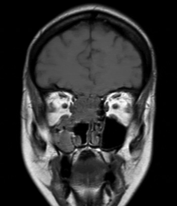
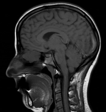
To aid pre-operative planning, magnetic resonance imaging (MRI) was performed. This confirmed the presence of a 3.2 × 2.6 × 2.3 cm mass with intense, homogeneous enhancement, involving the nasal septum, right posterior ethmoid cells, right superior sphenoid sinus and anterior skull base (Figures 3 and 4).
Coronal MRI, demonstrating glomangiopericytoma.

Sagittal MRI, demonstrating glomangiopericytoma.

Complete endoscopic excision of the lesion, including a modified Lothrop's procedure, was performed with the aid of an image-guided navigation system. Frozen section analysis performed during surgery confirmed the presence of clear margins. Intra-operative findings included extensive tumour involvement of the anterior skull base, with associated osseous dehiscence of the cribriform plate and extension of the tumour into the sphenoid sinus.
The nature and extent of surgery facilitated post-operative surveillance. Eighteen months after the resection, no evidence of recurrence was noted.
Discussion
Glomangiopericytomas are defined as sinonasal tumours demonstrating a perivascular myoid phenotype, and account for between 0.5 and 1 per cent of all sinonasal tumours.Reference Dandekar and McHugh4, Reference Higashi, Nakaya, Watanabe, Ikeda, Suzuki and Oshima5 These tumours can occur at any age; however, previous case series have suggested a peak incidence during the sixth and seventh decades.Reference Schlosser, Woodworth, Gillespie and Day8 The usual mode of presentation includes either nasal obstruction or epistaxis.Reference Bignami, Dallan, Battaglia, Lenzi, Pistochini and Castelnuovo10
Whilst corticosteroid use, hypertension and pregnancy have been proposed as aetiological factors in the development of glomangiopericytoma, this is not widely accepted.Reference Worden, Getz, Luo and Hwang3, Reference Higashi, Nakaya, Watanabe, Ikeda, Suzuki and Oshima5
Diagnostic investigation of suspected sinonasal tumours depends firstly on thorough endoscopic evaluation and secondly on imaging techniques including CT and MRI. The characteristic findings of these tumours on CT imaging include a soft tissue mass with enhancement following the administration of intravenous contrast, and gadolinium contrast in the case of MRI scanning.Reference Mosesson and Som11 The role of routine angiography in the management of glomangiopericytoma is not clearly defined. However, the majority of authors advocate angiography in larger tumours, in order not only to facilitate pre-operative planning but also to enable embolisation, which in turn significantly reduces the risk of intra-operative haemorrhage.Reference Schlosser, Woodworth, Gillespie and Day8, Reference Bignami, Dallan, Battaglia, Lenzi, Pistochini and Castelnuovo10, Reference Serrano, Coste, Percodani, Herve and Brugel12
Stout and Murray were the first to describe haemangiopericytoma, in 1942, as vascular soft tissue tumours of presumed pericytic origin.Reference Dandekar and McHugh4, Reference Bignami, Dallan, Battaglia, Lenzi, Pistochini and Castelnuovo10 The histological features of haemangiopericytoma consist of uniform, spindle-shaped cells with indistinct cytoplasm and large nuclei, distributed around vascular channels which typically exhibit a stag horn branching pattern.Reference Serrano, Coste, Percodani, Herve and Brugel12 The vascular channels are often more readily defined following reticulin staining, which clearly demonstrates the proliferation of tumour cells outside the vascular sheath.Reference Thiringer, Costantino and Houston2
However, since its initial description the characteristic histological features encountered in haemangiopericytoma have been found to be nonspecific and encountered in up to 15 per cent of unrelated soft tissue neoplasms.Reference Dandekar and McHugh4, Reference Higashi, Nakaya, Watanabe, Ikeda, Suzuki and Oshima5 This has led many authors to view haemangiopericytoma as a histological pattern rather than a distinct clinical entity.Reference Dandekar and McHugh4 Furthermore, haemangiopericytoma of the sinonasal cavity appears to have a lower malignant potential and metastatic potential, compared with haemangiopericytomas found elsewhere.Reference Schlosser, Woodworth, Gillespie and Day8 This has led some authors to conclude that sinonasal haemangiopericytomas are a separate pathological entity altogether; some have described them as ‘haemangiopericytoma-like’ tumours, whilst others have considered them to be a variant of glomus tumours.Reference Tse and Chan13 In the 2005 World Health Organization classification of head and neck tumours, it was recommended that sinonasal-type haemangiopericytoma be referred to as glomangiopericytoma, in light of the similarity with glomus tumours.Reference Worden, Getz, Luo and Hwang3
Glomangiopericytoma is a spindle cell neoplasm which exhibits haemangiopericytoma-like vessel patterns with frequent perivascular hyalinisation of the capillary-sized vessels. There is strongly positive immunohistochemical staining with vimentin and with actin, as demonstrated in our case. Staining for desmin, cluster of differentiation 34, 99 and 117 proteins, S100, and Bcl-2 typically yields negative results.Reference Dandekar and McHugh4 The histological differential diagnosis of glomangiopericytoma includes other spindle cell neoplasms, in particular solitary fibrous tumours; however, unlike glomangiopericytoma, solitary fibrous tumours demonstrate overlapping cells arranged in a more haphazard fashion, as well as the presence of coarse collagen bundles, not encountered in glomangiopericytoma. Furthermore, solitary fibrous tumours fail to show any immunohistochemical features of myoid differentiation, and therefore are almost always negative for desmin and cluster of differentiation 34 protein staining.Reference Dandekar and McHugh4, Reference Gengler and Guillou6
Some authors have advocated conservative management of glomangiopericytoma involving the anterior skull base, consisting of debulking and subsequent endoscopic surveillance.Reference Thiringer, Costantino and Houston2 However, the overwhelming weight of opinion supports the principles of an ‘oncological plane’ and en bloc excision with clear margins, in order to reduce the potential for recurrence, as these neoplasms are relatively radio-resistant.Reference Schlosser, Woodworth, Gillespie and Day8, Reference Bignami, Dallan, Battaglia, Lenzi, Pistochini and Castelnuovo10, Reference Serrano, Coste, Percodani, Herve and Brugel12
Traditionally, this has been achieved through a variety of approaches, including craniofacial resection (considered to be the ‘gold standard’), lateral rhinotomy and mid-facial degloving, all of which are associated with significant morbidity and major complications in up to 40 per cent of cases.Reference Thiringer, Costantino and Houston2, Reference Schlosser, Woodworth, Gillespie and Day8, Reference Leong, Citardi and Batra9
Endoscopic surgery of the nasal cavity and paranasal sinuses has evolved to the extent that it is now the preferred means of treatment for both benign and malignant neoplasms of the nasal cavity as well as the anterior skull base. The greatest benefits of endoscopic surgery lie in the enhanced visualisation it offers, as well as its facilitation of post-operative surveillance of the surgical site for recurrent disease.Reference Snyderman, Carrau, Kassam, Zanation, Predevello and Gardner7
Despite this, open surgery still has a role to play in cases where there is extensive involvement of the paranasal sinuses, invasion of the ocular structures and/or involvement of the superficial tissues.Reference Snyderman, Carrau, Kassam, Zanation, Predevello and Gardner7, Reference Serrano, Coste, Percodani, Herve and Brugel12
Major complications following endoscopic skull base surgery are relatively uncommon compared with the high rates reported for open approaches. The most commonly encountered major complication of endoscopic skull base surgery is cerebrospinal fluid leakage, which occurs in approximately 5 per cent of cases; in 95 per cent of such cases, this can be managed with either a lumbar spinal drain or further endoscopic surgery.Reference Snyderman, Carrau, Kassam, Zanation, Predevello and Gardner7
• Glomangiopericytomas constitute less than 1 per cent of sinonasal tumours
• Presenting symptoms include epistaxis and nasal obstruction
• Haemangiopericytoma-like vessel patterns are seen, with perivascular capillary hyalinisation
• Actin and vimentin staining is typically positive
• Traditional management involved open surgery
• Endoscopic surgery now enables resection with less morbidity and complications
The reported recurrence rate of glomangiopericytoma is quite varied, ranging from 7 to 50 per cent, with an average time to recurrence of six to seven years.Reference Thiringer, Costantino and Houston2 Whilst incomplete primary excision has been identified as the primary factor in recurrent disease, severe nuclear pleomorphism, osseous invasion, large tumour size (i.e. more than 5 cm) and a high mitotic to proliferation rate also appear to significantly increase the risk of recurrence.Reference Dandekar and McHugh4, Reference Bignami, Dallan, Battaglia, Lenzi, Pistochini and Castelnuovo10 Glomangiopericytomas exhibit low malignant potential and distant metastases are exceedingly rare; this is reflected in the positive prognosis associated with this tumour.Reference Bignami, Dallan, Battaglia, Lenzi, Pistochini and Castelnuovo10
Conclusion
Glomangiopericytomas are rare, vascular tumours which account for less than 1 per cent of all sinonasal tumours. They present most commonly in the sixth to seventh decades with nasal obstruction and epistaxis. Successful management depends on complete resection, traditionally achieved via an open approach. However, recent advances in endoscopic surgical approaches have enabled the complete endoscopic resection of these tumours, minimising morbidity and facilitating subsequent surveillance of the operative site.