Introduction
Documenting the vocal fold mobility status as paresis or paralysis may be subject to observational bias by the attending physician. Furthermore, as electromyography is not performed in most cases, a mechanical cause of vocal fold fixation or other mimicking conditions cannot be entirely ruled out. Other commonly used terms, such as vocal fold immobility, vocal fold hypomobility and vocal fold motion impairment, describe the qualitative physical examination vocal fold mobility findings without suggesting the aetiology.Reference Abu Ghanem, Junlapan, Tsai, Shih, Sung and Damrose1 In clinical practice, these terms are used interchangeably because of the awareness of other possible conditions that vocal fold paralysis mimics or the uncertainty of clinical diagnosis.
The utility and cost-effectiveness of computed tomography (CT) in the evaluation of idiopathic unilateral vocal fold paralysis are well established,Reference Paquette, Manos and Psooy2–Reference Ryu, Kwon, Kim, Kim and Park5 with a high diagnostic yield (35–62 per cent).Reference Terris, Arnstein and Nguyen6, Reference Ramadan, Wax and Avery7 However, its role in vocal fold motion impairment remains controversial. Paddle et al. and Badia et al. reported low diagnostic yields of 2.9 and 1.7 per cent, respectively, in the evaluation of idiopathic unilateral true vocal fold paresis.Reference Paddle, Mansor, Song and Franco8, Reference Badia, Hillel, Shah, Johns and Klein9 In a study by Chew et al., the diagnostic yield of CT was 11.5 per cent for vocal fold paresis and 29.1 per cent for vocal fold paralysis, with a 21 per cent overall diagnostic yield for both groups.Reference Chew, Goh and Tham10
Although the evidence is limited, cross-sectional CT or magnetic resonance imaging along the course of the vagus and recurrent laryngeal nerves is recommended to evaluate unilateral vocal fold immobility without a clear aetiology.Reference Kupfer and Meyer11 Nonetheless, Chen et al. reported a high negative rate of up to 94 per cent for the routine use of CT in the evaluation of vocal fold motion impairment without an apparent cause.Reference Chen, Young, Donovan and Ongkasuwan12 However, their study included a small sample size of 47 cases. To the best of our knowledge, the utility of CT in the aetiological assessment of patients with vocal fold motion impairment has not been established in a larger sample size.
This study aimed to examine the utility of CT, including accuracy and diagnostic yield, in the aetiological assessment of vocal fold motion impairment and to determine the underlying pathological causes of unexplained vocal fold motion impairment.
Materials and methods
Study design and population
This study was approved by the Human Research Ethics Unit of Prince of Songkla University (Approval number REC.66-160-7-4). The requirement for obtaining informed consent was waived owing to the retrospective design of the study. The authors assert that all procedures contributing to this work comply with the ethical standards of the relevant national and institutional guidelines on human experimentation (Human Research Ethics Unit, Prince of Songkla University) and with the Helsinki Declaration of 1975, as revised in 2008.
We retrospectively reviewed the electronic medical records of consecutive adult patients diagnosed with unilateral or bilateral unexplained vocal fold paralysis, vocal fold immobility or vocal fold hypomobility who underwent neck CT (from the skull base down to include the aorto-pulmonary window in the mid-chest) between June 2010 and March 2023. Patients aged under 15 years, those without final diagnosis and those diagnosed with idiopathic vocal fold motion impairment with less than 2 years of follow up were excluded from the study.
Computed tomography imaging
Images were obtained using the following devices: Philips CT Brilliance (64-slice; Milwaukee Wisconsin), Toshiba CT Aquilion Prime (160-slice; Zoetermeer netherland) and GE dual-energy CT Revolution. Following intravenous administration of contrast (concentration, 350–370 mg iodine/ml; dose, 1.5–2 ml/kg; injection rate, 2–3 ml/s), thin-slice axial scans were obtained from the skull base down to include the aorto-pulmonary window in the mid-chest, with the acquisition plane parallel to the hard palate. The imaging parameters were as follows: slice thickness, 1–2 mm; matrix, 512 × 512; field of view, 200–250 mm. Subsequently, reconstruction into sagittal and coronal views was performed.
Data collection and definitions
The following data were extracted from the electronic records: patient sex, age and clinical history; ENT specialist physical examination findings, including endoscopic documentation of vocal fold mobility, such as paralysis, paresis, immobility or hypomobility; clinical symptoms and CT findings. Vocal fold motion impairment was classified into two groups according to the endoscopic documentation: vocal fold paralysis and/or immobility, characterised by complete fixation with no observed vocal fold movement, and vocal fold paresis and/or hypomobility, defined as impaired but not absent vocal fold movement.
Computed tomography findings were correlated with further management and final diagnoses, and classified into four categories: true positive, false positive, true negative and false negative. True-positive cases were defined as those in which CT findings accurately diagnosed the underlying cause of vocal fold paralysis or correctly guided further diagnostic investigations to confirm a diagnosis. True-negative cases were those in which CT findings were negative or suggested idiopathic disease that was subsequently confirmed clinically. To diagnose idiopathic disease, patients had to have at least two years of follow up. False-negative and false-positive cases were defined as those in which CT failed to detect the underlying cause of vocal fold paralysis and those that provided an incorrect diagnosis, respectively.
Statistical analysis
Categorical data were summarised as numbers (percentages) and compared using univariate analysis (chi-square test). All statistical analyses were performed using R version 4.23 and p values less than 0.05 indicated statistical significance.
Results and analysis
Patient characteristics
Of the 271 patients screened, 177 (77 men and 100 women) were included in the study. The mean age was 58 years (range, 20–93 years). The laterality distribution was as follows: left vocal fold motion impairment, n = 107 (60.45 per cent); right vocal fold motion impairment, n = 55 (31.07 per cent); and bilateral vocal fold motion impairment, n = 15 (8.47 per cent). Overall, 145 patients had vocal fold paralysis and/or immobility, while 32 had vocal fold paresis and/or hypomobility.
Hoarseness was the most commonly reported symptom, with an overall incidence as high as 96.61 per cent. The rate of occurrence of hoarseness was similar in the vocal fold paresis and/or hypomobility (96.88 per cent) and vocal fold paralysis and/or immobility (96.55 per cent) groups. In contrast, most other symptoms, such as aspiration, dysphagia and stridor, were more common in patients with vocal fold paralysis and/or immobility than in those with vocal fold paresis and/or hypomobility (Table 1).
Symptom distribution

The final diagnosis was established by histopathological examination in 59 cases (33.33 per cent), CT in 89 cases (50.28 per cent), magnetic resonance imaging in 7 cases (3.95 per cent) and fibre-optic laryngoscopy and clinical judgment in 22 cases (12.43 per cent).
Diagnostic performance of computed tomography for vocal fold motion impairment aetiology detection
Of the 177 patients, 119 had positive CT findings that helped identify the cause of vocal fold motion impairment, corresponding to a diagnostic yield of 67.23 per cent. The distribution of true-positive, true-negative, false-positive and false-negative cases diagnosed by CT is shown in Table 2.
Distribution of true-positive, true-negative, false-positive and false-negative cases of vocal fold motion impairment diagnosed by computed tomography

The overall accuracy, sensitivity and specificity of CT in detecting the underlying causes of vocal fold motion impairment were 96.05 per cent (95 per cent confidence interval [CI], 92.02–98.40), 99.17 per cent (95 per cent CI, 95.44–99.98 per cent) and 89.47 per cent (95 per cent CI, 78.48–96.04 per cent), respectively (Table 3). In the two groups, the respective values were 95.86 per cent (95 per cent CI, 91.21–98.47), 99.08 per cent (95 per cent CI, 94.99–99.98 per cent) and 86.11 per cent (95 per cent CI, 70.50–95.33 per cent) for the vocal fold paralysis and/or immobility group (Table 4), and 96.88 per cent (95 per cent CI, 83.78–99.92), 100 per cent (95 per cent CI, 80.49–100 per cent) and 93.33 per cent (95 per cent CI, 68.05–99.83 per cent) for the vocal fold paresis and/or hypomobility group (Table 5).
Diagnostic performance of computed tomography in all patients with vocal fold motion impairment

*For these results, the chi-square test p value was <0.01.
Diagnostic performance of computed tomography in the vocal fold paralysis and/or immobility group

Diagnostic performance of computed tomography in the vocal fold paresis and/or hypomobility group

Vocal fold motion impairment aetiology
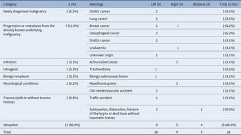
The aetiology of vocal fold motion impairment in the study population is presented in Table 6. The most common causes were malignancy (40.68 per cent) and idiopathic disease (27.68 per cent). Among the 46 patients with already known malignancy, 34 (73.91 per cent) developed vocal fold motion impairment due to disease progression or metastases, whereas 12 (26.09 per cent) developed vocal fold motion impairment due to other causes unrelated to the underlying malignancy (Table 7). Detailed information on vocal fold motion impairment aetiology across the vocal fold paralysis and/or immobility and vocal fold paresis and/or hypomobility groups are provided in Tables 8 and 9, respectively.
Aetiology of vocal fold motion impairment

Patients with known underlying malignancy who developed vocal fold motion impairment due to other unrelated causes

Aetiology of vocal fold motion impairment in the vocal fold paralysis and/or immobility group

Aetiology of vocal fold motion impairment in the vocal fold paresis and/or hypomobility group

Discussion
Contrary to the findings of previous studies, the present study demonstrated a high diagnostic yield of CT of up to 67.23 per cent for identifying the aetiology of unexplained vocal fold motion impairment, with high accuracy (96.05 per cent), sensitivity (99.17 per cent) and specificity (89.47 per cent). Moreover, high diagnostic yield and accuracy were both demonstrated for patients with vocal fold paralysis and/or immobility and vocal fold paresis and/or hypomobility. The most common cause of unexplained vocal fold motion impairment in our population was malignancy, followed by idiopathic disease.
Malignancies constituted up to 40.68 per cent of cases in this study. Among them, 19.21 per cent were due to recurrence or progression of already known malignancy, whereas 21.47 per cent were due to newly diagnosed malignancy. The three most common newly diagnosed malignancies causing vocal fold motion impairment were thyroid (7.34 per cent), lung (5.08 per cent) and oesophageal (3.95 per cent) cancers, which is consistent with previously reported findings.Reference Paquette, Manos and Psooy2, Reference Bando, Nishio, Bamba, Uno and Hisa13, Reference El Badawey, Punekar and Zammit-Maempel14 Among patients with already known malignancy, the most common primary cancer causing vocal fold motion impairment was breast cancer with nodal metastases (5.65 per cent).
Other less common primary cancers were oesophageal (2.82 per cent), lung (2.26 per cent) and thyroid (1.13 per cent), similar to the findings in the newly diagnosed malignancy group. Because up to 26.09 per cent of patients with already known malignancy developed vocal fold motion impairment as a result of other causes unrelated to the underlying disease, CT is still recommended for such patients with unexplained vocal fold motion impairment.
The second most common cause of vocal fold motion impairment was idiopathic disease, accounting for 27.68 per cent of cases. Idiopathic vocal fold motion impairment was more common among patients with vocal fold paresis and/or hypomobility than among those with vocal fold paralysis and/or immobility (46.88 per cent vs 23.45 per cent). In most of these cases, vocal fold motion impairment was unilateral (45 of 49 cases, 91.84 per cent), whereas bilateral idiopathic vocal fold motion impairment was found only in 4 of 49 cases (8.16 per cent). In prior studies, the reported prevalence was 2–41 per cent for unilateral and 3–13 per cent for bilateral idiopathic vocal fold paralysis.Reference Dworkin and Treadway15 Idiopathic vocal fold paralysis has been associated with viral infections, including herpes simplex, influenza and other pathogens affecting the upper respiratory tract.Reference Koç AE, Türkoğlu, Erol and Erbek16
Iatrogenic vocal fold motion impairment accounted for 11.30 per cent of cases in our study. The most common causes were mediastinal surgery, pulmonary lobectomy, endotracheal tube insertion, tracheostomy trauma and post-radiation changes. Although thyroid surgery is one of the most common causes of vocal fold motion impairment, no such cases were found in our study because vocal fold motion impairment occurrence was explained by the surgery in all these cases and additional CT was not requested.
The prevalence of traumatic vocal fold motion impairment resulting from fractures or dislocation and/or subluxation of the larynx was 8.47 per cent. Interestingly, in up to 73.3 per cent of these cases (11 of 15), no clinical history of injury was obtained before the diagnosis.
Abnormally dilated vascular structures in the mediastinum can compress the left recurrent laryngeal nerve, resulting in left vocal fold paralysis, a syndrome called cardiovagal or Ortner syndrome. In our study, these cases constituted only 2.82 per cent of all vocal fold motion impairment cases and were mostly caused by aortic aneurysms (4 of 5 cases, 80 per cent). One case (1 of 5 cases, 20 per cent) was due to pulmonary artery enlargement related to pulmonary hypertension and chronic pulmonary embolism. This syndrome was first described in 1897 by Norbert Ortner in a case series of patients with severe mitral valve stenosis and left vocal fold paralysis. Prompt correction of the underlying cardiovascular condition may result in left recurrent laryngeal nerve function recovery and hoarseness reduction.
Infection was responsible for a small number of vocal fold motion impairment cases in this study (2.82 per cent). Most of these (3 of 5 cases) were due to tuberculosis, either active or inactive. In patients with tuberculosis, the recurrent laryngeal nerve may be affected by the inflammatory process, direct invasion and/or damage, or extrinsic compression by a mediastinal lymph node.
The limitations of our study are mostly related to its retrospective nature. The severity of vocal fold motion impairment, as described in the medical records, could not be standardised and categorised. The indications for CT were also based on the judgment of each clinician, who had different clinical experiences. The proportion of patients diagnosed with idiopathic vocal fold motion impairment (27.68 per cent) might have been underestimated because many cases had to be excluded from the study population because of short follow up (<2 years).
• The role of computed tomography (CT) in the evaluation of idiopathic unilateral vocal fold paralysis is well established and cost-effective by a high diagnostic yield
• The role of CT in vocal fold motion impairment is controversial, with some studies reporting low diagnostic yield in the evaluation of idiopathic unilateral true vocal fold paresis and vocal fold movement impairment
• This study reports the high diagnostic yield and accuracy in the aetiology detection of unexplained vocal fold motion impairment in the largest sample size and uncontroversially suggests that CT is highly recommended in patients with unexplained vocal fold motion impairment
Conclusion
Computed tomography demonstrated high accuracy and diagnostic yield in the aetiological assessment of unexplained vocal fold motion impairment. We highly recommend contrast-enhanced neck CT, from the skull base down to include the aorto-pulmonary window in the mid-chest, for identifying the aetiology of unexplained vocal fold motion impairment. The most common cause of vocal fold motion impairment was malignancy, followed by idiopathic disease.
Competing interests
None declared.