The Rhynie and Windyfield cherts in the Rhynie outlier of Old Red Sandstone in NE Scotland (Fig. 1A) were originally deposited as siliceous sinters by Early Devonian hot springs. The cherts preserve in superb detail a diverse flora and fauna which formed a terrestrial and freshwater ecosystem on an alluvial floodplain close to active hot-spring vents (Trewin Reference Trewin1994; Trewin & Wilson Reference Trewin and Wilson2004). The hot springs were the surface expression of a precious-metal-bearing hydrothermal system (Rice et al. Reference Rice, Ashcroft, Batten, Boyce, Caulfield, Fallick, Hole, Jones, Pearson, Rogers, Saxton, Stuart, Trewin and Turner1995, Reference Rice, Trewin and Anderson2002) which formed near the marginal fault of a small trans-tensional basin (Rice & Ashcroft Reference Rice and Ashcroft2004). The Rhynie chert biota is world-renowned for early land plants (Edwards Reference Edwards2004) and arthropod faunas, containing the earliest known representatives of several groups (Anderson & Trewin Reference Anderson and Trewin2003; Fayers & Trewin Reference Fayers and Trewin2004). The preservation allows interpretation of interactions between plants, fungi and the fauna within the ecosystem.
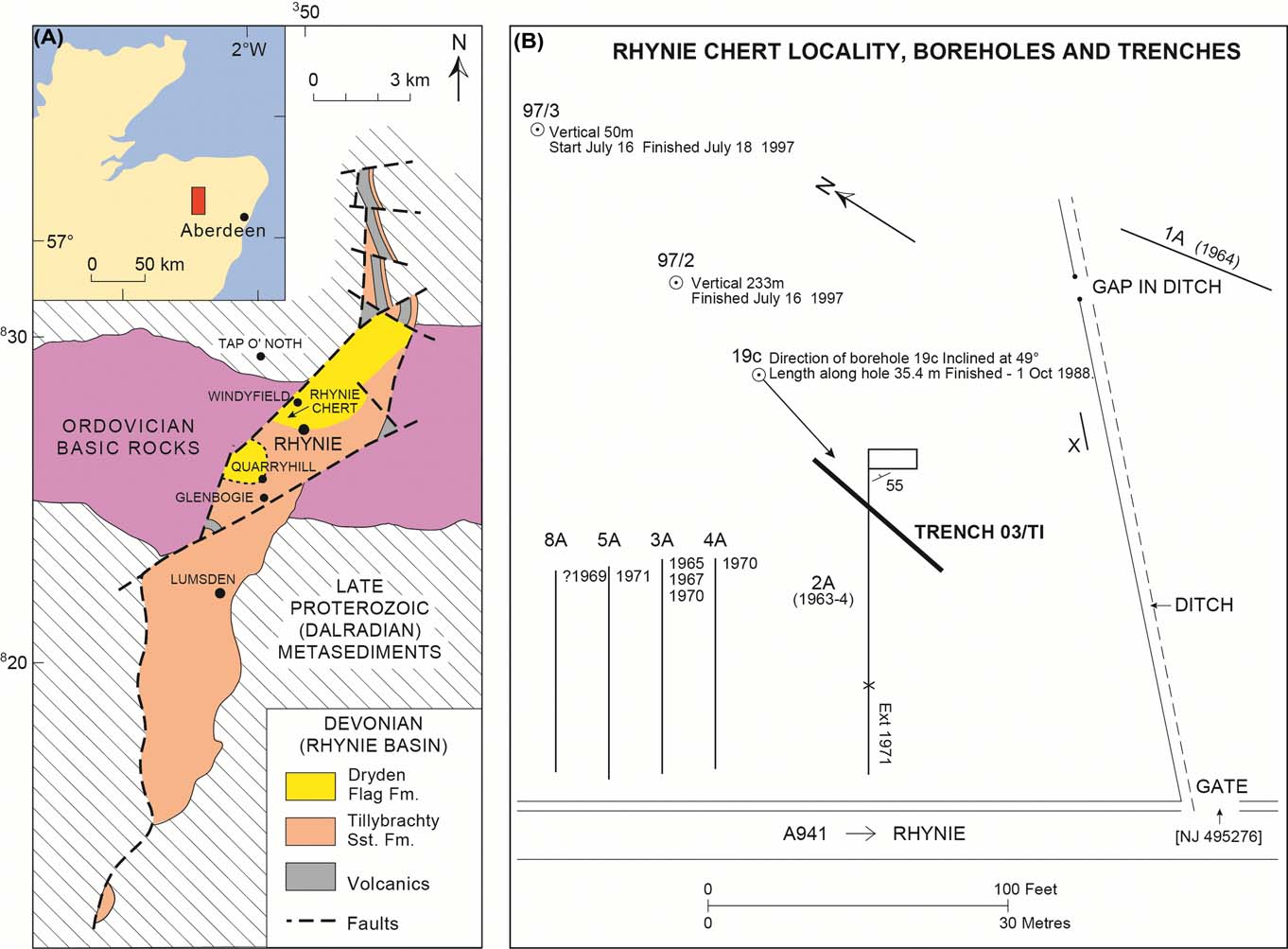
(A) Locality map. (B) Map showing the location of trench 03/T1 in relation to those excavated between 1963 and 1971, and recent boreholes (Modified from Trewin Reference Trewin2004).

The preservation state of plants within the Rhynie chert shows a range from perfect cellular preservation in 3D, with plants in original growth position, through to compacted carbonaceous films lacking any recognisable plant morphology. It is apparent that silicification by waters from the hot springs could occur at any stage in the natural cycle of growth and decay of the Rhynie plants. The effects of silicification are also variable, sometimes producing near-perfect per-mineralisation of plant axes, and preserving short-lived events such as spore germination and the release of sperm cells from antheridia. However, there is a gradation to more common material where silicification has been poor, and organic material lost.
Many features of the silicification have been described (Trewin Reference Trewin1994, Reference Trewin, Brock and Goode1996; Trewin et al. Reference Trewin, Fayers and Kelman2003), and compared with features seen in modern hot-spring areas such as those in Yellowstone National Park, USA and in North Island, New Zealand. Whilst many close comparisons can be made between textures seen in modern hot-spring sinters and those in the Rhynie chert, perfect modern analogues for all the preservation features seen at Rhynie have not been found. Modern studies generally rely on samples collected at the surface, and formed under oxidising conditions, whereas much of the Rhynie chert contains disseminated early pyrite, indicating reducing conditions during silicification. On a macro-scale, the silicification and plant preservation textures can be examined on cut and polished surfaces of chert blocks, and related to the textures of the original sinter. On a micro-scale, thin sections provide the most useful evidence relating to details of preservation.
New observations on silicification features at a macro-scale have been aided by the excavation in 2003 of a trench through the Rhynie chert sequence. This provided the first chance for 30 years to examine the chert in situ, to investigate lateral variation in beds and to take large samples through the full thickness of individual beds. Previous recent studies of the biota of individual beds has relied on the small specimens obtainable from borehole core, and poorly located trench material collected over 40 years ago. We report here the details of the succession and biota in the trench of 2003, and provide new information on the silicification processes, and depositional environments.
1. Methods and materials
The trench was excavated using a tractor equipped with a mechanical shovel. The autumn of 2003 was exceptionally dry, and no water was encountered during excavation. This was a very lucky circumstance, totally avoiding problems with mud and flooding. However, the mass of chert blocks cemented in dry mud above the outcrop was difficult to extract and, with large blocks present, there were initial difficulties in recognising true bedrock. The trench exposed bedrock to a width of over 1 m, and bedrock was encountered from 0.3 m to over 2 m below the field surface. Another result was that large blocks of chert lying loose above bedrock could be extracted without them breaking up, because cracks were sealed by dried mud. The mud-filled cracks became apparent when a large block was being sawn using a water-cooled rock saw at Robertson's Granite Works in Aberdeen. The block fell apart, luckily without damage to the saw. Subsequently, cracks in blocks were sealed with epoxy resin adhesive prior to cutting.
Individual beds from the trench were sampled and vertical slices 25 mm thick taken for surface examination and thin section making. Representative thin sections were prepared from each bed, and series of sections were made to cover the full thickness of particularly interesting beds. Sections were mostly made to 60 microns thickness for examination for textures and biota. Selected specimens were impregnated with blue-dyed resin to highlight porosity.
Surfaces of selected blocks were smoothed and polished for examination using a binocular microscope. However, a surprising amount of detail can be seen on wet sawn surfaces. The chert blocks and slides are stored in the Aberdeen University Geological Museum Collection. Specimen numbers quoted in this paper carry the prefix AUDG (University of Aberdeen Department of Geology).
2. Stratigraphy, sedimentology and the 2003 trench
2.1. Stratigraphy
The most recent interpretation of the stratigraphy following drilling and remapping of the area (Rice & Ashcroft Reference Rice and Ashcroft2004) places the Rhynie Cherts Unit within the Dryden Flags Formation near the top of the succession of Lower Old Red Sandstone in the Rhynie Basin. They consider that the Windyfield cherts are positioned higher in the Dryden Flags Formation than the Rhynie cherts, but the time gap is probably small, there being no significant differences in the biotas of the two chert occurrences. Recent palynological studies of trench and borehole material in the Dryden Flags Formation indicate that the entire sequence containing the Rhynie and Windyfield cherts belongs to the polygonalis–emsiensis Spore Assemblage Biozone of Richardson & McGregor (Reference Richardson and McGregor1986), indicating an early (but not earliest) Pragian to ?earliest Emsian age (Wellman Reference Wellman2006).
2.2. The 2003 trench
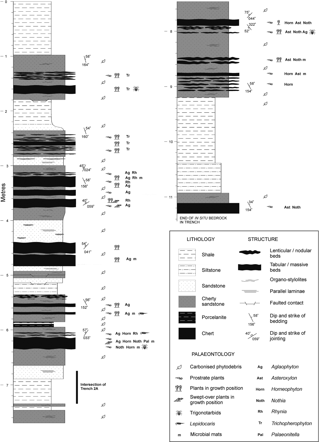
During September 2003, a trench was excavated through the Rhynie chert sequence (Trench: 03/T1), the first excavation undertaken within the area of the Site of Special Scientific Interest (SSSI) since Geoffrey Lyon's trenching programme finished in 1971 (Trewin Reference Trewin2004, fig. 7) (Fig. 1B) There were three reasons for excavating a new trench: first, to obtain a detailed sedimentological log through the sequence (Fig. 2). Previous excavations concentrated almost solely on the content of the plant-rich chert beds exposed, and the nature of interbed material was poorly recorded; secondly, to compare a trench sequence with previous data recorded from cores drilled in the vicinity of the SSSI (e.g. Powell et al. Reference Powell, Edwards, Trewin, Friend and Williams2000a; Trewin & Wilson Reference Trewin and Wilson2004); and thirdly, to extract oriented in situ chert beds. Three chert beds containing in situ plants preserved in growth position were recovered, each covering an area of over 1 m2. This material allows observation of rapid lateral changes in lithology and biota within individual beds. Such variation has previously only been hinted at from examination of blocks of chert float. For the purposes of the current article, we present a brief review of the sedimentology of trench 03/T1, together with a simplified log of the excavated sequence (Fig. 2).
Simplified lithological log through the Rhynie Chert Unit (sensu Rice et al. Reference Rice, Trewin and Anderson2002) excavated in trench 03/T1, showing preliminary observations of the biota and floral associations and structural information.

2.3. General observations
The trench was excavated trending NNE–SSW, close to the alignment of the inclined borehole 19c, cored through the chert sequence during 1988 (Powell Reference Powell1994; Trewin Reference Trewin2004) (Fig. 1B). Approximately 11.3 m (stratigraphic thickness) of in situ bedrock was exposed comprising a sequence of interbedded shales, siltstones, carbonaceous and micaceous sandstones, chert-cemented sandstones and cherts forming part of the Rhynie Cherts Unit (Fig. 2). Much of the sedimentary succession, particularly the mud-rocks and non-silicified sandstones, was found to be deeply weathered.
Approximately 6.7–7.4 m below the upper stratigraphic limit of in situ bedrock, the new trench cuts Trench 2A (Fig. 2). The latter was one of a series of trenches excavated by Geoffrey Lyon between 1963 and 1971 (see Trewin Reference Trewin2004 for details), and provided the chert material for the PhD Thesis of El-Saadawy (Reference El-Saadawy1966). Tantalisingly, it has not been possible to unequivocally correlate between the two trenches, given the very simplified lithological section through Trench 2A presented by El-Saadawy (Reference El-Saadawy1966). However, rapid lateral variation in chert beds also contributes to the lack of bed-to-bed correlation.
Below the lowest unequivocally in situ bedrock (a 25 cm-thick unit of massive sandy chert), a bed of chert approximately 40 cm thick is comprised of blocks which are slightly displaced, though still more-or-less in situ. Open joints and fissures between the displaced chert blocks are infilled by purplish-brown, unlithified alluvial gravel. These slipped slabs are probably the result of rock-creep at the edge of a post-glacial river terrace related to the nearby Easaiche Burn.
The bedding generally strikes NW–SE, with beds dipping to the northeast. The strike varies between 152° and 164°. The dip of the bedding is fairly consistent, typically ranging between 54° and 58°, though the lowest in situ chert and cherty sandstone beds have a dip of 34°. Jointing is strong, particularly within the chert beds. Joint orientations are variable, but generally two sets are recognised: a dominant joint set on a more-or-less NE–SW strike with dips to the northwest (strikes: 024°–059°; dips: 40°–75°); and a minor set on a strike sub-parallel to that of the bedding and dipping to the southwest (strike: 322°; dip: 52°). Faulting is evident, concordant with the dominant joint set, with some beds displaying very minor (mm- to cm-scale) normal displacement and associated in situ brecciation with occasional chert-cemented veins. A fault occurring below a chert/cherty sandstone unit at approximately 1.8 m below the top-most in situ bedrock (Fig. 2) is of uncertain displacement, though it truncates an underling shale unit such that in the trench, the latter thins from 55 cm to 35 cm. Generally, the strike of the faulting and the dominant joint set appears to run parallel with that of the fault zone forming the western margin of the Rhynie Basin.
2.4. Lithology and palaeoenvironments
2.4.1. Shales and siltstones
Shales and siltstones comprise approximately 34 % of the excavated sequence, forming units up to 1.7 m thick (Fig. 2). They are typically parallel to occasionally wavy laminated, and locally exhibit thin (mm-scale) micaceous sandstone laminae. Locally, these mud-dominated units exhibit general fining-upward trends, from sandy siltstones and sandy shales into cleaner shales with more carbonaceous material. Colouration is variable, ranging from light to dark greenish-grey at the bottom of the sequence to dark purplish-brown towards the top. These mud-dominated units compare with Lithofacies 1 of Powell et al. (Reference Powell, Edwards, Trewin, Friend and Williams2000a).
At least three thin beds of mud-rich sediments have been silicified to form a pale grey to white-coloured porcelanite (Fig. 2), and are typically associated with nodular chert and cherty sandstone horizons. These rare porcelaneous beds, comprising less than 2 % of the sequence, correspond with Lithofacies 4 of Powell et al. (Reference Powell, Edwards, Trewin, Friend and Williams2000a).
2.4.2. Carbonaceous, micaceous sandstones
Carbonaceous, micaceous sandstones are a relatively minor component, forming approximately 9.5 % of the sequence. These sandstones are typically very fine to fine-grained, parallel laminated and cemented mainly by clays, but may locally contain small lenses of chert cement (Fig. 2). The detrital mineralogy is dominated by sub-rounded to sub-angular quartz grains, with minor feldspars and varying amounts of mica. The sandstones occur as relatively thin tabular beds (up to 40 cm in thickness) and are closely associated with cherty sandstone units (see below). Their organic content is variable, but comprises mainly unidentifiable carbonaceous phytodebris. Colouration is mainly dark grey, but appears substantially darker to almost black where there is a high organic content. Occasionally, laminae with a ferruginous content may be oxidised, creating orange/brown streaks. These sandstones equate to Lithofacies 2 of Powell et al. (Reference Powell, Edwards, Trewin, Friend and Williams2000a).
2.4.3. Cherty sandstones
Cherty sandstones comprise over 30 % of the sequence (Fig. 2). In terms of detrital mineralogy, these sandstones are practically identical to those described above, with varying amounts of carbonaceous matter (primarily unidentifiable phytodebris and spores) and mica. These sandstones are pervasively chert-cemented, displaying little in the way of primary sedimentary structures, but a common pseudo-wavy lamination is created by carbonised plant debris forming microstylolitic contacts with the cemented sediment. These chert-cemented sandstones form units up to 120 cm thick and are closely associated with nodular and tabular beds of chert. Small chert nodules and lenses within these cemented sandstones occasionally exhibit rhizomes and degraded plant litter. The cherty sandstones correspond with Lithofacies 3 of Powell et al. (Reference Powell, Edwards, Trewin, Friend and Williams2000a).
2.4.4. Chert beds
Chert beds comprise over 26 % of the excavated sequence, ranging from nodular horizons within the cherty sandstones to single and composite tabular beds (Fig. 2). Internally, beds variably display the massive, vuggy, lenticular, nodular and locally brecciated textures described by Trewin (Reference Trewin1994). None of the beds within the logged sequence exhibit the laminated stromatolitic chert textures observed higher in the Rhynie chert succession (Powell et al. Reference Powell, Edwards, Trewin, Friend and Williams2000a; Trewin & Wilson Reference Trewin and Wilson2004) or in the Windyfield chert (Fayers Reference Fayers2003; Fayers & Trewin Reference Fayers and Trewin2004).
The biota of the chert beds is highly variable in terms of composition, associations, autochthony and preservation/decay (see below and Fig. 2). Most beds are plant-bearing, though the majority contain mainly prostrate axes and plant litter (Fig. 2). Numerous horizons contain plants preserved in growth position. However, in the majority of these, particularly in the very thin and nodular beds, only rhizomatic axes are well preserved, indicating that silicification was confined to the litter layer (for example, the chert beds towards the top of the sequence containing the embryophyte Trichopherophyton). The thicker, tabular and occasionally composite beds of massive and vuggy chert sometimes contain in situ plants where the aerial axes are also preserved in upright growth position (e.g., the chert beds at depths of 3.2–3.8 m, 4.4–4.8 m and 6.2–6.3 m). The majority of the plant-bearing chert beds contain an exclusively terrestrial biota. Remains of terrestrial arthropods, namely trigonotarbid arachnids, have been recorded from three beds (Fig. 2). A few beds, however, also contain the charophyte alga Palaeonitella, the crustacean Lepidocaris, and chert textures indicative of an aquatic environment (e.g. the chert beds at depths of 5.7 m and 6.1–6.3 m). The latter beds represent areas of terrestrial plant growth which were inundated and remained in standing water prior to silicification. Draped microbial laminae observed between plant axes in at least four chert beds (e.g., those at approximately 3.2 m, 4.7 m, 6.2 m, 8.5 m and 8.8 m depth) (Figs 2, 3) also attest to the presence of shallow standing water among growing stems prior to silicification.
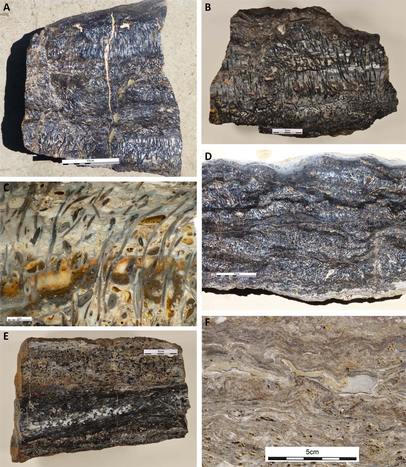
Macro textures in large chert blocks: (A) upright Rhynia axes in several layers within a chert bed. Above the basal development of upright axes, axes are preserved swept over into an inclined orientation. Block R68, AUDG12603; (B) chert bed with upright Rhynia and horizontal layers representing microbial sheets. Oval rounded white areas between axes are chert-filled gas bubbles which were trapped in the microbial mats. Block R33, AUDG12482; (C) detail of slice from AUDG12482 showing internal detrital sediment (brown) part-filling pore space and showing geopetal texture; (D) lensoid texture representing silicification of clonal colonies of Rhynia, with axes partly preserved in growth position. Darker material comprises mixed detrital sediment with carbonaceous debris. AUDG12602; (E) chert bed with part of blueish chert lens in lower part containing well-preserved, but compacted, axes of Aglaophyton (see Fig. 5D). The upper part of the bed is extensively brecciated, with silicified plants and detrital material. The initial silicification failed to produce a load-bearing structure. AUGD12604; (F) chert bed showing irregularly laminated chert formed from microbial sheets. Block R21, AUDG12605. Scale bars=10 cm (A, D); 5 cm (B, E, F); 1 cm (C).

2.5. Discussion of trench material
Shales and siltstones are primarily overbank deposits on floodplains and, possibly, in ephemeral floodplain lakes. Periodic flood events introduced thin, occasionally graded laminae of sand and silt.
Carbonaceous, micaceous and chert-cemented sandstone units represent rapidly deposited levée and crevasse splay sediments, indicating periods of flooding and avulsion of the local river system. We envisage that the Rhynie vegetation growing beside watercourses would have been sufficient to trap sediment and form levées, so encouraging ponding of overbank flood water. Plant colonisation resulted in the incorporation of carbonaceous matter, particularly evident below plant-bearing chert horizons. The cherty sandstones represent areas of silica precipitation, where silica-bearing hydrothermal fluids were percolating laterally and vertically through porous sands in the shallow subsurface.
Chert beds were deposited as siliceous sinters; originally highly porous and deposited as non-crystalline silica phases, they were converted to chert during burial. The abundance of plant-bearing chert units, often containing a diverse terrestrial and locally aquatic biota, suggests formation in an area relatively distal to hot-spring activity. Trewin & Wilson (Reference Trewin and Wilson2004) suggest such beds were deposited on the distal, often marshy regions of a hot-spring outwash apron. Alluvial sand and mud deposited during periodic flooding of the local river system interrupted sinter deposition. Relatively rapid temporal variations in terms of hot-spring activity/inundation by hydrothermal fluids and alluvial flooding events meant that areas of terrestrial plant growth probably did not have sufficient time to develop climax communities (Trewin & Wilson Reference Trewin and Wilson2004). Similarly, there was no time for the development of recognisable soil profiles between sedimentation events.
The features seen in the trench strengthen the conclusions of Trewin & Wilson (Reference Trewin and Wilson2004) that the Rhynie Cherts Unit is laterally extensive, but that individual beds cannot be correlated with confidence between boreholes c.20 m apart. In the trench, most of the thicker chert beds could be traced over the 1–2 m width of the trench. However, changes in bed thickness, chert texture and flora could be such that it would not be possible to correlate on the basis of borehole cores. Some beds, such as the bed crowded with Aglaophyton at 4.5 m, continue for distances in excess of 2 m but, on the basis of borehole records, do not extend for distances of 20 m. Thin beds, particularly those of nodular chert within cherty sandstone, are no more than thin lenses only tens of centimetres in lateral extent. Furthermore, it is frequently not possible to match blocks of chert which occur in the float above bedrock, to specific beds in the logged sequence; a further illustration of the rapid lateral variability of chert beds. We conclude that the fossilised areas of plant growth are of limited extent, pointing to the presence of small-scale streams and marshes. The area may have resembled a small-scale version of the Tangled Creek outwash system described by Trewin et al. (Reference Trewin, Fayers and Kelman2003) from Yellowstone National Park. In the Yellowstone example, the system is depositing silica, and preserving plants 2–3 km from the main hydrothermal vent; but at Rhynie, the preservation appears to have taken place about 200 m from the inferred vent position at the western bounding fault of the Rhynie Basin.
Caution should be exercised regarding plant cover and distribution. In trenches and boreholes, we are recording lateral continuity in chert beds; this may well reflect lateral extent of silicification, rather than the lateral extent of plant growth. Cherts frequently die out laterally into cherty sandstone, where plants are only preserved as carbonaceous stylolitised laminae. Clearly, the Rhynie sequence was deposited in a rapidly subsiding area, and sediment input from river floods prevented the build-up of a sinter terrace; a formation typical of deposition from hot-springs in areas undergoing erosion.
It is pertinent to note that palynological analyses of the host mudrocks and siltstones of the Dryden Flags Formation indicate a greater diversity in floral taxa than that identified to date within the Rhynie and Windyfield cherts (Wellman Reference Wellman2004, Reference Wellman2006). The plethora of palynological species identified in the host sediments, together with those identified in the cherts, are largely comparable to assemblages found elsewhere in other Old Red Sandstone continental basins of similar age. This would suggest that the more limited plant species present in the chert were simply more tolerant of the hydrothermal setting in the Rhynie area, rather than necessarily being adapted to such an environment.
3. Chert textures and plant preservation
3.1. Macro textures seen in chert beds
Most chert beds show a rapid gradation to, or a sharp irregular contact with, cherty sandstone. Preservation becomes poor and compaction effects such as stylolitisation increase at bed boundaries. Best preservation is normally seen in the central part of the bed, where plants may be preserved in growth position. Preserved rhizomes, particularly of Horneophyton, Asteroxylon and Nothia, indicate silicification of the substrate on which the plants grew. This substrate comprises a mix of plant material of variable preservation, together with detrital sand. There is no evidence that soils with a developed profile had been produced. When upright aerial axes are present, there is usually evidence of microbial material in sheet or network form filling spaces between axes.
Rhynia is the plant most frequently preserved with aerial axes in growth position, one block showing such preservation through 50 cm (Fig. 3A) Another block shows that a large amount of porosity remained after the plants and microbial sheets had been silicified. This porosity is revealed by internal sediments, both of silica and detrital material (Fig. 3B, C), and cavities with an oval cross-section which represent gas bubbles that were trapped in microbial mats. Preservation of the delicate silicified plant and bacterial framework depended on continuing silica deposition to produce a load-bearing structure. In many cases this did not happen, and only fractured remains are preserved. Laminated sinter of probable microbial origin seldom dominates chert beds, and no deposits resembling those of the thick sinter terraces of modern hot-spring areas have been found. A thin development of laminated sinter is illustrated in Figure 3F.
The silica fill of the framework is variable, consisting of both pore-lining silica and geopetal silica. Many of the structures seen have a close similarity to those seen in agates from vesicles in lavas. The pore-filling process usually ended with crystalline quartz, but by this stage some pores had become isolated from circulating fluids and remain as open quartz-lined pores.
In some blocks, a distinct lensoid texture is seen (Fig. 3D), which appears to result from preferential silicification of individual clonal plant colonies, partly in growth position, set in more compacted plant debris with abundant detrital material. Rhizomes seem to be preferentially preserved in this type of chert.
Preservation can be highly variable within individual beds. Figure 3E contains a lens of blueish chert with well-preserved but compacted axes, whereas the top part of the bed contains brecciated silicified plants in a sandy matrix, the silicified plants having been crushed in early compaction.
Some blocks of the Windyfield chert (Fayers & Trewin Reference Fayers and Trewin2004) show evidence for subaerial exposure and oxidation of the top of the bed following silicification. Rusty brown sand fills empty plant straws of Ventarura, which can be traced down into the chert where cellular structure is well preserved. Such early exposure may also be responsible for the oxidation of pyrite.
The colour of Rhynie chert is highly variable on cut surfaces. Very dark brown to black chert contains abundant preserved, but compacted, organic matter. Well-preserved plants are grey to black on cut surfaces and chert matrix is blue to white, with varying degrees of transparency. Chert with rusty brown colours, as is common in the Windyfield chert, has undergone oxidation, affecting diagenetic framboidal pyrite.
3.2. Preservation of plants
The micro aspects of plant taphonomy are studied in thin sections and on cut chert slabs. Preservation is exceptionally variable within the Rhynie Cherts Unit. Taxonomic descriptions of the plants have generally been based on exceptionally well preserved material which shows complete silicification of all cellular structure and full three-dimensional preservation. Furthermore, the plants were living or very recently dead and tissue structure had not broken down prior to the onset of silicification. It is important to stress that, in the majority of the chert, the silicification is less than perfect, and the degree and style of preservation may not only differ between different types of plant tissue (e.g., callose vs. sporopollenin), but also preservation of the same tissue may also differ within the same plant specimen (Wellman et al. Reference Wellman, Kerp and Hass2006). The plants in the chert may exhibit variable states of decay. Decay was due to both bacteria and fungi, the latter displaying a great variety, including saprophytic forms (Taylor et al. Reference Taylor, Hass, Krings, Klavins and Kerp2004). Whilst bacteria, for the most part, are not preserved in the chert, larger forms, including cyanobacteria, have been recorded within plant axes (Kidston & Lang Reference Kidston and Lang1921; Croft & George Reference Croft and George1959; Edwards & Lyon Reference Edwards and Lyon1983), as have possible forms within stromatolitic laminated cherts (Fayers & Trewin Reference Fayers and Trewin2004). Nematodes (Poinar et al. Reference Poinar, Kerp and Hass2008) also exploited decaying plants. The following three stages control the preservation of plants seen in the cherts:
• the condition of the plant material at the onset of silicification;
• the silicification process and extent of silicification; and
• the post-silicification diagenetic history.
3.2.1. The condition of the plant material prior to silicification
Many features seen in the chert relate to the state of plant material prior to silicification (Fig. 4). Most studies of the plants concentrate on material with good 3D cellular preservation (Fig. 5A), indicating that the plant was rapidly silicified whilst fresh. In fact, the silicification in many instances probably killed the plants which are preserved in growth position; though there is evidence in some chert blocks of plants surviving numerous flooding events, where plant axes are encased and supported by successive generations of microbial mats displaying trapped gas bubbles (Fig. 3B, C). These studies give a false impression of general preservation in the chert. The majority of beds contain material with a significant decomposition history prior to silicification, giving information on the environment in the region of the hot springs. Information is also revealed which relates to the structures in the various higher land plants and their ease of preservation. Plant axes are the most common fossils in the chert and provide the material for this study.
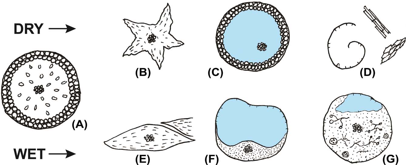
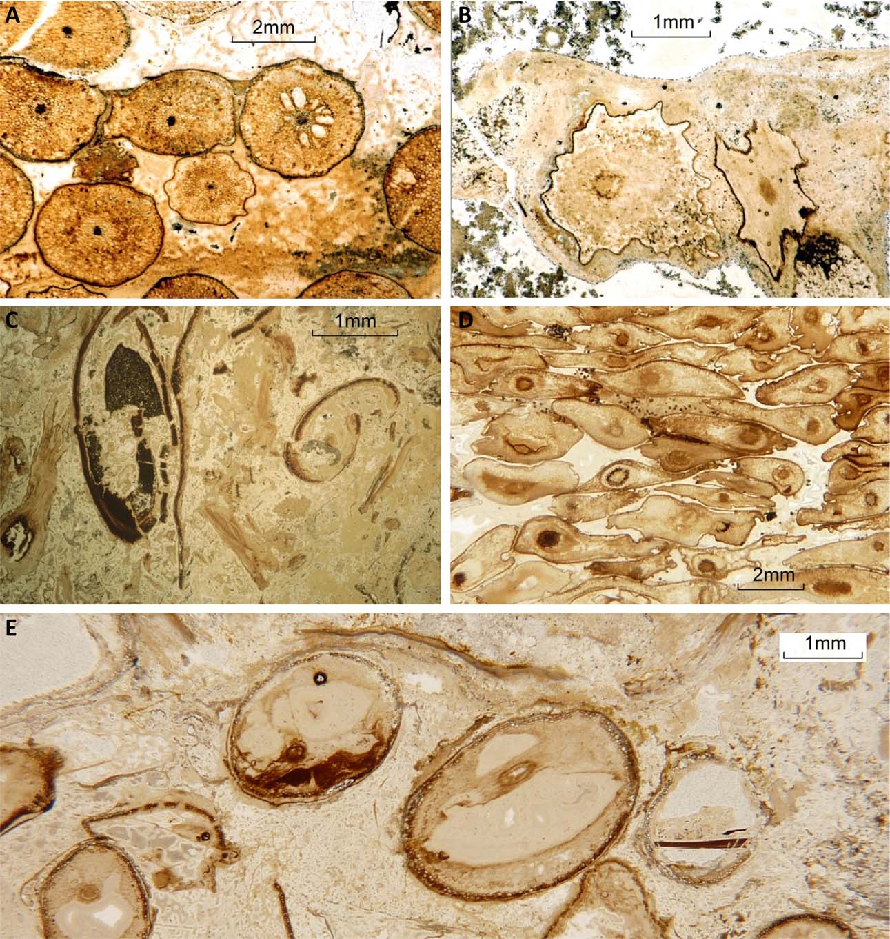
Sketch (not to scale) of some characteristic features of plant material prior to silicification in dry and wet environments: (A) axis as in life; (B) shrunken dried axis; (C) axis preserved as a straw with marginal robust cells and xylem strand preserved, blue is porosity; (D) dried fragments, including xylem strands and curled cuticle; (E) compacted axes; (F) decay resulting in amorphous residue, displaced xylem strand and porosity; (G) internal decay with fungal hyphae and cysts.

Pre-silicification plant features: (A) good cellular preservation with only minor shrinkage of axes; (B) shrunken dried axes with microbial coating which post-dates shrinkage; (C) fragmentary plant debris, including curled cuticle and fractured sporangium; (D) axes of Aglaophyton compacted in wet conditions prior to silicification; (E) axes preserving overall shape, but internally decayed with xylem strand displaced.

Two end members can be identified for conditions in which plant degradation took place: dry and wet (Fig. 4).
3.2.1.1. Dried plants and debris
Evidence for the drying in air of plant axes is commonly seen, normally as shrivelled axes which have dried to a star-shaped cross-section (Figs 4, 5B). Internally, cells may still be visible, but are collapsed. Both Rhynia and Aglaophyton show this feature, and the fact that the dried axes are frequently associated with well-preserved axes may indicate that these are axes of seasonal growth from a previous year. Indeed, other clonal taxa from the chert, namely Horneophyton and Nothia, also indicate seasonality (Hans Kerp pers. comm. 2015). Similar features can be seen in the modern naked fern Psilotum. The structure of some axes, notably Ventarura, which includes a middle cortex of thick-walled cells that do not collapse during drying, results in a hollow straw following the drying process; possibly still retaining the xylem strand, but all soft tissue is lost. Such straws can be preserved containing geopetal features due to washed-in sediment, or later silica deposits. Further breakdown of dried plant axes results in isolated xylem strands, curled cuticle fragments and cellular debris (Fig. 5C).
For preservation of air-dried material to occur before it is oxidised in the atmosphere, it must be immersed in hot-spring waters either at the surface through flooding, or by transport to a wet area. It may also be preserved by permeation of the substrate by hot-spring water. Thus dried material will normally undergo further changes in wet conditions at the time of silicification.
3.2.1.2. Wet plants and debris
In water, plants remain more pliable and are subjected to organic breakdown by bacteria, fungi and animals. Fresh axes can also compact under low pressure, very close to the surface. Some chert beds contain plants which display compaction prior to silicification to give lensoid cross-sections to closely-packed axes (Figs 4, 5D). Internally, cells are distorted, but not decayed. This feature represents mats of fallen axes in a waterlogged situation, and compacting under their own weight. This is a very local feature, and undistorted axes can occur in close proximity both above and below, and within the same chert bed. Thus, this is not a progressive compaction with depth as might be seen in a peat. The end member of the compaction process, in the absence of silicification, is to reduce the axes to carbonaceous compression fossils.
As with dried material, it is the softer phloem which decays first, usually leaving a mushy residue. The cuticle and xylem strand may still be present, with the xylem strand displaced from its central position (Fig. 5E). This decay can continue during silicification, following initial coating of plant axes with silica. This results in a variety of textures where cuticle detaches from the silica coating, leaving voids that may or may not be filled during further silicification.
3.2.1.3. Influence of microbial coatings
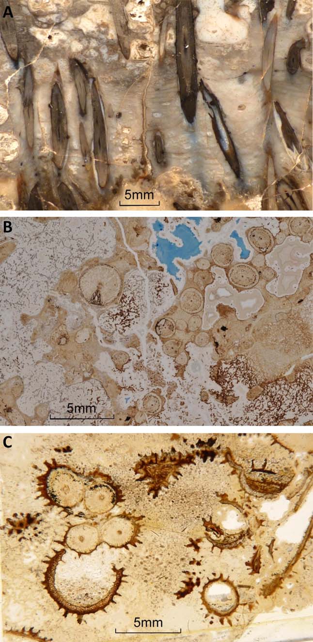
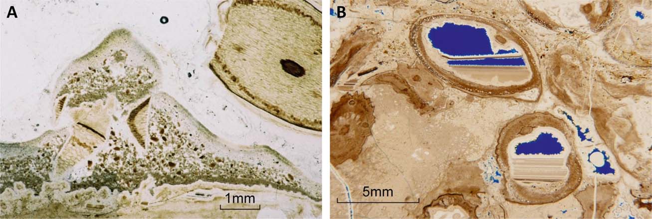
Textures in the chert show that, locally, plants were coated by a microbial layer prior to silicification (Fig. 6). This is a common feature of vegetation seen submerged in modern hot springs. The coating may be bacterial and/or cyanobacterial and include filamentous growths. Silicification of the coating aids support of the plants and forms an initial framework. Voids in the framework may be partly filled with filaments, probably of fungal origin, which become coated with silica (Fig. 6B).
(A) Microbial sheet support between Rhynia axes. (B) Silicified plant axes and microbial coatings, with extensive remaining porosity filled with microbial meshworks. Remaining porosity (filled with blue resin) is lined with late-stage quartz. (C) Microbial micro-stromatolites on fragments of Ventarura. Geopetal features formed from organic-rich pond debris, with abundant small arthropod coprolites. Windyfield chert.

Filamentous microbial sheets develop in modern hot-spring pools and various types form mats which coat substrate, and also grow at the air/water interface. Those on the surface are commonly buoyed up by gas bubbles. Surface sheets can be silicified due to evaporation from the mat surface, whilst water remains below the surface mat. If plants are present in shallow water, filamentous sheets grow between plant axes, and can form a supporting framework. In the Rhynie chert, aerial axes of plants which are preserved in growth position are frequently supported by laminated microbial sheets (Fig. 6A). The silicification of the microbial framework facilitated the preservation of upright plant axes. Thus, the specimens showing upright axes are due to shallow flooding of an area of plant growth, and the development of microbial mats which supported the plants.
In some chert beds, microbial sheets also form wavy laminated textures with features such as oval voids after gas bubbles, and folded laminae which were created prior to silicification (Fig. 3F). The delicate laminae were frequently crushed after silicification and show brittle fractures. In the Windyfield chert, microbial growths on submerged Ventarura axes developed into micro-stromatolitic forms (Fig. 6C).
3.2.2. The silicification process and extent of silicification
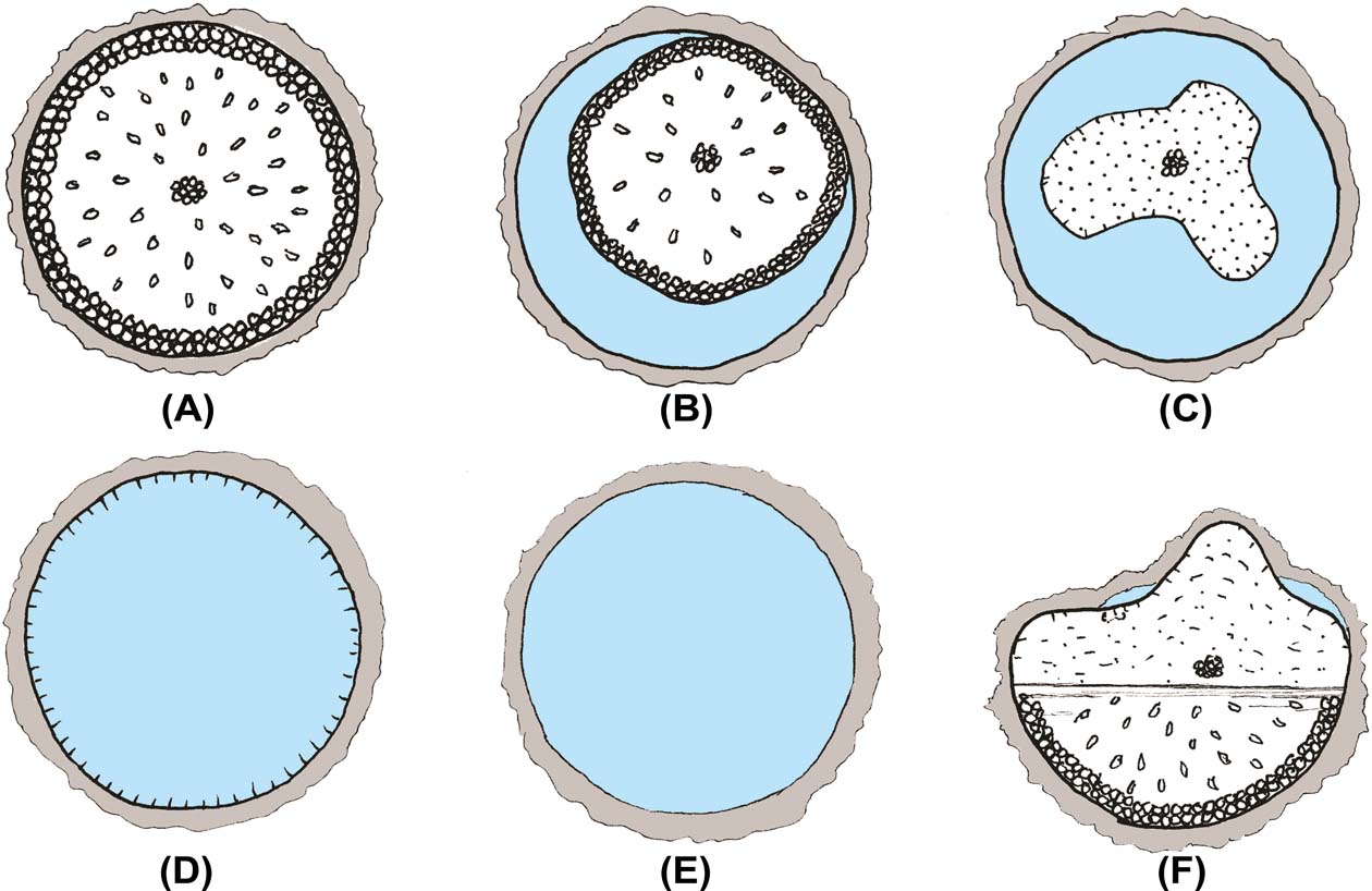
The effects of silicification are highly variable within the cherts (Fig. 7), reflecting factors such as temperature, Si concentration and pH of the hot-spring water. The site of silicification is also critical. In subaqueous settings, and under reducing conditions, plant material is slow to decay; whereas at the surface, in an oxygenated environment, organic decay will be more rapid. Where the water temperature is above 70°C, organic matter is destroyed; thus the cherts resulting from sinters deposited in very hot water lack plant fossils, or at best they are represented by ghosts, lacking organic matter. Silicification of plant material, with organic preservation, is probably most effective at 20–35°C. Evidence for the salinity of the hot-spring fluids and its effects on the biota and silicification in the cherts is tentative. However, the diverse freshwater biota observed within examples of Rhynie and Windyfield chert deposited in ponded aquatic settings are considered to be broadly representative of contemporary freshwater environments elsewhere, and not specifically adapted to saline or hydrothermal conditions (Fayers & Trewin Reference Fayers and Trewin2004).
Sketch (not to scale) of silicification features of plants: (A) good cellular preservation, with microbial coating on epidermis; (B) shrinkage of axis produces porosity, but cellular structure is preserved; (C) cuticle has shrunk, and softer phloem decayed, xylem strand still visible; (D) only cuticle remains, other structures not preserved; (E) a hollow straw, no organic material remains; (F) lower part of axis preserved but upper part decayed; probably indicating a fluid level below which initial silicification took place, whilst the upper part of the axis continued to decay prior to later silicification.

3.2.2.1. Early silicification
The silicification of plants in the chert normally commenced with a thin coating of silica on external surfaces. This could be by direct precipitation onto cuticle, or by the silicification of a bacterial coating which may also have included fungal and cyanobacterial material (Fig. 6). This coating provided some rigidity to support axes in an upright position, but support was greatly aided by microbial mats which bound the stems, and were themselves silicified. Microbial filamentous meshworks grew in open porosity and were also coated by silica (Fig. 6B). Filaments were mostly only 1–2 µm in diameter, but became coated by opal-A to produce structures which formed a template for continued cementation. Modern examples are illustrated by Jones et al. (Reference Jones, Renault and Rosen2004), and show bulbous growths with a very finely laminated internal structure encrusting filaments. Thus, an early framework could be built that gave some physical support; but the framework was highly porous and contained large voids through which silica-rich waters could flow.
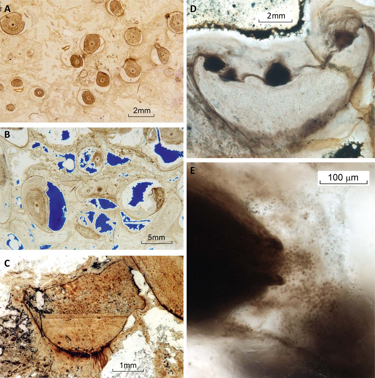
Silicification continued with permeation of the cell structure of the plant. Perfect cellular preservation resulted when the intercellular spaces and cell lumina of the plant tissue were rapidly filled with silica. This process was not always complete before shrinkage of the plant axis commenced, producing voids where the epidermis shrunk away from the initial silica coatings (Fig. 8A, C). It is highly likely that the plant axis was undergoing dehydration due to immersion in a silica-rich medium. In the absence of the original external silica coating, this reduction in volume could go undetected, since preservation is still excellent, but cells have been reduced in size.
Silicification features: (A) Rhynia axes showing variable shrinkage during silicification, but preserving cellular structure; (B) variable decay and shrinkage of axes, with cuticle pulled away from coating and leaving porosity. Coated filaments, probably fungal, grew in porosity after shrinkage and decay of axis at lower centre; (C) two stages of silicification: the lower part of an Aglaophyton rhizome bearing delicate rhizoids is well preserved, and sharply separated by a geopetal surface from the distorted and partially decayed upper part; (D) well-preserved fertile axis of the male gametophyte Lyonophyton, bearing at least four antheridia; (E) close-up of the left-most antheridium in (D), showing released gametes trapped within a pale mucilaginous coating.

The voids created may also become filled, or partly filled, with silica. This deposit usually displays a botryoidal texture and relict fibrous structure, representing chalcedony.
Incomplete silicification of plant axes allows decay to continue during the silicification process, and there can be a sharp divide, even within a single plant axis, between different states of preservation (Fig. 8B). Incomplete silicification can also result in situations where the plant tissue is variably impregnated with silica, the central part being left as a void. In a few cases, it can be seen that the epidermis alone remained within the initial silica coating, and has subsequently shrunk, prior to later filling of the void (Fig. 7).
In surface situations around modern geysers, or on sinter terraces, plants are frequently trapped in sinter. This situation does not result in good silicification of the plant tissue, and the organic material generally decays or is oxidised, to leave a hollow mould in the sinter. In general, plants encased in sinter above the water table will not be silicified. This feature is present in the Windyfield chert, where well preserved plant axes can be traced to the top of a bed, where they become empty straws filled with sediment and geopetal silica.
3.2.2.2. Speed of silicification of organic material
There are numerous instances of superb preservation in the cherts which require silicification to proceed very rapidly; indeed, virtually instantaneously. Preserved delicate details such as germinating spores (Kerp et al. Reference Kerp, Trewin and Hass2004), the various cell division stages of the photobiont of Winfrenatia (Taylor et al. Reference Taylor, Hass and Kerp1997), fungal infections in Nothia (Krings et al. Reference Krings, Taylor, Hass, Kerp, Dotzler and Hemsen2007), book lungs in Palaeocharinus (Claridge & Lyon Reference Claridge and Lyon1961) and nematode worms (Poinar et al. Reference Poinar, Kerp and Hass2008), together with the life cycle of short-lived mycorrhiza arbuscles in Aglaophyton (Remy et al. Reference Remy, Taylor, Hass and Kerp1994), are remarkable; but the examples of the antheridia of the gametophytes Lyonophyton and Remyophyton preserved in the act of ejecting sperm cells (Fig. 8D, E) (Kerp et al. Reference Kerp, Trewin and Hass2004, figs 5F, 9E) require special consideration. In each case, the cloud of sperm cells is fossilised with the appearance of a puff of smoke from a chimney; in detail, the released gametes commonly appear ‘fixed’ within pale-coloured cumuli, locally containing other very finely disseminated organic matter, forming an irregular, patchy mucilaginous coating to the surface of the plant (Fig. 8E). It is impossible for this ejection of sperm cells to have taken place into low viscosity water; they would have been washed away as nature intended. It must be concluded that ejection was into a stagnant high-viscosity medium; a silica gel is a strong possibility, as is a deposit of opal-A microspheres. Both might be expected to rapidly stop the progress of the sperm cells and, furthermore, these media could both have been wet enough to trigger the release of the sperm cells. In recent mosses, such as Physcomitrella, mature antheridia release packages of sperm cells as soon as they come into contact with water. Initially, the sperm cells are coalesced and bound by mucilage; within a matter of minutes, individual cells become released and the package of cells is completely dispersed after around 15 minutes (Hans Kerp pers. comm. 2015). Such mucilaginous material may form a component of the ‘cumuli’ in which the released gametes of Lyonophyton are preserved; nevertheless, the preservation of released sperm packages clearly demonstrates the rapidity and detail of silica preservation that may be observed in the Rhynie chert.
This evidence possibly also has a bearing on the preservation of other delicate organic structures, such as delicate plant axes in growth position within chert where they have no visible means of support. Maybe they were enclosed in silica gel or precipitate. Silicification of delicate features and soft tissue must have been virtually instantaneous.
The experimental work of Channing & Edwards (Reference Channing and Edwards2004) in modern natural hot-spring environments illustrates the development of silica (Opal-A) coatings on plant tissue, and the filling of voids in cellular tissue with opal-A microspheres less than 0.5 µm in diameter. Partial mineralisation of plant tissue took place over a period of months (330 days). Samples were immersed in a hot-spring vent, and it may be that the silicification process was slow due to the relatively constant vent temperature, rather than to the cooling that decreases silica solubility and promotes precipitation, and occurs as waters flow away from a vent. Thus, whilst the features observed by Channing & Edwards (Reference Channing and Edwards2004) can be recognised in the Rhynie chert, the rate of silicification must have been much faster (minutes to a few hours) to preserve some features seen in the Rhynie chert. Conversely, recent experimental work on the processes of silicification of wood in hydrothermal and volcanogenic settings (Ballhaus et al. Reference Ballhaus, Gee, Bockrath, Greef, Mansfeldt and Rhede2012; Läbe et al. Reference Läbe, Gee, Ballhaus and Nagel2012; Hellawell et al. Reference Hellawell, Ballhaus, Gee, Mustoe, Nagel, Wirth, Rethemeyer, Tomaschek, Geisler, Greef and Mansfeldt2015) has demonstrated that silicification may take place very rapidly; far more in keeping with observations made in the Rhynie chert.
In modern marsh environments in hot-spring outwash areas, both diatoms and grasses take up and fix silica from the water. In the early Devonian, silica concentrations could have been much higher in waters flowing into hot-spring marshes; so promoting direct silica precipitation and rapid silicification of the flora. Although possible, there is no unequivocal evidence for the Rhynie chert plants taking up and fixing silica during life.
3.2.2.3. Later stages of silicification
There is clear evidence that silicification of plant axes was completed whilst large voids still remained in the palaeo-sinter. There are examples of internal sediment infiltrating down into voids and being deposited with ‘snow-roof’ textures, indicating a degree of cohesion of the deposit. The deposits are silica, but include fragments of silicified plants displaying brittle fracture, and originating from within the framework of the bed (Fig. 9A). The exact nature of the silica at the time of deposition is not known, due to crystallisation to chert, but opal-A microspheres are a possibility, as demonstrated by Channing & Edwards (Reference Channing and Edwards2004). The fills of cavities within plants include both banded geopetal fills and radial growth from cavity walls, indicative of silica deposition in differing forms. Several spirit-level fills may be present, sometimes showing changes in tilt (Fig. 9B). Rarely, silica bridges cross cavities, possibly a record of fluid surfaces. The resemblances of the geopetal cavity fills to agates found in vesicles in lavas are remarkable.
(A) ‘Snow roof’ texture in internal sediment. Fragments of silicified plant axes showing brittle fracture are included in the internal sediment, indicating that plants were silicified prior to introduction of the internal sediment. Block R33 (see also Fig. 3B, C). (B) Decayed axes with displaced xylem strands and showing geopetal structures, including a ‘bridge’ with later tilt. Late-stage pore-lining quartz part fills remaining porosity (blue resin-impregnated).

The later stages of silicification were also important for preservation of the biota. The open fabric of the beds needed to be filled with enough silica to provide strength during burial. This was not always the case and examples of crushing and brittle fracture of silicified plants and microbial laminae are common. The final stages involved introduction of silica from fractures, deposited as quartz crystals lining cavities and in veins. This took place at higher temperatures, certainly over 70°C, since organic material can be burnt out adjacent to veins. Oxygen isotopic analysis of the chert gives temperatures of formation of around 100°C, representing not the depositional temperature, but that of crystallisation. Late quartz as pore-lining prismatic crystals, and introduced from fractures, is localised to the vicinity of the fracture, and adjacent pores may lack late quartz. This indicates that the chert was impermeable at time of late-quartz introduction (e.g., Fig. 8C).
3.2.3. The post-silicification diagenetic history
Several post-silicification events have affected preservation in the cherts. Where silicification was incomplete and the organic material was subject to oxidation, voids may be present, representing the position of organic matter. These voids may be filled with sediment prior to deposition of the overlying bed (Fig. 3C). This can also occur on a micro-scale, when cell walls are removed and remain as void space. This void space can later be filled with other minerals, as in the case of Ventarura from the Windyfield chert (Powell et al. Reference Powell, Trewin and Edwards2000b), where the brown colour of the cell walls is not organic, but iron hydroxide.
There are many instances in which silicified plants have been subsequently brecciated. This can occur at an early stage and the breccia can be resealed by a later phase of silica deposition. It can also occur with a mix of silicified plant fragments and detrital material which has a chert cement.
Silica cements continued to be deposited from hot-spring fluids permeating the sediment in early burial. By comparison with modern systems (e.g., Campbell et al. Reference Campbell, Buddle and Browne2004), a sequence of opal-A to opal-CT to opal-C, followed by crystallisation to microquartz, would be expected. Relict radial fibrous textures are seen preserved in coatings on organic matter and cavity walls, which probably represent chalcedony. Phase transformations frequently reduce or destroy preservational detail. The Rhynie chert is remarkable in that organic material was preserved intact and in great detail through these transformations. Still later, brecciation and resealing of the chert at depth during burial was due to structural deformation and faulting. The brittle chert beds within soft shale and sandstone were susceptible to fracturing during deformation of the Rhynie Basin.
4. Conclusions
Trench material from the Rhynie Cherts Unit, together with loose blocks of chert from float, reveal distinctive textures relating to the taphonomy of the fossil plants and the processes and timing of silicification. The best preservation of plant tissue was achieved when fresh plant material became immersed in waters in the distal regions of hot-spring outwash. Aquatic microbial mats gave support to plant axes in growth position, and silicification of plant tissue and microbial mats was rapid. Extensive remaining porosity could then be filled with both pore-lining and geopetal silica, and also invaded by internal clastic sediment which filtered down through remaining porosity.
The key to ultimate preservation was the establishment of a load-bearing framework which survived burial. In many cases this did not occur and the framework collapsed after silicification of the plants, resulting in chert beds with silicified plant fragments, disoriented, showing brittle fractures and set in later silica cements.
Silicification was also effective at the terrestrial surface, where silica-rich waters permeated plant litter and detrital sand. Here, the rhizomes of the plants, and delicate single-celled rhizoids, are preferentially preserved. This is commonly seen in the preservation of Horneophyton, Nothia, Asteroxylon and Aglaophyton. Fungi are particularly common in this situation, and a wide range of plant preservation is present, with all states of pre-silicification plant decay.
In some cases, silicification was very rapid, preserving organic phases which lasted only hours, and exceptionally delicate structures such as germinating spores and soft-bodied nematodes. The preservation of sperm cells emerging from an antheridium, and frozen like a puff of smoke, indicates that a viscous silica medium trapped the cells. This was probably a silica gel, or an aggregate of opal lepispheres.
Silicification appears to have taken place under reducing conditions, as evidenced by the presence of pyrite. Exposure to the atmosphere, or even to oxygenated water, caused plant decay. Thus, the preserved plants were probably only safe from destruction once the porous silica phases had been converted to chert, probably at a temperature of around 100°C.
The depositional environment surrounding the hot-spring vents, and in the outwash areas, was laterally variable on a sub-metre scale, and included terrestrial surfaces with growing plants and aquatic environments such as small pools and outwash streams. Rapid subsidence and sedimentation in the Rhynie basin buried the proto-Rhynie cherts and, where a generally reducing environment prevailed, silicification of organic matter continued. Those beds, or parts of beds, which acquired an early load-bearing silica framework before plants decayed or were compacted, provide the best three-dimensional preservation.
5. Acknowledgments
The authors thank the Lyon Bequest to the University of Aberdeen for funding the excavation and support for SRF; Scottish Natural Heritage for permission to excavate the trench at the Rhynie SSSI; and A. & J. Robertson (Granite) Ltd. for their care in slabbing blocks of Rhynie chert. Thanks are also due to Professor Hans Kerp (University of Münster) and an anonymous referee for their helpful comments and suggestions to improve the manuscript. Hans Kerp is also thanked for kindly allowing us to include the photographs of the gametophyte Lyonophyton and mature antheridia illustrated in Figs 8D and 8E.