Images in congenital heart disease
Cardiogenic unilateral pulmonary oedema is an uncommon finding seen in severe mitral regurgitation with an eccentric jet usually directed towards the right upper or middle pulmonary veins, resulting in localised pulmonary oedema. In adults, the most common causes are degenerative heart disease and myocardial ischaemia with subsequent prolapse of the posterior leaflet of the mitral valve.Reference Attias, Mansencal and Auvert 1
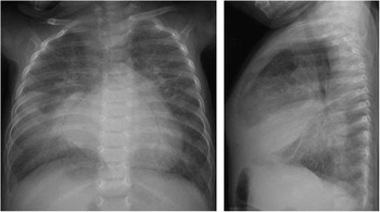
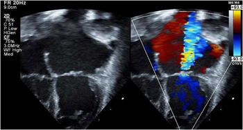
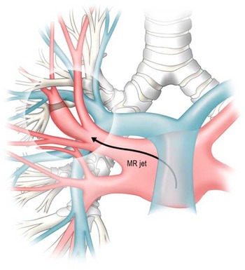
In this study, we describe the case of a four-month-old infant with severe juxtaductal coarctation of the aorta, hypoplastic transverse aortic arch, and moderate mitral stenosis, with residual severe mitral regurgitation following mitral commissurotomy and aortic arch repair, who developed unilateral pulmonary oedema and subsegmental atelectasis. She had multiple admissions for respiratory distress, and was empirically treated for pneumonia as her chest radiograph demonstrated a persistent right middle lobe opacity and atelectasis (Fig 1). A transthoracic echocardiogram showed poor coaptation of the mitral valve leaflets with severe mitral regurgitation directed towards the right-sided pulmonary veins, which were significantly dilated (Fig 2). We postulated that in our patient the mitral regurgitant jet directed towards the right middle pulmonary vein caused increased transudative pressure in this lung segment with resultant focal pulmonary oedema, and the dilation of the pulmonary vein resulted in extrinsic compression of a subsegmental bronchus leading to atelectasis (Fig 3).
Chest radiograph prior to mitral valve replacement demonstrating diffusely prominent lung markings, right middle lobe opacity, and subsegmental atelectasis.

Apical 4-chamber view with two-dimensional echocardiography and color Doppler demonstrating severe mitral regurgitation directed towards the right-sided pulmonary veins, which are dilated.

An eccentric mitral regurgitant (MR) jet directed towards the right middle pulmonary veins, caused increased transudative pressure in this lung segment with resultant focal pulmonary edema. The dilated right pulmonary veins caused extrinsic compression of a subsegmental bronchus leading to subsegmental atelectasis.

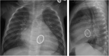
At six months of age, she underwent a mechanical mitral valve replacement. The postoperative transthoracic echocardiogram demonstrated a well-functioning mechanical valve with no evidence of mitral regurgitation or stenosis. The postoperative chest radiograph (Fig 4) demonstrated near resolution of the cardiogenic unilateral pulmonary oedema and atelectasis in the absence of mitral insufficiency.
Chest radiograph one week after mechanical mitral valve replacement, demonstrating resolution of the unilateral edema and atelectasis.

Recognition of cardiogenic unilateral pulmonary oedema is clinically important, particularly in patients presenting with respiratory distress. Patients can be misdiagnosed with pneumonia, leading to a delay in diagnosis, a potential delay in surgical repair, and unnecessary investigations or treatments such as chest computed topography, bronchoscopy, or antibiotics.
Acknowledgements
The authors acknowledge Nancy Heim, Medical Illustrator (Fig 3).
Financial Support
The study was funded by Andrew King Research Award from Colin’s Kids Inc., Columbia University Medical Center.
Conflicts of Interest
None.