Introduction
Studies of post-larval stages and growth series of ophiuroid species have been considered of great relevance for species identification, particularly for cryptic species and those in early life stages, in addition to contributing to the resolution of phylogenetic relationships and taxonomic uncertainties (Schoener, Reference Schoener1967; Monteiro et al., Reference Monteiro, Reis and Pardo1992; Sumida et al., Reference Sumida, Tyler, Gage and Nørrevang1998; Stöhr, Reference Stöhr2005; Borges et al., Reference Borges, Alitto and Amaral2015; Martynov et al., Reference Martynov, Ishida, Irimura, Tajiri, O'Hara and Fujita2015; Humara-Gil et al., Reference Humara-Gil, Granja-Fernández, Montoya-Márquez and López-Pérez2022). Due to the difficulties associated with the identification of juvenile stages, these studies are mostly restricted to brooding species, whose assignment to the species level is unambiguous (Clark, Reference Clark1914; Hendler, Reference Hendler1988; Stöhr, Reference Stöhr2005; Falkner and Byrne, Reference Falkner and Byrne2006; Borges et al., Reference Borges, Alitto and Amaral2015). Nevertheless, as only a small number of ophiuroid species are brooders (Clark, Reference Clark1914; Schoener, Reference Schoener1967; Stöhr, Reference Stöhr2005; Borges et al., Reference Borges, Alitto and Amaral2015), most identification keys do not routinely include juvenile characters, and images and detailed descriptions of early life stages are lacking in the literature (Sumida et al., Reference Sumida, Tyler, Gage and Nørrevang1998; Stöhr, Reference Stöhr2005).
Despite the importance of studying and describing juvenile specimens, this field is still under-explored. Of the approximately 2000 living ophiuroid species, early growth stages have been described for only 76 (3.5%) species so far (Ludwig, Reference Ludwig1898, Reference Ludwig1899; Clark, Reference Clark1914; Mortensen, Reference Mortensen1936; Schoener, Reference Schoener1967, Reference Schoener1969; Hendler, Reference Hendler1978, Reference Hendler1988; Webb and Tyler, Reference Webb and Tyler1985; Turner and Miller, Reference Turner, Miller, Burke, Mladenov, Lambert and Parsley1988; Vadon, Reference Vadon1990; Monteiro et al., Reference Monteiro, Reis and Pardo1992; Tyler et al., Reference Tyler, Patterson, Sibuet, Guille, Murtons and Segonzac1995; Sumida et al., Reference Sumida, Tyler, Gage and Nørrevang1998; Borges et al., Reference Borges, Monteiro and Amaral2002, Reference Borges, Alitto and Amaral2015; Stöhr, Reference Stöhr2005; Falkner and Byrne, Reference Falkner and Byrne2006; Olbers and Samyn, Reference Olbers and Samyn2012; Martynov et al., Reference Martynov, Ishida, Irimura, Tajiri, O'Hara and Fujita2015; Alitto et al., Reference Alitto, Bueno, Guilherme, Di Domenico, Christensen and Borges2018; Humara-Gil et al., Reference Humara-Gil, Granja-Fernández, Montoya-Márquez and López-Pérez2022; da Silva et al., Reference da Silva, Prata and Christoffersen2023). Among the species with unstudied juvenile stages are those belonging to the genus Ophiocoma L. Agassiz, 1836. Species of Ophiocoma are known to be challenging to identify, especially juveniles, since their main diagnostic characteristics, such as the number of arm spines, change throughout growth and may overlap with other species at different stages of development. This problem is intensified by the lack of structures to distinguish the species and the fact that some of them can change their body colour from day to night (Hendler, Reference Hendler1984; Benavides-Serrato and O'Hara, Reference Benavides-Serrato and O'Hara2008; Stöhr et al., Reference Stöhr, Conand and Boissin2008). Although morphological information on post-larval developmental transformations could contribute to circumventing the existing limitations, the knowledge on growth changes in Ophiocoma is very limited. Grave (Reference Grave1898) provided information on the embryo stages of Ophiocoma echinata (Lamarck, Reference Lamarck1816), but not on post-larval development. Devaney (Reference Devaney1968) showed that the number of spines and tentacle scales change with growth in Ophiocoma erinaceus Müller & Troschel, 1842, Ophiocoma aethiops, and Ophiocoma anaglyptica Ely, 1944. Devaney (Reference Devaney1970) detected that a particular disc size was correlated with the first occurrence of granules in O. aethiops, O. anaglyptica, O. echinata, O. erinaceus, and Ophiocoma scolopendrina (Lamarck, Reference Lamarck1816) and discussed how this could help to identify these species within Ophiocoma. The correlation between the disc size and granule development was also studied by Price and Rowe (Reference Price and Rowe1996), but only for O. erinaceus. H.L. Clark (Reference Clark1933) and A.H. Clark (Reference Clark1939) analysed a juvenile of O. echinata (3 and 4.5 mm of disc diameter, respectively), but both only reported that the specimens had already developed disc granules, two tentacle scales in the first arm segments, and swollen dorsalmost arm spines.
Considering the importance of information on post-larval developmental transformations to further taxonomic and life history research, the present study aims to describe and illustrate the main morphological modifications during the growth of two species of Ophiocoma, O. echinata and Ophiocoma trindadensis Serrano et al., Reference Serrano, Damiano, Alitto, Guilherme, Cerqueira, Mendonça, Tavares and Borges2023, selected due to their close morphological similarity. The study of numerous juvenile and adult specimens with the aid of morphometrical analysis and scanning electron microscopy (SEM) imagery revealed some structures that could be useful in distinguishing O. echinata and O. trindadensis from each other from the early stages of growth.
Materials and methods
Study sites and data collection
The studied specimens come from the collections of the Museum of Biological Diversity – MDBio, Zoology Area of the State University of Campinas (ZUEC – UNICAMP), Museum of Zoology, University of São Paulo (MZUSP – USP), and Paulo Young Invertebrate Collection of the Department of Systematics and Ecology, Federal University of Paraíba (CIPY/DSE – UFPB). Ophiocoma echinata has been recorded from different locations at the Brazilian coast, especially the northeastern region, whereas Ophiocoma trindadensis is endemic to the remote oceanic archipelago Trindade and Martin Vaz (Serrano et al., Reference Serrano, Damiano, Alitto, Guilherme, Cerqueira, Mendonça, Tavares and Borges2023 and references therein) (see Supplementary Tables S1 and S2).
Morphological analysis
The studies of the growth series were conducted using 189 specimens (94 of O. echinata and 95 of O. trindadensis), from the smallest juveniles to the largest adults of both species. After observations and analyses of external morphological characters, specimens of each species were classified according to morphological variations of the structures into young early stage (juveniles), intermediate stage, and adult stage (da Silva et al., Reference da Silva, Prata and Christoffersen2023) (see Supplementary Tables S3 and S4). The separation was based mostly on the shape of dorsal arm plates, the number of tentacle scales, the shape and number of arm spines, and the presence of granules ventrally.
Ophiocoma echinata
2.9–4.1 mm disc diameter (dd): young early stage;
4.2–9.5 mm dd: intermediate stage;
9.6–22.7 mm dd: adult stage.
Ophiocoma trindadensis
1.7–3.9 mm dd: young early stage;
4.0–9.5 mm dd: intermediate stage;
9.6–19.4 mm dd: adult stage.
Juveniles were defined as individuals lacking fully developed adult characters, differing, for example, in the number of tentacle scales or the shape of microstructures. Specimens in intermediate stages were considered those that presented some structures similar to juveniles and others similar to adults. The adult morphological characters of O. echinata and O. trindadensis were traced back to the smallest individuals available and compared with the original descriptions of both species. Species identification was aided by taxonomical keys and descriptions (Lamarck, Reference Lamarck1816; Tommasi, Reference Tommasi1970; Albuquerque, Reference Albuquerque1986; Hendler, Reference Hendler2018; Borges and Alitto, Reference Borges, Alitto, Steiner, Amaral and Borges2021; Serrano et al., Reference Serrano, Damiano, Alitto, Guilherme, Cerqueira, Mendonça, Tavares and Borges2023).
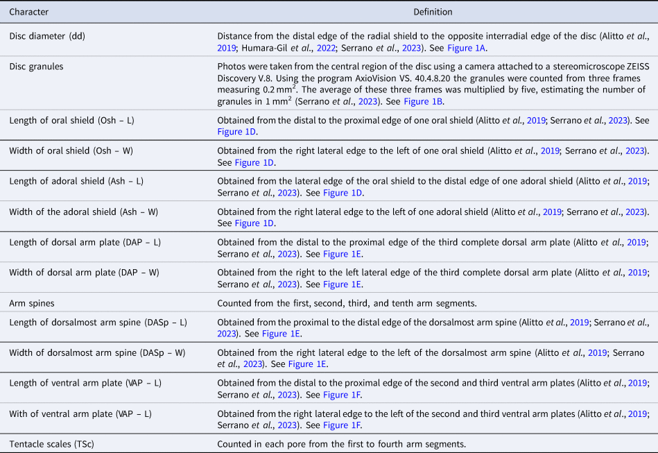
Structures such as the disc, dorsal arm plates, ventral arm plates, and arm spines were measured following Serrano et al. (Reference Serrano, Damiano, Alitto, Guilherme, Cerqueira, Mendonça, Tavares and Borges2023) (Figure 1, Table 1). All the samples were measured using a camera attached to a stereomicroscope ZEISS Discovery V.8 and the program AxioVision VS. 40.4.8.20 (Carl Zeiss Microscopy, Oberkochen, Germany) for specimens and structures smaller than 10 mm dd and/or with a digital caliper for specimens larger than 10 mm dd.
Schematic image with the structures measured and analysed: (A) dorsal view; (B) disc granules; (C) ventral view – detail of the papillae; (D) ventral view – detail of the oral and adoral shields; (E) detail of dorsal view of the arm; (F) detail of ventral view of the arm; (G) dorsal arm plate; (H) lateral arm plate – outer view; (I) vertebra – proximal view; (J) vertebra – distal view; (K) oral plate – abradial; (L) ventral arm plate; (M) dental plate; (N) vertebra – dorsal view; (O) vertebra – ventral view; (P) oral plate – adradial. 2° AdShSp, secondary adoral shield spine; AdSh, adoral shield; AdShSp, adoral shield spine; AdSh – L, length of adoral shield; AdSh – W, width of adoral shield; d, dorsal; DAP, dorsal arm plate; DAP – L, length of dorsal arm plate; DAP – W, width of dorsal arm plate; DASp, dorsalmost arm spine; DASp – L, length of arm spine; DASp – W, width of arm spine; dd, disc diameter; ddi, dorso-distal; di, distal; dp, dorso-proximal; IPa, infradental papilla; LyOs, Lyman's ossicle; Osh, oral shield; Osh – L, length of oral shield; Osh – W, width of oral shield; p, proximal; TPa, tooth papilla; TSc, tentacle scale; v, ventral; VAP, ventral arm plate; VAP – L, length of ventral arm plate; VAP – W, width of ventral arm plate; vdi, ventro-distal; vp, ventro-proximal. Scale bars: (A and B) 1 mm; (C–F) 0.5 mm; (G) and (I–P) 500 μm; (H) 200 μm.

Definition of the external features measured and analysed

The data with continuous variables were analysed in a linear discriminant analysis (LDA), which was useful for determining morphological differences between the established groups (species and life stages). The LDA analysis was conducted in R version 4.0.4 using the software packages MASS (Venables and Ripley, Reference Venables and Ripley2002; R Development Core Team, 2018), ggord (Beck, Reference Beck2022), vegan (Oksanen et al., Reference Oksanen, Blanchet, Friendly, Kindt, Legendre, McGlinn, Minchin, O'Hara, Simpson, Solymos, Stevens, Szoecs and Wagner2019), and tidyverse (Wickham et al., Reference Wickham, Averick, Bryan, Chang, McGowan, François, Grolemund, Hayes, Henry, Hester, Kuhn, Pedersen, Miller, Bache, Müller, Ooms, Robinson, Seidel, Spinu, Takahashi, Vaughan, Wilke, Woo and Yutani2019).
One individual of each stage of O. echinata and O. trindadensis was examined using a JEOL JSM5800LV scanning electron microscope at the Electron Microscopy Laboratory of the Institute of Biology at the State University of Campinas – UNICAMP. The internal and external microstructures present in the arm were taken for examination from the fifth to the tenth segments (Figure 1). For internal microstructures of the buccal armature, the entire disc or a fragment was used (Figure 1). The fragments of the arms and the disc were immersed in regular household bleach (NaClO) until the tissues were removed and the ossicles were released (Stöhr et al., Reference Stöhr, Conand and Boissin2008). The ossicles were then washed in distilled water, air-dried, arranged on aluminium stubs, gold coated, examined by SEM, and photographed.
The terminology and abbreviations used in all descriptions followed Thuy and Stöhr (Reference Thuy and Stöhr2011, Reference Thuy and Stöhr2016), Stöhr et al. (Reference Stöhr, O'Hara and Thuy2012), Alitto et al. (Reference Alitto, Bueno, Guilherme, Di Domenico, Christensen and Borges2018), Hendler (Reference Hendler2018), O'Hara et al. (Reference O'Hara, Stöhr, Hugall, Thuy and Martynov2018), and Goharimanesh et al. (Reference Goharimanesh, Stöhr, Mirshamsi, Ghassemzadeh and Adriaens2021).
Abbreviations
Morphological structures
2° AdShSp – secondary adoral shield spine;
AdSh – adoral shield;
AdShSp – adoral shield spine;
DAP – dorsal arm plate;
DASp – dorsalmost arm spine;
IPa – infradental papilla;
LAP – lateral arm plate;
LyOs – Lyman's ossicle;
Osh – oral shield;
TPa – tooth papilla;
TSc – tentacle scale;
VAP – ventral arm plate.
Results
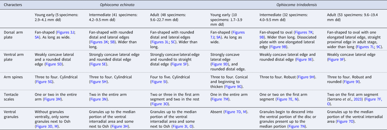
Our study demonstrates that Ophiocoma echinata and Ophiocoma trindadensis can be distinguished during their growth, especially, due to differences in the shape of the VAP and DASp, the number of TSc, and the presence of granules ventrally. The main differences observed during the growth of the species and between them are highlighted in Table 2; the morphometrical analysis and the descriptions of the species are presented below.
Characters that differ between the two studied species of Ophiocoma

Osh, oral shields.
Ventral arm plates were described considering only dissociated plates.
Morphometry
A total of 94 specimens of O. echinata and 87 specimens of O. trindadensis were measured and separated into young early, intermediate, and adult stages. Damaged specimens of O. trindadensis were excluded from this analysis. The LDA using 14 morphological characters (see Supplementary Tables S3 and S4) was effective in separating the species, and partially separating the stages of development (Figure 2).
LDA analysis based on 14 morphological variables. Ash.L, length of adoral shield; Ash.W, width of adoral shield; DAP.L, length of dorsal arm plate; DAP.W, width of dorsal arm plate; DASp.L, length of dorsalmost arm spine; DASp.W, width of dorsalmost arm spine; dd, disc diameter; disc.granule, granules of disc; Osh.L, length of oral shield; Osh.W, width of oral shield; VAP1.L, length of first ventral arm plate; VAP1.W, width of first ventral arm plate; VAP2.L, length of second ventral arm plate; VAP2.W, width of second ventral arm plate.

The width of the second VAP and the width of the DASp (VAP2.W and DASp.W) were the morphological characters with the highest positive coefficients, and the width of the first VAP and length of the Osh (VAP1.W and Osh.L) presented the highest negative coefficients in the first discriminant axis (LD1 – 67.53%) indicating a contribution of these characters in separating O. echinata from O. trindadensis (specimens in all stages). The second discriminant axis (LD2 – 27.5%) separated the adult and intermediate stages of both species from the young early stages with a high positive value in the width of the DASp (DASp.W), width of DAP (DAP.W), and width of the second VAP (VAP2.W), and, as negative coefficients, the width of Ash (Ash.W) and length of DASp (DASp.L) (Figure 2).
Growth series
Order OPHIACANTHIDA O'Hara, Hugall, Thuy, Stöhr & Martynov, 2017
Family OPHIOCOMIDAE Ljungman, 1867
Genus Ophiocoma L. Agassiz, 1836
Ophiocoma echinata (Lamarck, Reference Lamarck1816)
Type locality. Antilles (Lamarck, Reference Lamarck1816)
Distribution. Florida, Bermuda, Bahamas, Tortugas, St. Thomas, Antilles, Haiti, Puerto Rico, Antigua and Barbuda, Bonaire, Aruba, Curaçao, Grenada, Tobago, Panama, Venezuela (Cumana Bay), Liberia, and Brazil (Lyman, Reference Lyman1865; Rathbun, Reference Rathbun1879; Verrill, Reference Verrill1899; Koehler, Reference Koehler1914; Clark, Reference Clark1921; Clark et al., Reference Clark, Rathbun, Boone, Shoemakae and Clark1921; Engel, Reference Engel1939; Tommasi, Reference Tommasi1970; Albuquerque, Reference Albuquerque1986). In Brazil, there are records in Ceará, Paraíba, Pernambuco, Alagoas, and Bahia (Rathbun, Reference Rathbun1879; Verrill, Reference Verrill1899; Tommasi, Reference Tommasi1970; Albuquerque, Reference Albuquerque1986; Manso, Reference Manso1993; Manso et al., Reference Manso, Alves and Martins2008; Lima and Fernandes, Reference Lima and Fernandes2009; Miranda et al., Reference Miranda, Lima, Sovierzoski and Correia2012).
Material examined. 94 specimens (dd: 2.9–22.7 mm). See Specimen Information (Supplementary Table S1).
Disc. Pentagonal to rounded, covered by sparse, rounded granules (Figure 3A–C). Number of granules per mm2 of disc varies and decreases as disc size increases.
External features of Ophiocoma echinata (Lamarck, Reference Lamarck1816): (A, D, J, and M) young early specimen (3.7 mm dd – ZUEC OPH 3220); (G) young early specimen (3.8 mm dd – UFPB. ECH-2700); (B, E, H, K, and N) intermediate (7.3 mm dd – ZUEC OPH 3219); (C, F, I, L, and O) adult (19.5 mm – ZUEC OPH 3072). (A–C) Dorsal view; (D–F) ventral view; (G–I) detail of the jaw; (J–L) detail of dorsal arm; (M–O) detail of ventral arm. 2° AdShSp, secondary adoral shield spine; AdSh, adoral shield; AdShSp, adoral shield spine; DAP, dorsal arm plate; DASp, dorsalmost arm spine; IPa, infradental papilla; LyOs, Lyman's ossicle; Osh, oral shield; TPa, tooth papilla; TSc, tentacle scale; VAP, ventral arm plate; VG, ventral granules. Scale bars: (A, B, D, E, J, K, M, and N) 500 μm; (C) 2 mm; (F, L, and O) 1 mm; (G) 0.2 mm; (H and I) 0.5 mm.

Mouth. At each half jaw: TPa followed by IPa, 2° AdShSp, AdShSp, and LyOs (Figure 3D–I). Osh approximately rectangular with rounded edges (Figure 3D–I). AdSh triangular, wider than long (Figure 3D–I). Juveniles without granules ventrally, only some granules next to Osh (Figure 3G, M). Granules up to median portion of ventral interradial area and some next to Osh in intermediate and adult stages (Figure 3H, I, O).
Arms. DAP fan-shaped, as long as wide or almost as long as wide in juveniles (Figure 3J); DAP fan-shaped to oval in intermediate and adult stages (Figure 3K, L). VAP approximately square with rounded distal edge (Figure 3M–O). Three to four arm spines in juveniles and intermediate stage, three to five in adults up to 10th arm segment. DASp cylindrical, robust, and elongated (Figure 3J–L). One or two TSc on each pore in juvenile stage, two in intermediate stage, two to three in first arm segment, and two in remaining segments in adults (Figure 3M–O).
Morphological variations: One juvenile specimen (3.8 mm dd) presented some granules in two interradii, besides those next to the Osh. Three specimens in the intermediate and two in the adult stages showed the ventral granules extending up to the Osh as a fine line. In two specimens in the intermediate stage, three tentacle scales were observed in the first arm segment. In two specimens in the adult stage, three tentacle scales were observed in the third and fourth arm segments.
Microstructure of ossicles (Figures 4–6)
Specimens selected for description: 3.7 mm (ZUEC OPH 3220) – young early; 7.3 mm (ZUEC OPH 3219) – intermediate stage; 19.3 mm (ZUEC OPH 1538) – adult stage.
Oral ossicles of O. echinata (Lamarck, Reference Lamarck1816): (A, D, and G) young early specimen (3.7 mm dd – ZUEC OPH 3220); (B, E, and H) intermediate (7.3 mm dd – ZUEC OPH 3219); (C, F, and I) adult (19.3 mm dd – ZUEC OPH 1538). (A–C) Dental plate – outer side; (D–F) oral plate – abradial; (G–I) oral plate – adradial. d, dorsal; di, distal; hds, horizontal and diagonal striations; hr, horizontal ridge; maa, muscle attachment area; p, proximal; pk, protruding knobs; sck, sockets; sp, septum; ts, tooth sockets; v, ventral. Scale bars: (A) 100 μm; (D and G) 200 μm; (B, C, E, F, H, and I) 500 μm.

Arm plates and arm spines of O. echinata (Lamarck, Reference Lamarck1816): (A, D, G, and J) young early specimen (3.7 mm dd – ZUEC OPH 3220); (B, E, H, and K) intermediate (7.3 mm dd – ZUEC OPH 3219); (C, F, I, and L) adult (19.3 mm – ZUEC OPH 1538). (A–C) Dorsal arm plate – outer side; (D–F) ventral arm plate – outer side; (G–I) dorsalmost arm spine; (J–L) lateral arm plate – outer side. d: dorsal; ddi: dorso-distal; di: distal; dp: dorso-proximal; p: proximal; sa, spine articulation; v: ventral; vdi: ventro-distal; vp: ventro-proximal. Scale bars: (A, D, and J) 100 μm; (B, C, E–H, and K) 200 μm; (I and L) 500 μm.

Vertebrae of O. echinata (Lamarck, Reference Lamarck1816): (A, D, G, and J) young early specimen (3.7 mm dd – ZUEC OPH 3220); (B, E, H, and K) intermediate (7.3 mm dd – ZUEC OPH 3219); (C, F, I, and L) adult (19.3 mm dd – ZUEC OPH 1538). (A–C) Vertebrae ossicle – proximal surface; (D–F) vertebrae ossicle – distal surface; (G–I) vertebrae ossicle – dorsal surface; (J–L) vertebrae ossicle – ventral surface. d, dorsal; di, distal; DGr, dorsal groove; kb, knobs; p, proximal; v, ventral; zd, zygocondyles; zp, zygosphene. Scale bars: (D) 100 μm; (A, E, G, H, J, and K) 200 μm; (B, C, F, I, and L) 500 μm.

Dental plate. Outer side: Dorsalmost portion with two tooth sockets, only one with thin septum in young early stage; dorsalmost portion with three tooth sockets, all with thin septum in intermediate stage; dorsalmost portion with two tooth sockets, all with thin septum in adult stage (Figure 4A–C). Series of horizontal ridges in median portion and sockets for tooth papillae in ventralmost portion, tooth sockets surrounded by protruding knobs (Figure 4B, C). Protruding knobs, horizontal ridges, and sockets for tooth papillae still developing in young early stage (Figure 4A).
Oral plate. Abradial: Still-developing muscle flange with soft horizontal and diagonal striations in young early stage (Figure 4D), large and well-defined muscle flange with horizontal and diagonal striations in intermediate and adult stages (Figure 4E, F). Adradial: Muscle attachment area arranged vertically in middle region, long, tapering, spoon-shaped (Figure 4G–I).
Dorsal arm plates. General outline: Fan-shaped with rounded distal and lateral edges, straight proximal edge (Figure 5A–C). DAP with asymmetrical lateral edges in intermediate stage (Figure 5B), and symmetrical in adult stage (Figure 5C).
Ventral arm plates. General outline: Pentagonal, distal portion wider than proximal, convex distal edge, proximal edge ‘V’ like (Figure 5D–F). Lateral edges with weak incisions in young early stage (Figure 5D), become strongly concave in larger specimens (Figure 5E, F).
Dorsal arm spines. Cylindrical, with approximately the same width in distal and proximal portions (Figure 5G–I).
Lateral arm plates. General outline: Arched shape. Ventral portion projecting ventro-proximally (Figure 5J–L). LAP decreases in width as disc size increases (Figure 5J–L). Spine articulations: Three to four, on elevated portion not bordered proximally by ridge (Figure 5J–L), restricted to central portion of distal edge and equidistant in young early and intermediate stages (Figure 5J, K), arranged over entire distal edge, and distance between spine articulation dorsalwards increasing in adult stage (Figure 5L).
Vertebrae. Zygospondylous articulation. Proximal side: Groove dorsally (Figure 6A–C). Distal side: Zygosphene fused with pair of parallel zygocondyles (Figure 6D–F). Well-developed muscle attachment area with knobs around dorsalmost lateral edges in adult stage (Figure 6I). Dorsal side: Dorsal groove not projecting beyond zygocondyles (Figure 6G–I). Ventral side: All specimens with ‘V’ cavities in proximal–ventral edge, zygosphene projecting beyond ventral edge of zygocondyles with projecting part shorter than zygocondyles (Figure 6J–L).
Ophiocoma trindadensis Serrano, Damiano, Alitto and Borges, Reference Serrano, Damiano, Alitto, Guilherme, Cerqueira, Mendonça, Tavares and Borges2023
Type locality. Trindade and Martin Vaz Oceanic Archipelago, Espírito Santo, Brazil (Serrano et al., Reference Serrano, Damiano, Alitto, Guilherme, Cerqueira, Mendonça, Tavares and Borges2023).
Distribution: Only known from its type locality (Serrano et al., Reference Serrano, Damiano, Alitto, Guilherme, Cerqueira, Mendonça, Tavares and Borges2023).
Material examined: 95 specimens (dd: 1.7–19.4 mm). See Specimen Information (Supplementary Table S2).
Disc. Rounded, covered by sparse, rounded granules (Figure 7A–C). Number of granules per mm2 of disc decreases as disc size increases.
External features of Ophiocoma trindadensis Serrano et al., Reference Serrano, Damiano, Alitto, Guilherme, Cerqueira, Mendonça, Tavares and Borges2023: (A, D, G, J, and M) young early specimen (1.7 mm dd – ZUEC OPH 3092); (B, E, H, K, and N) intermediate (8.2 mm dd – MZUSP 2784); (C, F, I, L, and O) adult (14 mm dd – ZUEC OPH 3097). (A–C) Dorsal view; (D–F) ventral view; (G–I) detail of the jaw; (J–L) detail of dorsal arm; (M–O) detail of ventral arm. 2° AdShSp, secondary adoral shield spine; AdSh, adoral shield; AdShSp, adoral shield spine; DAP, dorsal arm plate; DASp, dorsalmost arm spine; IPa, infradental papilla; LyOs, Lyman's ossicle; Osh, oral shield; TPa, tooth papilla; TSc, tentacle scale; VAP, ventral arm plate; VG, ventral granules. Scale bars: (D, G, J, and M) 0.2 mm; (E, F, H, I, L, and O) 1 mm; (B and K) 2 mm; (C) 10 mm; (A and N) 0.5 mm.

Mouth. At each half jaw: TPa followed by IPa, 2° AdShSp, AdShSp, and LyOs (Figure 7D–I), papillae still developing in the smallest juvenile (1.7 mm dd) (Figure 7G). Osh approximately rectangular with rounded edges (Figure 7D–I). AdSh triangular, wider than long (Figure 7D–I). Young early stage without granules ventrally (Figure 7D, M). Granules begin descending into ventral portion of disc or granules present up to median portion of ventral interradial area in intermediate stage (Figure 7N). Granules up to median portion of ventral interradial area in adult specimens (Figure 7O).
Arms. DAP fan-shaped, as long as wide or almost as long as wide in young early stage (Figure 7J), DAP fan-shaped to oval, wider than long in intermediate stage (Figure 7K), DAP oval in adult stage, wider than long (Figure 7L). VAP approximately square with rounded distal edge (Figure 7M–O). Three to four arm spines up to 10th arm segment. DASp conical and begins to thicken in young early stage (Figure 7J), robust and rounded in intermediate and adult stages (Figure 7K, L). Number of TSc in first segment varies: one in young early stage (Figure 7M), one or two in intermediate stage (Figure 7E, N), two in adult stage (Figure 7F, O), and one in remaining arm segments.
Morphological variations. The highest number of granules per mm2 of disc (78–98) was observed at 1.7 and 4.5 mm dd, while the lowest (4) was observed at 19.4 mm dd. One specimen in the young early stage (3.4 mm dd) presented two TSc at most of the tentacle pores of the first arm segment. In one adult specimen (17.9 mm dd) a single arm segment with five arm spines was observed.
Microstructure of ossicles (Figures 8–10)
Specimens selected for description: 1.7 mm (ZUEC OPH 3092) – young early; 8 mm (MZUSP 2772) – intermediate stage; 14 mm (ZUEC OPH 3097) – adult stage.
Oral ossicles of O. trindadensis Serrano et al., Reference Serrano, Damiano, Alitto, Guilherme, Cerqueira, Mendonça, Tavares and Borges2023: (A, D, and G) young early specimen (1.7 mm dd – ZUEC OPH 3092); (B, E, and H) intermediate (8 mm – MZUSP 2772); (C, F, and I) adult (14 mm dd – ZUEC OPH 3097). (A–C) Dental plate – outer side; (D–F) oral plate – abradial; (G–I) oral plate – adradial. d, dorsal; di, distal; hds, horizontal and diagonal striations; hr, horizontal ridge; maa, muscle attachment area; p, proximal; pk, protruding knobs; sck, sockets; sp, septum; ts, tooth sockets; v, ventral. Scale bars: (A) 50 μm; (C, F, and I) 500 μm; (D and G) 100 μm; (B, E, and H) 200 μm.

Arm plates and arm spines of O. trindadensis Serrano et al., Reference Serrano, Damiano, Alitto, Guilherme, Cerqueira, Mendonça, Tavares and Borges2023: (A, D, G, and J) young early specimen (1.7 mm dd – ZUEC OPH 3092); (B, E, H, and K) intermediate (8 mm dd – MZUSP 2772); (C, F, and I) adult (14 mm dd – ZUEC OPH 3097); (L) adult (19.3 mm dd – ZUEC OPH 3095). (A–C) Dorsal arm plate – outer side; (D–F) ventral arm plate – outer side; (G–I) dorsalmost arm spine; (J–L) lateral arm plate – outer side. d, dorsal; ddi, dorso-distal; di, distal; dp, dorso-proximal; p, proximal; as, spine articulation; v, ventral; vdi, ventro-distal; vp, ventro-proximal. Scale bars: (A, D, G, and J) 100 μm; (B, E, H, and K) 200 μm; (C, F, I, and L) 500 μm.

Vertebrae of O. trindadensis Serrano et al., Reference Serrano, Damiano, Alitto, Guilherme, Cerqueira, Mendonça, Tavares and Borges2023: (A, D, G, and J) young early specimen (1.7 mm dd – ZUEC OPH 3092); (B, E, H, and K) intermediate (8 mm dd – MZUSP 2772); (C, F, I, and L) adult (14 mm – ZUEC OPH 3097). (A–C) Vertebrae ossicle – proximal surface; (D–F) vertebrae ossicle – distal surface; (G–I) vertebrae ossicle – dorsal surface; (J–L) vertebrae ossicle – ventral surface. d, dorsal; di, distal; DGr, dorsal groove; kb, knobs; p, proximal; v, ventral; zd, zygocondyles; zp, zygosphene. Scale bars: (A, D, G, and J) 100 μm; (B, E, H, and K) 200 μm; (C, F, I, and L) 500 μm.

Dental plate. Outer side: Dorsalmost portion with tooth socket without septum, median portion with small tooth socket with septum in young early stage (Figure 8A). Dorsalmost portion with two tooth sockets with thin septum in intermediate and adult stages (Figure 8B, C). Series of horizontal ridges in median portion and sockets for tooth papillae in ventralmost portion, tooth sockets surrounded by protruding knobs (Figure 8B, C). Horizontal ridges, sockets for tooth papillae, and protruding knobs weakly developed in young early stage (Figure 8A).
Oral plate. Abradial: Developing muscle flange without horizontal and diagonal striations in young early stage (Figure 8D), large and well-defined muscle flange with horizontal and diagonal striations in intermediate and adult stages (Figure 8E, F). Adradial: Muscle attachment areas arranged vertically in middle region, long, tapering, spoon-shaped (Figure 8G–I).
Dorsal arm plates. General outline: Fan-shaped. DAP with rounded distal and lateral edges, proximal edge ‘V’ like in young early stage (Figure 9A). DAP with rounded distal and lateral edges, with one elongated lateral edge, straight proximal edge in intermediate stage (Figure 9B), fan-shaped to oval with one elongated lateral edge, straight proximal edge in adult stage (Figure 9C).
Ventral arm plates. General outline: Pentagonal, distal portion wider than proximal, convex distal edge, proximal edge ‘V’ like (Figure 9D–F). Lateral edges strongly concave in young early stage (Figure 9D), becoming shallower incisions with growth (Figure 9E, F).
Dorsal arm spines. Conical and serrated in young early stage (Figure 9G). Becomes robust and cylindrical or rounded in intermediate stage (Figures 7K and 9H) and robust and rounded (almost circular) in adult stage (Figure 9I).
Lateral arm plates. General outline: Arched shape. Approximately rectangular in young early stage (Figure 9J). Ventral portion projecting ventro-proximal in intermediate and adult stages (Figure 9K, L). LAP decreases in width as disc size increases. Spine articulations: Three to four, on elevated portion not bordered proximally by ridge (Figure 9J–L), restricted to central portion of distal edge and equidistant in young early stage and intermediate stages (Figure 9J, K), arranged over entire distal edge and distance between spine articulation dorsalwards increasing in adult stage (Figure 9L).
Vertebrae. Zygospondylous articulation. Proximal side: groove dorsally (Figure 10A–C). Distal side: Zygosphene fused with pair of parallel zygocondyles (Figure 10D–F). Well-developed muscle attachment area with knobs around dorsalmost lateral edges in intermediate and adult stages (Figure 10E, F). Dorsal side: Dorsal groove not projecting beyond zygocondyles (Figure 10G–I). Ventral side: All specimens with ‘V’ cavities in proximal–ventral edge, zygosphene projecting beyond ventral edge of zygocondyles with projecting part shorter than zygocondyles (Figure 10J–L).
Discussion
Young early stage of Ophiocoma echinata and O. trindadensis have never been described previously, although adults of both species are common in shallow waters from northeastern Brazil and the remote oceanic archipelago Trindade and Martin Vaz, respectively (Rathbun, Reference Rathbun1879; Tommasi, Reference Tommasi1970; Albuquerque, Reference Albuquerque1986; Manso, Reference Manso1993; Manso et al., Reference Manso, Alves and Martins2008; Lima and Fernandes, Reference Lima and Fernandes2009; Miranda et al., Reference Miranda, Lima, Sovierzoski and Correia2012; Serrano et al., Reference Serrano, Damiano, Alitto, Guilherme, Cerqueira, Mendonça, Tavares and Borges2023). Ophiocoma echinata and O. trindadensis are morphologically similar, particularly at smaller sizes. However, the study of their growth series proved to facilitate the identification of young early, intermediate stages, and adults.
Schoener (Reference Schoener1967, Reference Schoener1969), Monteiro (Reference Monteiro1987), and da Silva et al. (Reference da Silva, Prata and Christoffersen2023) showed that some structures in brittle stars are not deeply affected during ontogeny, while others suffer important changes depending on the species. In Ophiocoma echinata and O. trindadensis, the number of granules per mm2 and shape of DAP varied between young early stage and adults, whereas disc shape and type of dorsal coverage remained essentially unchanged during growth, as did the general shape of dental and oral plates. It has also been documented that the number of TSc is usually smaller or even absent in smaller specimens (Monteiro, Reference Monteiro1987; Stöhr and Martynov, Reference Stöhr and Martynov2016; Humara-Gil et al., Reference Humara-Gil, Granja-Fernández, Montoya-Márquez and López-Pérez2022) and the number of arm spines is lower in young early stage (Vadon, Reference Vadon1990; Sumida et al., Reference Sumida, Tyler, Gage and Nørrevang1998; Stöhr, Reference Stöhr2005; Borges et al., Reference Borges, Alitto and Amaral2015; Humara-Gil et al., Reference Humara-Gil, Granja-Fernández, Montoya-Márquez and López-Pérez2022). Similarly, a small number of TSc and arm spines were observed in the young early stage of the species herein studied. The number and position of the mouth papillae can also change during development (Vadon, Reference Vadon1990; Sumida et al., Reference Sumida, Tyler, Gage and Nørrevang1998; Borges et al., Reference Borges, Monteiro and Amaral2002; Stöhr, Reference Stöhr2005; Borges et al., Reference Borges, Alitto and Amaral2015). In this study, a lower number of mouth papillae was observed only in the smallest young early specimen of O. trindadensis (1.7 mm dd), while the smallest specimen of O. echinata analysed (2.9 mm dd) already had a similar number of mouth papillae to the adult.
As summarized in Table 2, the main differences between the two species are related to the ventral arm plate, dorsalmost arm spines, the number of tentacle scales, and the presence of granules ventrally. The young early stage of Ophiocoma echinata has a shallow incision in the lateral edges of the ventral arm plate and a strong incision in the intermediate and adult stages, while O. trindadensis has a strong incision in young early stage and shallow in the intermediate and adult stages. The shape of dorsalmost arm spines also differentiated these species: in O. echinata it is cylindrical and always longer than wide. In O. trindadensis it is conical in young early specimens, cylindrical to robust in the intermediate stage, and robust and rounded in the adult stage, and it can be as long as wide or almost as long as wide. At first look, some dorsalmost arm spines of adults of O. echinata can seem similar in size to those of O. trindadensis, but the proportion between length and width is usually smaller in O. trindadensis. The young early specimens of O. echinata have one or two tentacle scales in the proximal arm segments, specimens in the intermediate stage have two in almost the entire arm, and adults have two or three in the first arm segment and two in the rest. In contrast, young early specimens of O. trindadensis have one tentacle scale in all arm segments, one and two in the first arm segment in the intermediate stage, two in the first arm segment and one in all other segments in the adult stage. In young early specimens of O. echinata, the ventral granules are absent, but there are some granules next to the oral shields, and in the intermediate and adult stages granules occur up to the median portion of the ventral interradial area and there are some granules next to the oral shields. The ventral granules are also absent in the young early specimens of O. trindadensis, and in the intermediate and adult stages granules occur up to the median portion of the ventral interradial area and do not reach the oral shields.
Devaney (Reference Devaney1970) reported that as the development of the granules on the disc can change species specifically according to disc size in Ophiocoma, it may help to identify the species. In his study, the author reports disc granules in specimens of O. echinata with 2.3 mm dd (Devaney, Reference Devaney1970), whereas the present study reports disc granules in a specimen with 1.7 mm dd of O. trindadensis.
The LDA analysis confirmed the separation of the species from each other. Serrano et al. (Reference Serrano, Damiano, Alitto, Guilherme, Cerqueira, Mendonça, Tavares and Borges2023) indicated that the main morphological characters that separate the two species are the width of the second ventral arm plate, the length of the first ventral arm plate, the length of the oral shield, and the oral diameter. The present study carried out the LDA analysis with more specimens including different stages of development of both species, and confirmed that the width of the second ventral arm plate and length of the oral shield are important for distinguishing the species. Additionally, the width of the dorsalmost arm spines and the width of the first ventral were also important in their separation. The length of the first ventral arm plate, despite not having presented the highest positive coefficient on the LD2 axis, was also shown in the analysis, especially to separate the adult and intermediate stages of the two species from the young early stage of development. The oral diameter was not analysed in this study. Adults and intermediate specimens are easily separated from young early specimens mainly by the width of the dorsalmost arm spines, the width of the dorsal arm plate, the width of the second ventral arm plate, the width of the adoral shield, and the length of dorsalmost arm spines. To better separate young early specimens between species, more specimens in this stage need to be analysed in further studies.
The results obtained here reinforced that morphometrical analysis and SEM imagery may help to morphologically distinguish species from the early stages of growth. In addition, they illustrate the importance of growth series studies in increasing our knowledge about Ophiocoma, and additional progress can be made in the taxonomy of the group with more studies on the morphological changes during ontogeny and the correlation of young early specimens and adults in other species of the genus.
Supplementary material
The supplementary material for this article can be found at https://doi.org/10.1017/S0025315424001164.
Data
All data generated or analysed during this study are included in this published article and its Supplementary materials.
Acknowledgements
We are grateful to the Paulo Young Invertebrate Collection of the Federal University of Paraíba, especially Jéssica Prata, for granting access and donation of specimens of Ophiocoma echinata for comparative studies. We also acknowledge the access to equipment and assistance provided by the Electron Microscope Laboratory (LME/UNICAMP). We are grateful to the researcher Dr Pablo D. B. Guilherme for the assistance with the statistical analysis.
Financial support
This study was funded by the São Paulo Research Foundation (FAPESP, Process no. 2019/01620-9; Process no. 2019/05997-0), and CAPES (Coordenação de Aperfeiçoamento de Pessoal de Nível Superior – Brasil) – Finance Code 001. This work was also supported by the BIOTA-Collections Project (Process no. 2018/10313-0), and the ProTrindade Project (Process no. 443278/2019-9). We thank the Brazilian Navy (1st District) and SECIRIM (Interministerial Secretariat for Marine Resources) for all the support provided in Trindade and Martin Vaz. This is the contribution number 18 of the ProTrindade Marine Invertebrates Project.
Competing interests
None.
Ethical standards
The study was registered at the National System for the Management of Genetic Heritage and Associated Traditional Knowledge – SisGen (Sistema Nacional de Gestão do Patrimônio Genético e do Conhecimento Tradicional Associado), according to Brazilian legislation Law number 13.123/2015 and Decree 8772/2016. The approval ID for this study was A3014E0.