INTRODUCTION
There are interesting superficial similarities of some Antarctic littorinoideans with the Naticidae. Several samples have been identified as so in collections, and two cases are herein disclosed. In the literature, there are the examples of the genera Frovina Thiele 1912 and Prolacuna Thiele 1913, which were described as Naticidae. The Antarctic littorinoideans that resemble naticids and other families were studied by Warén & Hain (Reference Warén and Hain1996), resulting in the erection to a new family – Zerotulidae – which is so far restricted to the south-glacial, Antarctic region (Aldea & Troncoso, Reference Aldea and Troncoso2008). However, the analysis on this naticid similarity is scanty, as no detailed comparison of both groups has been published.
Developing a comparative study on the family of predator moon-snails Naticidae, partially published in Simone (Reference Simone2011), some Antarctic samples came to attention identified as such, including supposed samples of the genus Amauropsis Mörch 1857, and Kerguelenatica Powell, Reference Powell1951. Despite the remarkable similarities of shells and opercula, the study of the morphology and internal anatomy revealed that those samples actually do not belong to naticoideans, rather they belong to a branch better placed in the Littorinoidea-Rissooidea clade of the Caenogastropoda (sensu Simone, Reference Simone2006, Reference Simone2011), and one of them is better allocated in the Zerotulidae (Warén & Hain, Reference Warén and Hain1996).
As the wide study (Simone, Reference Simone2011) is mainly based on phenotypic-morphological features, the foremost objective of this paper is to discuss the noteworthy similarities between these Antarctic species and naticoideans, both in external aspects and in the internal structures, exploring the convergences and differences between them and true naticids. Additionally, a taxonomic treatment is performed on the studied samples, and their insertion in a wide phylogenetic study, in order to better understand their relationships, as the basis for a stronger supported discussion. The presence of a zerotulid in Brazilian deep waters is also reported.
The Littorinidae genus Laevilacunaria Powell, Reference Powell1951 (type species Pellilitorina bransfieldensis Preston, Reference Preston1916; OD) is restricted to the Antarctic region, encompassing six species, and sometimes referred as synonym of Macquariella Finlay 1926 (Reid, Reference Reid1989). Samples belonging to this genus were collected by the Brazilian Antarctic Project – PROANTAR, and are herein included, extending its description to the anatomy. Likewise, a sample belonging to an undescribed littorinoidean genus is also included, which has been previously published as possible naticids (Simone, Reference Simone2011). The new genus is tentatively allocated in the above-mentioned Zerotulidae. Another sample collected by the RV ‘Marion-Dufresne’ French Expedition (MD55) in the central Brazilian coast (May 1987) appears to belong to the same new genus. This extends the previously endemic family from Antarctica northwards, to the middle Atlantic.
MATERIALS AND METHODS
The samples studied here were collected by the Brazilian Antarctic Project – PROANTAR (Cirm, 2007) – and deposited in the Museu de Zoologia da Universidade de São Paulo (MZSP) collection. The shells were additionally processed by Zeiss V12 autofocus microscope for illustration. The preserved specimens were extracted from their shells, and dissected by standard techniques. All drawings were obtained with the aid of a camera lucida. Digital photos of all dissecting steps were also obtained. Some hard parts, such as radula and jaws, were also examined in a scanning electron microscope (SEM) provided by the Laboratório de Microscopia Eletrônica do Museu de Zoologia da USP (Zeiss DSM 940). The material MD55 refers to that collected by the RV ‘Marion-Dufresne’, operated by Terres Australes et Antarctiques Françaises. It was a joint project of Muséum National d'Histoire Naturelle, in Paris (MNHN), and Universidade Santa Ursula, in Rio de Janeiro (USU); Philippe Bouchet, José H. Leal and Bernard Metivier were the malacologists on board (Simone & Cunha, Reference Simone and Cunha2012). The material was sent for further taxonomic study.
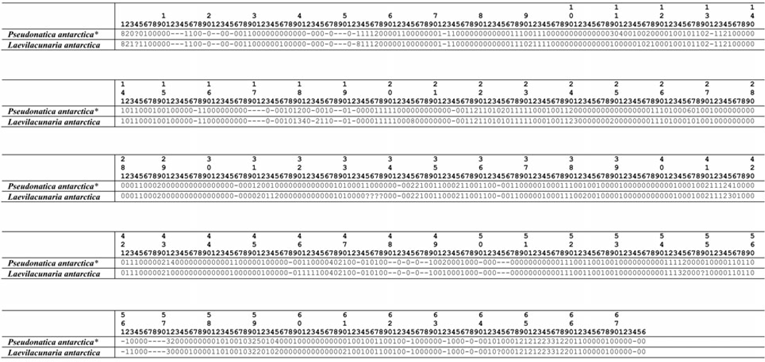
The two species herein anatomically studied had their characters coded in a character-matrix (Table 1), under the criteria and methodologies described in Simone (Reference Simone2011: 163–175). The two lines of the present matrix (Table 1) were added to the matrix of that paper (Simone, Reference Simone2011: 267–308) and the resulting cladogram is shown here, focusing the allocation of the cladogram on the results of the presently studied taxa and neighbouring taxa (Figures 62 & 63), as the resulting cladogram is exactly the same of that paper (Simone, Reference Simone2011: figure 20). The set of synapomorphies of each node is also shown here in the same way as that paper (Simone, Reference Simone2011: 309–317). As no new character was added to those already presented in Simone (Reference Simone2011), the number of non-homoplastic synapomorphies are presently few. All these are properly debated in the Discussion section. The additional intention is to display the present results in a wider scenario. Despite the existence of important papers that include some anatomy of Littorinidae (e.g. Reid, Reference Reid1989) and Zerotulidae (e.g. Warén & Hain, Reference Warén and Hain1996), the data of the species included there are not complete enough (e.g. odontophore) to be included in the present analysis.
Matrix of characters of examined specimens which is inserted in Simone's (Reference Simone2011) matrix.

* Coded as ‘Amauropsis rossiana’ in Simone (Reference Simone2011).
As the available samples were not properly preserved, and are relatively old, efforts of sequencing the material were not fruitful.
In the figures, the following abbreviations are used: aa, anterior aorta; af, afferent gill vessel; ag, albumen gland; an, anus; ap, female pore; au, auricle; bg, buccal ganglion; br, subradular membrane; ce, cerebral-pleural ganglia; cg, capsule gland; cm, columellar muscle; cv, ctenidial vein; dd, duct to digestive gland; df, dorsal fold of buccal mass; dg, digestive gland; ec, oesophageal crop; ef, oesophageal fold; eg, oesophageal gland; ep, epipodium; es, oesophagus; ey, eye; fp, female pore; ft, foot (mesopodium); gi, gill or gill filament; gp, pleural ganglion; he, head; hg, hypobranchial gland; in, intestine; ir, insertion of m4 in tissue on radula (to); is, insertion of m5 in subradular membrane; jw, jaw; ki, kidney; m1 to m14, extrinsic and intrinsic odontophore muscles; mb, mantle border; mj, jaws, buccal, and oral tube muscles; mo, mouth; ne, nephrostome; ng, nephridial gland; nr, nerve ring; oa, opercular pad; oc, odontophore cartilage; od, odontophore; of, osphradium satellite fold; om, ommatophore; op, operculum; os, osphradium; ov, pallial oviduct; pc, pericardium; pd, penis duct; pe, penis; pg, pedal glands anterior furrow; pp, penis papilla; pr, propodium; pt, prostate; pu, pedal ganglion; rm, retractor muscle of snout; rn, radular nucleus; rs, radular sac; rt, rectum; rv, renal efferent vessel; sa, salivary gland aperture at buccal cavity; sc, subradular cartilage; sd, salivary duct; se, septum between oesophagus and odontophore in buccal mass; sg, salivary gland; sn, snout; st, stomach; su, suboesophageal ganglion; sy, statocyst; te, cephalic tentacle; tg, integument; to, tissue on middle region of radula preceding buccal cavity; vd, vas deferens; ve, ventricle; vg, vaginal atrium; vo, visceral oviduct.
Institutional abbreviations: MNHN, Museum National d'Histoire Naturelle, Paris, France; MZSP, Museu de Zoologia da Universidade de São Paulo, Brazil; NMW, National Museum Wales, UK; Proantar, Programa Antártico Brasileiro, Comissão Interministerial para os Recursos do Mar (CIRM) http://www.cnpq.br/web/guest/proantar; ZMB, Zoological Museum Berlin, Germany.
SYSTEMATICS
Order Caenogastropoda Cox, 1960
Superfamily RISSOOIDEA Gray, 1847
Family ZEROTULIDAE Warén & Hain, Reference Warén and Hain1996
Genus Pseudonatica new genus
Urn: lsid:zoobank.org:act:7E598BD5-ACDE-487F-B1EF-63600EDC19AC
DIAGNOSIS
Shell somewhat thick walled; spire bluntly pointed, with ~1/2 of body-whorl height. Periostracum smooth, opaque, lacking colours, tending to be eroded. Propodium relatively simple, restricted to anterior region. Tentacle with ommatophore at outer base. Pair of jaw plates narrow and short. Odontophore pair m7 fused. Oesophageal gland entirely attached to oesophagus (sessile); septa divided in middle by longitudinal septum. Distance between cerebro-pleural ganglia and pedal ganglia somewhat wide.
TYPE SPECIES
Pseudonatica antarctica n. sp.
LIST OF INCLUDED TAXA
Type species; P. ampullarica n. sp.
Gender: Feminine.
Etymology: The generic name is an allusion to the similarity of the species with the current naticids, in such Natica is the type species; added to the Greek word pseudo, meaning lie, false, i.e. a false-Natica.
Pseudonatica antarctica new species
Figures 1–8, 14–25
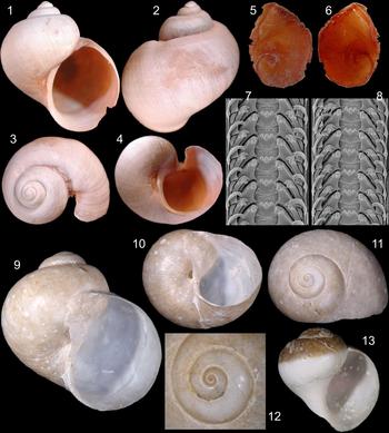
1–8: Pseudonatica antarctica n. sp. Holotype MZSP 113345. (1) Apertural view (L 20.4 mm). (2) Dorsal view. (3) Apical-slightly right view. (4) Inferior-slightly apertural view. (5) Operculum, outer view. (6) Same, inner view. (7–8) Radula in SEM, scale = 50 µm. 9–13. Pseudonatica ampullarica n. sp. types: (9) Holotype MNHN 26866, frontal view (W 25.3 mm). (10) Same, umbilical view. (11) Same, apical view. (12) Same, detail of apex, apical view. (13) Paratype MZSP 105126 (inferior region broken) (W 12.0 mm).

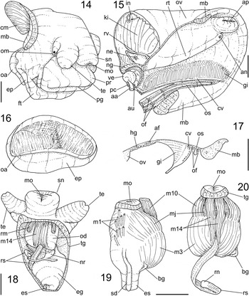
Pseudonatica antarctica n. sp. anatomy. (14) Head-foot, female, right-slightly anterior view. (15) Pallial cavity hoof and reno-pericardial structures, female, ventral view. (16) Foot, detail of posterior region, dorsal view. (17) Pallial cavity hoof, transverse section on middle level of osphradium. (18) Head and haemocoel, ventral view, foot removed. (19) Buccal mass, right view. (20) Odontophore, ventral view, radular sac uncoiled. Scales = 2 mm.

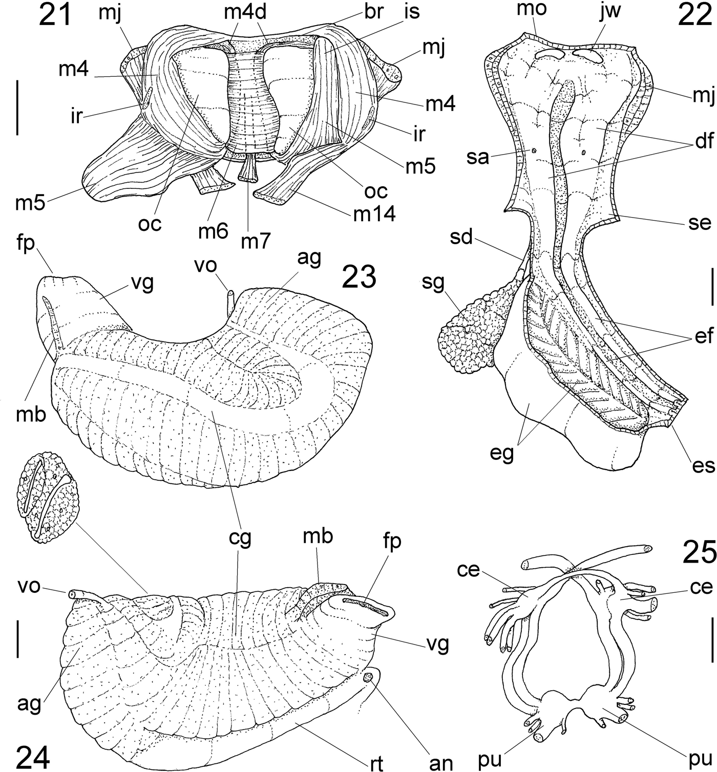
Pseudonatica antarctica n. sp. anatomy. (21) Odontophore, dorsal view, radula removed, both odontophore cartilages (oc) deflected, left m5 deflected downwards. (22) Foregut, ventral view, odontophore removed, remaining tubes opened longitudinally to show inner structures. (23) Pallial oviduct, outer view, mantle removed. (24) Same, inner-ventral view, some adjacent structures also shown. (25) Nerve ring, ventral view. Scales = 1 mm.

‘Amauropsis’ rossiana: Simone, Reference Simone2011: 167, 212, 214, 221, 269, 276, 283, 290, 297, 304, 321 (non Smith 1907).
TYPE MATERIAL
Holotype MZSP 113345.
TYPE LOCALITY
ANTARCTICA. Antarctic Peninsula; King George Island; Admiralty Bay, 62°08′S 58°26′W, 60 m depth (Proantar; iii/2000).
Urn: lsid:zoobank.org:pub:17418B18-B7D4-4725-B4BE-677B1CDBC7EB
DIAGNOSIS
Shell ~20 mm, naticiform-turbiform; ~120% taller than wide. Umbilicus closed, totally covered by narrow callus. Sculpture deep, well-marked; subsutural cord narrow and low; surface opaque, with growth lines only, pale beige. Protoconch low, dome-shaped, of 1.5 whorls. Spire angle ~100°. Head with ommatophore separated from tentacle. Propodium not overlapping mesopodium. Operculum attachment with constriction in middle region. Satellite fold surrounding entire ridge-like osphradium. Radula with rachidian tricuspid, lacking basal cusps; lateral and inner marginal teeth also tricuspid, lateral with basal cusp and outer flap covering inner marginal; outer marginal tooth bicuspid and slender. Pallial oviducts with albumen and capsule glands spirally mixed; terminal wide female papilla. Pair of cerebral and pleural ganglia fused with each other, both far separated from pedal ganglia.
DESCRIPTION
Shell (Figures 1–4): Length of ~20 mm; height ~120% of width; general form naticiform-turbiform. Colour opaque reddish beige; periostracum thin pale brown in peri-umbilical area (Figure 4). Protoconch simple, dome-shaped, of 1.5 flattened whorls (Figure 3); surface smooth, glossy; diameter ~0.8 mm, occupying ~5% of shell surface in apical view. Teleoconch of ~3.0 whorls; suture well-marked, deep, by angle ~110° (Figures 1 & 2); spire angle ~100°. Sculpture absent except for growth lines; weak subsutural cord, located 1/6 whorl inferiorly to suture (Figures 2 & 3), clearer in last whorl. Aperture elliptic-almost circular, occupying ~64% of shell height and ~56% of shell width; outer lip thin, cutting-edged, weakly prosocline (Figure 4), simple (presence of anal slit possibly due to injury, absent in previous growth lines – Figures 3 & 4); inner lip slightly less concave than outer lip (Figure 1), superior third lacking callus, inferior 2/3 as thick apertural edge (Figures 1 & 4). Umbilicus narrow, occluded by inner lip.
Head-foot (Figures 14 & 18): Not pigmented. Head slightly protruded, pair of tentacles (te) stubby, broad, tip somewhat bifid; eyes located at tip of ommatophores in outer base od tentacles (om), ommatophores ~3 times longer than wide, with ~1/5 tentacles’ size; tentacles very laterally positioned. Snout (sn) broad, ~1/10 of head-foot volume, somewhat cylindrical; ventral surface bluntly bifid; mouth (mo) central, longitudinal. Foot broad, of ~1/2 whorl; propodium (pr) from foot anterior edge up to head ventral base, with same adjacent mesopodium extension; mesopodium (ft) with thick edges, sole simple; furrow of pedal glands (pg) restricted to anterior edge, deep, flanked by thick edges. Opercular pad (Figure 16: oa) with thin edges, slightly narrower than mesopodium; attachment with operculum as 2 barely elliptical regions, separated by transverse detached area as shown in Figure 16. Columellar muscle (cm) wide, thick, of ~¼ whorl. Haemocoel elliptical, occupying ~1/4% of head-foot volume, mostly fulfilled by foregut (Figure 18). Diaphragmatic septum (di) thin, compressed by oesophageal gland.
Operculum (Figures 5 & 6): Semi-circular, thin, corneous, flexible, occupying entire aperture. Nucleus in middle of inner-inferior quadrant; spiral sculpture with 2 whorls, low, easily visible; low undulations parallel to inner edge. Edges flexiclaudent. Inner surface smooth, glossy; scar elliptical (twice wider than long), closer to inner edge, occupying ~40% of inner surface.
Mantle organs (Figures 15 & 17): Pallial cavity triangular, of ~1/2 whorl, greatly compresses by right structures, mainly pallial oviduct. Osphradium (os) ridge-like, narrow, with 80% of pallial cavity length; weakly curved to left, almost straight. Osphradium satellite fold (of) surrounding it entirely at short distance from it; width ~half that of osphradium. Gill (gi) with ~1/4 pallial hoof width and 95% its length, wider region between anterior and middle areas; each filament triangular, about as tall as wide; apex central, sharp pointed, weakly curved to right. Ctenidial vein (cv) simple, narrow. Afferent gill vessel (af) relatively wide, flanking gill at some distance. Hypobranchial gland (hg) very thin, white, fulfilling area between gill and rectum/pallial oviduct. Rectum and pallial oviduct running in left-ventral region; oviduct very wide (described below); rectum (rt) relatively narrow, edging left margin of oviduct; anus (an) simple, sessile, located between middle and anterior quarters of cavity.
Visceral mass (Figure 15): Not seen in details (damaged). Kidney and pericardium as anterior structures, kidney (ki) encroaching right-posterior region of pallial cavity; occupying ~1/6 of pallial cavity volume. Pericardium (pc) with ~1/3 of kidney volume, located in opposed side, slightly more posteriorly. Digestive gland pale beige, fulfilling most of visceral volume.
Circulatory and excretory systems (Figure 15): Pericardium just posterior and right to gill posterior end, with very small portion dorsal to gill. Auricle (au) small and short, connected both to ctenidial vein (cv) and kidney anterior-left side. Ventricle (ve) with ~1/2 of pericardium volume, rounded and simple. Aortas in postero-left side of ventricle; anterior aorta (aa) ~3 times wider than posterior aorta. Kidney mostly solid, white, dorsal and right regions entirely fulfilled by solid lobe, ventral-left region narrowly hollow; efferent renal vessel (rv) narrow. Nephridial gland (ng) narrow, located between kidney and pericardium. Nephrostome as slit in left side of membrane between kidney and pallial cavity.
Digestive system (Figures 18–22): Snout (Figure 14: sn) short, described above. Pair of retractor muscles (rm) in ventro-lateral region of snout base, originating in lateral-anterior region of haemocoel floor running convergent medially, towards ventro-lateral mouth region. Buccal mass spherical, as wide as snout, protruding posteriorly beyond it in retracted condition (Figure 18). Pair of jaw plates (Figure 22: jw) very small, thin, occupying ~1/10 of dorsal surface of oral cavity; each plate elliptical, ~4 times wider than long. Pair of dorsal folds of buccal cavity (df) wide, running close from each other keeping relatively deep dorsal chamber. Pair of salivary glands clustering posteriorly to nerve ring (Figure 22: sg), yellow; ducts (sd) narrow, running thought nerve ring attached to anterior oesophagus; opening in middle region of dorsal folds (Figure 22: sa). Odontophore spherical (Figures 18: od, 19), occupying same volume of snout. Odontophore muscles (Figures 19–21): mj, pair of peri-buccal and jaw muscles, thick, originating in lateral and anterior surface of odontophore cartilages, running anteriorly splaying in oral tube; m1, several pairs of lateral protractor jugal muscles (Figure 19), more concentrated in lateral-dorsal surface of buccal mass, connecting inner surface of snout with buccal mass; m2, pair of retractor muscles absent; m3, pair of superficial tensor muscle, splaying throughout odontophore postero-ventral surface, mostly with transverse fibers (Figures 19 & 20); m4, pair of thick dorsal tensor muscles of radula (Figure 21), originating in outer surface of odontophore cartilages just posterior and medial to origin of mj, surrounding entire ventral edge of cartilages, tensioning subradular membrane (br), additionally inserting in tissue on radula preceding oral cavity (to) (Figure 21: ir); m5, pair of wide and long auxiliary dorsal tensor muscles of radula (Figure 21), originating part in posterior end of odontophore cartilages and part on posterior region of m4, running towards anterior and medial, inserting on radula in its portion preceding oral cavity, length same as of cartilage length; m6, horizontal muscle, connecting both ventro-medial edges of odontophore cartilages along almost their entire length, width ~3/4 of cartilages width (Figure 21); m7, pair of muscles originating inside central wall of radular sac, fused with each other in single bundle (Figure 21), running anteriorly covering ventrally posterior end of m6, inserting in subradular membrane (br) in median and middle level of m6; m10, pair of small and thin odontophore protractor muscles (Figures 19 & 20), originating in lateral region of mouth, inside snout, running towards posterior covering lateral edges of oral tube, inserting in latero-anterior surface of odontophore; m14, narrow pair of auxiliary protractor muscles of odontophore, originating in ventral edge of mouth (Figure 20), running towards posterior close to median line along almost entire buccal mass length, penetrating inside odontophore in region close to radular sac, inserting on posterior region of pair m4 surrounding pair m5 (Figure 21). Odontophore non-muscular structures (Figure 21): oc, pair of odontophore cartilages, somewhat elliptical, anterior region slightly wider than posterior region, ~twice longer than wide, ~3 times wider than thick; to, tissue on radula occluding radular sac in region preceding oral cavity; sc, subradular cartilage, transparent, thin but strong, running all along radula holding teeth, expanding in oral cavity forming cover to odontophore, protecting br; br, subradular membrane, covering internally subradular cartilage and building it. Radula (Figures 7 & 8): rachidian about as long as wide, occupying ~1/4 of radular width, rounded, strongly bent anteriorly, 3 sub-terminal bluntly pointed cusps strongly turned inwards, being median cusp ~twice larger than lateral cusps, 2 pairs of minute secondary cusps as continuation from 3 main cusps; lateral tooth strongly turned medially, performing arc reaching level of preceding rachidian laterally, ~double rachidian width, 3 terminal cusps somewhat similar to those of rachidian, single basal cusp as angulation touching anterior edge of preceding rachidian, inferior third of lateral edge with flap covering outer edge of inner marginal tooth; inner marginal tooth similar to lateral tooth in form and length, but slightly narrower, terminal cusps wider than base, base possessing single basal cusp turned inwards; outer marginal tooth hook-like, same length as but ~3-times narrower than inner marginal, 2 terminal blunt cusps, inner cusps about half of outer cusp, base somewhat wider than middle region. Radular sac relatively narrow, almost twice odontophore length (Figure 20: rs). Anterior oesophagus originating just posterior to odontophore (Figure 19: es), ~same odontophore length, ~1/4 odontophore width; inner surface simple, with pair of dorsal folds as narrower continuation of dorsal folds of oral cavity (Figure 22). Middle oesophagus as base of oesophageal gland, ~same anterior oesophagus length. Oesophageal gland as wide ventral expansion of mid oesophagus (Figures 18 & 22: eg), anterior and posterior ends similar sized, rounded; size ~1/4 of haemocoel volume (Figure 18); internally fulfilled by 2 rows of transverse glandular septa separated medially by longitudinal, similar-shaped septum (Figure 22); remaining (dorsal) oesophageal inner surface with narrow pair of folds continuation from those of anterior oesophagus (Figure 22: ef). Posterior oesophagus as long and wide as anterior oesophagus; inner surface with pair of folds coming from middle oesophagus, gradually (Figure 22: es) disappearing posteriorly. Midgut not seen. Rectum and anus described above (Figure 15).
Genital system: Male: Not seen (only female available).
Female (Figures 15, 23 & 24): Visceral structures not seen. Pallial oviduct broadly bulging into pallial cavity (Figure 15: ov), occupying ~1/3 of this cavity. Pallial oviduct glands not clearly separated from each other, albumen and capsule glands (Figures 23 & 24: ag, cg respectively) mixed, performing an N-shaped loop, becoming gradually broader towards anterior. Internal lumen wide and greatly flattened (Figure 24: upper detail). Capsule gland thick, somewhat transversally folded; inner lumen gradually becoming thinner walled anteriorly, connecting to terminal atrium in vaginal tube (Figures 23 & 24: vg), with ~1/6 of oviduct length and ~1/3 its width; walls non-glandular and weakly muscular. Female pore (Figure 24: fp) as transverse, simple slit.
Central nervous system (Figure 25): Located just posterior to buccal mass (Figure 18: nr). Cerebral ganglia (ce) spherical, each one with ~1/3 of adjacent oesophageal width; cerebral commissure narrow, with ~twice each ganglion length. Pleural ganglia partially fused with adjacent cerebral ganglia, with their approximate same size. Pedal ganglia (pu) spherical, ~30% larger than cerebral ganglia; pedal commissure narrow, with ~1/3 each ganglion length. Cerebro-pedal and pleuro-pedal connectives relatively thick, similar-sized, with ~3 times cerebral ganglion length.
Measurements (in mm): Holotype: 20.4 long by 17.2 wide.
Habitat: Sandy gravel bottoms, 60 m depth.
Etymology: The specific epithet is derived from the place of occurrence, the Antarctica.
TYPE MATERIAL
Holotype: MNHN 26866. Paratype: MZSP 105126 from type locality.
TYPE LOCALITY
BRAZIL. Rio de Janeiro; off Cabo Frio, 23°04′S 40°19′W, 2370–2380 m depth (MD55 sta. CP01; Bouchet, Leal & Métivier col, 08.v.1987).
Urn: lsid:zoobank.org:pub:17418B18-B7D4-4725-B4BE-677B1CDBC7EB.
DIAGNOSIS
Shell almost as tall as wide; whorls rounded. Umbilicus narrow, opened and simple. Sculpture absent except for growth lines. Protoconch of 1 simple whorl.
DESCRIPTION
Shell (Figures 9–13): Diameter of ~25 mm; height ~95% of width; general form broadly turbiform. Colour pale brown. Protoconch flattened (Figures 11 & 12), of 1 rounded, simple whorl; surface smooth, shining; suture somewhat deep; transition with teleoconch unclear, orthocline; diameter ~0.9 mm, occupying ~3% of superior shell surface (Figure 11). Teleoconch of ~4 whorls; suture deep and well-marked (Figures 9 & 13); spire angle ~100°. Sculpture absent except by smooth growth lines (Figures 9–11). Aperture white, rounded, wide, occupying ~71% of shell height and ~54% of shell width (Figures 9 & 13); outer lip simple, fragile, slightly prosocline; inner lip widely concave, superior half covered by thin, translucent callus, inferior half with thin edge (Figure 10). Umbilicus narrow, opened, flanked by middle portion of inner lip.
Measurements (width and height in mm): holotype: 25.3 by 24.0; paratype MZSP 105126 (12.0 by 12.7.
Habitat: 2370–2380 m depth, grey muddy bottoms (no living specimen).
Etymology: The specific epithet is an allusion to the similarity of the shell with the ordinary shell of the freshwater genus Ampullaria Lamarck, 1799 (Simone, Reference Simone2004).
TAXONOMIC DISCUSSION
Pseudonatica mostly resembles the naticids genus Amauropsis Mörch 1857 (Dell, Reference Dell1990; Pastorino, Reference Pastorino2005), and Pseudamauropsis Egorova, Reference Egorova2007 (more specific for the southern taxa) by its globose shell and paucispiral operculum (Egorova, Reference Egorova2007; Engl, Reference Engl2012). However, the anatomy is totally incompatible with that of the Naticidae. As already stated in Simone (Reference Simone2011), Pseudonatica antarctica, provisionally identified that time as ‘Amauropsis rossiana’, has already shown those differences in its place in the resulting cladogram – after the node 39 (p. 214, figure 20), while the naticids are in the node 97, and the true Amauropsis in the node 98 (same fig.).
Pseudonatica is allocated in Zerotulidae based mainly on the following characters (Warén & Hain, Reference Warén and Hain1996; Aldea & Troncoso, Reference Aldea and Troncoso2008): featureless, barely turbiform shell; weak spiral sculpture. Protoconch large, simple, smooth, paucispiral. Osphradium placed between ciliated ridges. Radula with rachidian possessing a terminal ridge projecting above cusps; lateral tooth lacking littorinoid notch; very slender outer marginal, with a separation from inner marginal by a bare zone of subradular membrane (Figures 7 & 8) (Powell, Reference Powell1951; Dell, Reference Dell1990). Absence of bursa copulatrix in the pallial oviduct (Figures 23 & 24). On the other hand, Pseudonatica lacks pair of oesophageal pouches and posterior pedal gland, commonly found in zerotulids. However, these features are not unanimous, lacking in some zerotulids, and the relative large size of Pseudonatica explains the absence of a posterior pedal gland, normally found only in very small snails (Fioroni, Reference Fioroni1966; Simone, Reference Simone2011).
Pseudonatica has not got such a developed foot, despite having a clear propodium; the head possesses well-developed eyes, inside an ommatophore separated from the tentacles. There is no proboscis, but, rather, a simple non-retractile snout, neither has an ABO (accessory boring organ) been found (Figures 14 & 18: sn). The osphradium is ridge-like (Figure 15: os), instead of being bipectinate as in current naticids. Besides the shell and operculum being similar-shaped to those of the naticids, the constitution of the oesophagus (Figure 22), and the shape of the pallial oviduct (Figures 15, 23 & 24), are similar to the same structures in the naticids (Reid, Reference Reid1989; Dell, Reference Dell1990; Kabat, Reference Kabat1991; Simone, Reference Simone2011).
Despite having no known soft parts, Pseudonatica ampullarica has clear similarities with P. antarctica, allowing the generic attribution. Pseudonatica ampullarica occurs far north, but in deep waters, which guarantees cold temperatures. Pseudonatica ampullarica differs from P. antarctica by the darker brownish shell, wider umbilicus, wider aperture (aperture 64% of the length in P. antarctica against 71% of P. ampullarica) and blunter spire. Besides, the shell surface is simpler in P. ampullarica, possessing only growth lines (Figures 9–11), while that of P. antarctica has a subsutural, low carina and more developed axial undulations (Figures 1–4). The fragility of the shell walls, the relatively tall spire with deep suture, and the simple umbilicus, lacking thick callus and central folds, are the main characters at the shell that easily differentiate Pseudonatica from the current naticids (Dell, Reference Dell1990; Pastorino, Reference Pastorino2005; Egorova, Reference Egorova2007; Simone, Reference Simone2011).
There is no Antarctic species that can be confused with both Pseudonatica presently described (Engl, Reference Engl2012). They barely resemble Amauropsis prasina (Watson, Reference Watson1881) and A. rossiana (Smith, Reference Smith1907), but differ in having more acuminate spire, suture shallower and narrower body whorl. Related to the littorinoideans, Pseudonatica barely resemble species of the genus Frovina Thiele 1912 (Warén & Hain, Reference Warén and Hain1996), but differs by the wider body whorl and, mainly, by the much larger size, as Frovina is in the range of 2–3 mm, Pseudonatica is ~10 times larger. However, a broader similarity of Pseudonatica shell can be attributed to Pellilitorina Pfeiffer 1886, which also has glacial habitat; Pseudonatica differs in lacking hairy periostracum, by the wider protoconch, in lacking a flange leading from the edge of the foot up towards the right side of the head, in lacking head dark pigmentation, in lacking square rachidian tooth, by reduction of the jaws, by salivary gland restricted to posterior region of nerve ring (Figure 22) (Arnaud & Riedel, Reference Arnaud and Bandel1978; Reid, Reference Reid1989). On the other hand, P. antarctica has the eyes borne at the short peduncles (Figure 14), a condition so far only found in Pellilitorina amongst the littorinoidean-rissoidean branch (Ponder, Reference Ponder1988; Reid, Reference Reid1989; Simone, Reference Simone2006), showing some closure between both genera.
Family LITTORINIDAE Children, 1834
Genus Laevilacunaria Powell Reference Powell1951
Laevilacunaria antarctica (Martens Reference Martens1885)
Figures 26–61
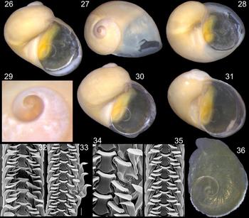
Laevilacunaria antarctica hard parts (MZSP 115506). (26) Specimen ♀1, apertural view. (27) Same, apical-slightly left view. (28) Same, umbilical view (W 7.1 mm). (29) ♂2, detail of apex (W 2 mm). (30) ♂3, apertural view (W 6.4 mm). (31) ♂2, apertural view (W 5.8 mm). (32) Radula in SEM, Scale 50 µm. (33) Same, other portion, Scale 50 µm. (34) Same, detail of central region, Scale 20 µm. (35) Same, another specimen, Scale 50 µm. (36) Operculum of ♀1, outer view.

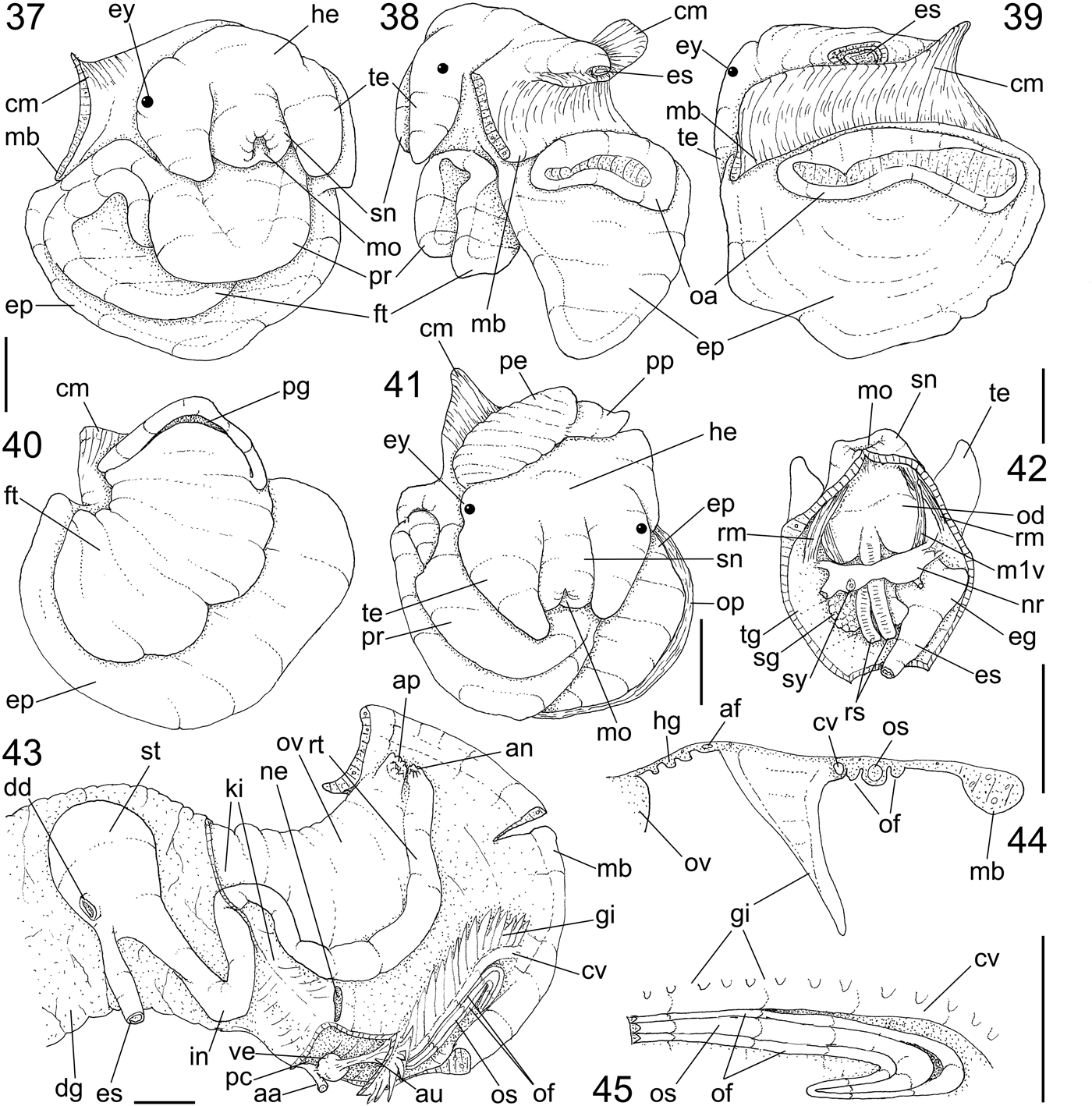
Laevilacunaria antarctica anatomy. (37) Head-foot, female, anterior view. (38) Same, left view. (39) Same, postero-dorsal view. (40) Same, ventral view; (41) Head-foot, male, anterior view. (42) Head and haemocoel, ventral view, foot removed, snout opened longitudinally. (43) Pallial cavity hoof and anterior coil of visceral mass, ventral view, ventral wall of pericardium opened and deflected to left (downwards), digestive gland portion ventral of stomach removed. (44) Pallial cavity hoof, female, transverse section on middle level of osphradium. (45) Detail of middle and anterior region of osphradium, ventral view, adjacent border of gill also shown. Scales = 1.0 mm.

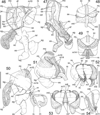
Laevilacunaria antarctica anatomy. (46) Foregut, ventral view, odontophore partially removed and deflected to right, most structures uncoiled, radular sac only partially shown, anterior portion of salivary glands and buccal ganglia shown by translucence. (47) Same, odontophore removed, oesophagus and oesophageal gland opened longitudinally, posterior region of this gland transversely sectioned. (48) Odontophore, dorsal view. (49) Same, ventral view. (50) Foregut as in situ, right view. (51) Buccal mass, right-slightly ventral view, radular sac only partially shown. (52) Odontophore, dorsal view, radular sac and radula deflected to left, both odontophore cartilages deflected, right m5 also deflected downwards. (53) Same, radular structures removed, most structures deflected to show cartilages. (54) Right odontophore cartilage, ventral view, only origin of intrinsic muscles shown. Scales = 1 mm.

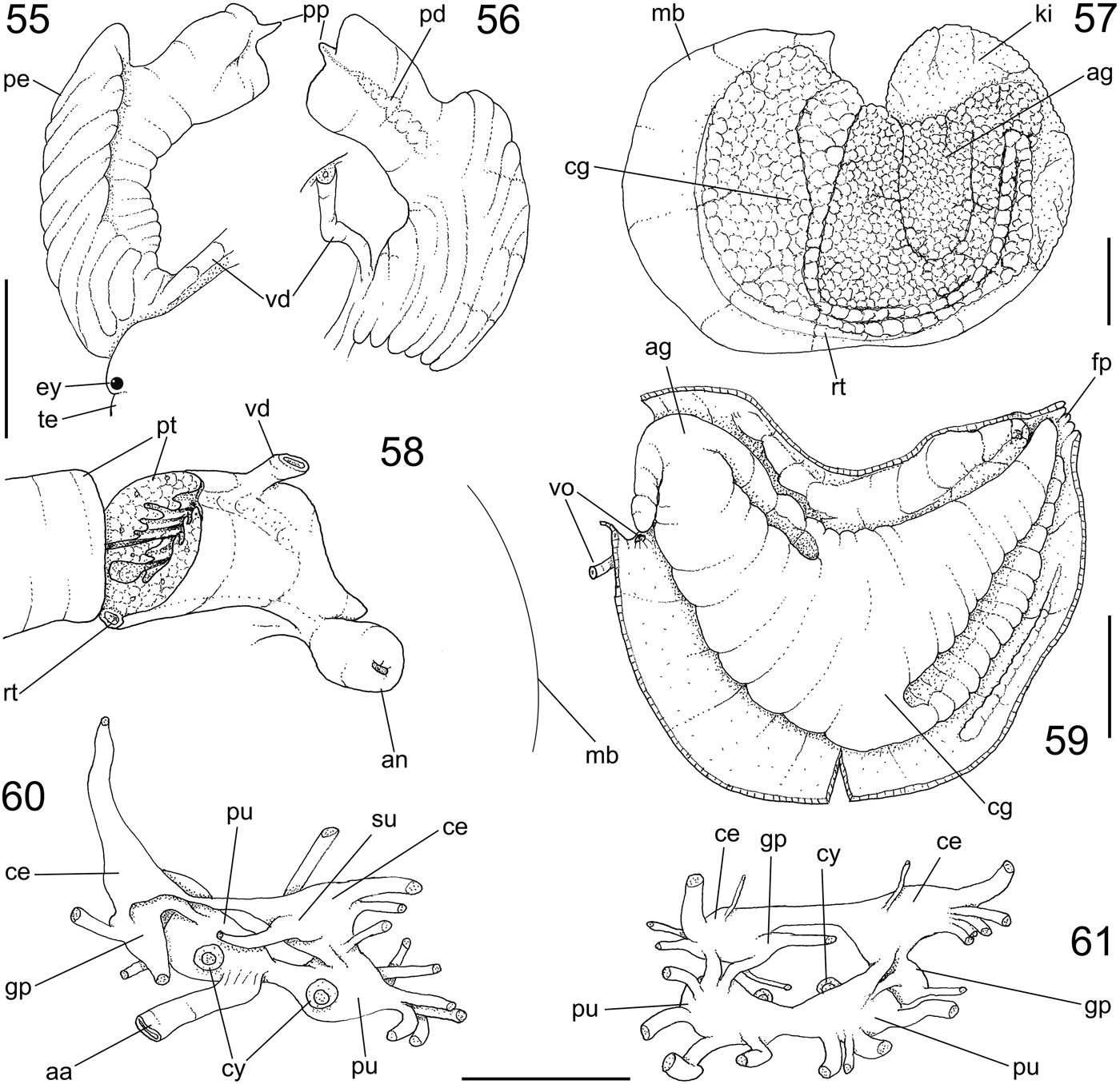
Laevilacunaria antarctica anatomy. (55) Penis, ventral view, adjacent portion of right tentacle also shown. (56) Same, dorsal view. (57) Pallial oviduct, outer-dorsal view, some adjacent structures also shown. (58) Pallial vas deferens, detail of anterior region, ventral view, transverse section artificially done, some adjacent structures and topology of mantle border also shown. (59) Pallial oviduct, ventral view, ventral wall sectioned longitudinally and deflected. (60) Nerve ring, dorsal view. (61) Same, ventral view. Scales = 1 mm.

Lacuna antarctica Martens, Reference Martens1885: 92.
Lacunella antarctica: Strebel, Reference Strebel1908: 52, 95, 96, 99.
Pellilittorina bransfieldensis Preston, Reference Preston1916: 271 (pl. 13, Figure 5; Fig. I29).
Laevilacunaria antarctica: Powell, Reference Powell1951: 58, 108; Picken, Reference Picken1979a: 71–78 (figures 2 & 3), 1979b: 125–127; Brey & Hain, Reference Brey and Hain1992: 224; Iken, Reference Iken1999: 133–145; Zelaya, Reference Zelaya2005: 118 (figure 20); Chenelot & Konar, Reference Chenelot and Konar2007: 114; Engl, Reference Engl2012: 101 (pl. 30, figure 1A–F); Bouchet, Reference Bouchet2013.
Laevilitorina (Macquariella) antarctica: Reid, Reference Reid1989: 92.
? Laevilitorina (Macquariella) bransfieldensis: Reid, Reference Reid1989: 92 (in syn.).
TYPE MATERIAL
Five syntypes in ZMB Moll 37473 (more details in Engl, Reference Engl2012). P. bransfieldensis: holotype NMW 1955.158.12890.
TYPE LOCALITIES
L. antarctica: South Georgia. P. bransfieldensis: Bransfield Strait, off Deception Island, South Shetland Islands.
DESCRIPTION
Shell (Figures 26–31): Diameter of ~7 mm; height ~87–94% of width; general form broadly turbiform. Walls thin, fragile, somewhat translucent. Colour pale brown to beige; periostracum thick. Protoconch flattened (Figure 29), of 1 rounded, simple whorl; surface smooth, opaque; suture somewhat deep; transition with teleoconch unclear, orthocline; diameter ~0.7 mm, occupying ~7% of superior shell surface (Figure 27). Teleoconch of ~2.5 whorls; suture shallow but well-marked (Figures 26, 30 & 31); spire angle ~115–125°. Sculpture absent except for smooth growth lines; some spiral ribs barely visible, uniformly distributed in last whorls, ~6 in penultimate whorl (Figures 26 & 30). Aperture white, rounded, wide, occupying ~66–77% of shell height and ~56% of shell width (Figures 30 & 31); outer lip simple, fragile, slightly prosocline (Figure 28); inner lip widely concave, almost straight, superior third covered by thin, translucent callus, inferior 2/3 with thin edge. Umbilicus wide, opened, flanked by middle portion of inner lip, occupying ~15% of ventral area (Figure 28).
Head-foot (Figures 37–42): Main characters similar to those of Pseudonatica antarctica, except for following features: most colour of exposed areas dark brown, almost black; gradually becoming unpigmented in hidden areas. Pair of tentacles (te) with tip simple, not bifid; eyes located at outer side of tentacles base, ommatophores absent (Figures 37 & 38). Snout (sn) smaller, ~1/15 of head-foot volume. Foot propodium (pr) broad, flap-like, ~50% wider than same adjacent mesopodium extension; mesopodium (ft) with thick edges, but ~50% smaller than propodium-epipodium flap (Figures 37 & 40); furrow of pedal glands (pg) along anterior edge, deep. Propodium (pr) and epipodium (ep) as continuation from each other, forming wide flap covering mesopodium (ft) (Figures 37, 38 & 40); epipodium extending longer beyond mesopodium, looking like additional operculum (Figure 38). Opercular pad (Figures 38 & 39: oa) located at base of epipodium, occupying ~30% of its area; attachment with operculum as transverse, narrow, central region, with constant form as shown in Figure 39. Columellar muscle (cm) of ~1/6 whorl.
Operculum (Figures 26, 30, 31 & 36): Similar to Pseudonatica antarctica, except in being thinner and translucent; with 0.5 more whorl, and wider flexiclaudent edges.
Mantle organs (Figures 43–45): General characters similar to those of Pseudonatica antarctica, main distinctive features following: Osphradium (os) also ridge-like, with 70% of pallial cavity length; anterior 1/4 strongly curved to left, anterior end curved backwards (Figures 43–45). Osphradium satellite fold (of) with width ~3/4 that of osphradium (Figures 44 & 45). Gill (gi) with ~1/5 pallial hoof width and 90% its length; each filament about twice taller than wide; apex central, sharp pointed (Figure 44: gi). Hypobranchial gland (hg) very thin, white, with some pallial reinforcements as oblique low folds immersed in glandular tissue (Figure 44: hg).
Visceral mass (Figure 43): Of 3 whorls posterior to pallial cavity. Digestive gland and gonad pale beige in colour. Stomach spherical, located half whorl posterior to pallial cavity, closer to dorsal and right side. Reno-pericardial structures as those described for preceding species; kidney (ki) transversally elongated; occupying ~1/6 of pallial cavity volume; pericardium (pc) with ~1/5 of kidney volume.
Circulatory and excretory systems (Figure 43): Organization similar to that of Pseudonatica antarctica, except in being ~30% smaller. Heart particularly small as described above; differing in having auricular subterminal connection to ctenidial vein. Ventral portion of pericardium located dorsal to posterior region of gill.
Digestive system (Figures 46–54): Most characters similar to those of Pseudonatica antarctica, with following main distinctions: Pair of snout retractor muscles (Figure 42: rm) is more laterally positioned. Pair of jaw plates (Figure 46: jw) relatively wide, but very thin, occupying ~1/3 of dorsal surface of oral cavity; each plate squared, about as wide as long. Pair of salivary glands elongated, each one separated from each other (except some posterior connection, resembling a ring), ~10 times longer than wide (Figures 46 & 50: sg), pale cream; lying in dorsal surface of buccal mass, totally free from nerve ring; ducts (Figure 46: sd) narrow, running attached to anterior oesophagus; opening in middle region of dorsal folds (Figures 46 & 47: sa). Odontophore muscles distinctions (Figures 48–54): mj, pair of peri-buccal and jaw muscles considerably thicker (Figure 49); m3, not developed (Figures 50 & 51); m5, pair of auxiliary dorsal tensor muscles of radula (Figures 52 & 53) somewhat narrower; m6, horizontal muscle slightly shorter, width ~3/4 of cartilages length (Figures 52–54); m7, separated from each other as two narrow bundles (Figures 52 & 53); m10, pair of thin odontophore protractor muscles, slightly ticker and covered by m14 pair (Figures 50 & 51); m14, pair of auxiliary protractor muscles of odontophore with origin more superficial and slightly more lateral (Figure 51). Radula (Figures 32–35): rachidian about 1.5 times longer than wide, occupying ~1/5 of radular width, rectangular, cutting edge strongly bent inside, mostly smooth except for 2 lateral, small pointed cusps; pair of basal and lateral reinforcements, barely resembling cusps (Figure 34); lateral tooth similar-shaped as rachidian, except in being obliquely deformed, slightly turned medially, cutting edge as that of rachidian, base possessing single basal cusp turned inwards, basal-lateral, triangular projection articulating with inner marginal tooth (Figures 33 & 34); inner marginal tooth similar to lateral tooth except in lacking lateral small cusp, and by narrower, triangular, pointed base; outer marginal tooth similar to inner marginal tooth, but with pointed cutting edge, by terminal, small cusp and wider, rounded base (Figure 33) (other radular details in Arnaud & Bandel, Reference Arnaud and Bandel1978: 222–224). Radular sac relatively narrow, almost 3-times odontophore length (Figure 50: rs). Anterior oesophagus with same odontophore length; interior surface with pair of dorsal folds; pair of oesophageal crops (Figures 46 & 47: ec), each one sac-like, connection with oesophagus slightly narrower than distal region, with approximately same adjacent oesophageal width, inner surface smooth. Middle oesophagus ten-times shorter than anterior oesophagus, serving as base of oesophageal gland. Oesophageal gland as blind sac, as wide as anterior oesophagus, as long as buccal mass (Figures 46 & 47: eg), posterior end slightly broader than basal region; internally fulfilled by single row of transverse glandular septa in ventral 3/4 region, dorsal 1/4 with narrow pair of longitudinal folds continuing from those of anterior oesophagus (Figure 47: ef). Posterior oesophagus as long as anterior oesophagus, ~half its width, originating at ventral base of oesophageal gland; inner surface with 4–5 longitudinal, low, wide folds. Stomach (st) spherical, with oesophageal insertion and intestinal origin in left side (intestine ~twice larger than adjacent oesophagus) (Figure 43: es, in); single duct to digestive gland (dd) located in ventral region in front of oesophageal insertion; inner gastric surface simple, glandular, with some longitudinal, low folds converging to intestinal origin. Rectum and anus described above (Figure 43).
Genital system: Male (Figures 55, 56 & 58): Testis lying along columellar surface of digestive gland, of the same colour. Seminal vesicle small, zigzagging in last whorl of visceral mass ventral side. Vas deferens gradually narrowing in right region of pallial cavity. Prostate gland (Figure 58: pt) abruptly expanding in right side of pallial cavity; wall thick muscular, lumen containing 5–8 relatively tall longitudinal folds; prostate size ~80% of pallial cavity length and 1/5 its width. Pallial vas deferens originating sub-terminally in right side of prostate, in its region preceding its anterior, blind-sac end (Figure 58); anterior prostate end bluntly pointed, located closed to anus. Pallial vas deferens running weakly coiling on pallial floor surface up to penis base (Figure 56: vd). Penis with ~1/5 head-foot size, located just posterior to right cephalic tentacle, lying to left (Figure 41: pe); basal region occupying 2/3 of penis length, longitudinally-weakly obliquely folded, about twice longer than wide, ventral region with blunt crest, slightly projected distally (Figures 55 & 56); distal 1/3 about half narrower than basal region, smooth, cylindrical; tip broad, slightly broader than its base. Penis papilla (Figures 55 & 56: pp) with ~1/15 of penis length and ~1/6 of distal portion width, located in middle region of blunt penis tip. Penis duct entirely closed (tubular), running along middle penis region slightly coiling; in region preceding papilla gradually becoming narrower than its preceding region, opening at papilla tip (Figure 56: pd).
Female (Figures 57 & 59): Visceral structures similar to equivalent ones of male. Pallial oviduct similar positioned and organized as preceding species (Figure 43: ov). Albumen (= jelly) and capsule glands (Figure 57: ag, cg respectively) mixed, performing a U-shaped loop. Internal lumen greatly flattened, U-shaped (Figure 59); ventral area covered by thin ventral membrane. Capsule gland thick, smooth; some longitudinal furrows as shown in Figure 59; glandular portion reaching female pore (Figure 59: fp). Bursa and seminal receptacle not found, but possibly represented by some cavity within pallial oviduct glands. Female pore (fp) as transverse, simple slit, with walls non-muscular; located close to anus (Figure 43: ap).
Central nervous system (Figures 60 & 61): Located just posterior to buccal mass (Figure 42: nr). Cerebral ganglia (ce) spherical, each one with ~1/3 of adjacent oesophageal width; cerebral commissure narrow, ~as long as each ganglion. Pleural ganglia almost totally fused with adjacent cerebral ganglia, approximately half of cerebral ganglion size. Pedal ganglia (pu) spherical, ~as large as cerebro-pleural ganglia; pedal commissure narrow, with ~1/3 each ganglion length. Cerebro-pedal and pleuro-pedal connectives relatively thick, similar-sized, with about same length of each cerebral ganglion.
Measurements (width and height in mm): MZSP 105506♀1: 7.1 by 6.5; ♂2: 5.8 by 4.8; ♂3: 6.4 by 6.1.
Habitat: Rocky bottoms, 0–74 m, eating algae (Picken, Reference Picken1979a; Iken, Reference Iken1999).
MATERIAL EXAMINED
ANTARCTICA. Antarctic Peninsula; King George Island; Admiralty Bay, 62°08′S 58°26′W, 60 m depth, MZSP 115506, 16 specimens (Passos et al. col; Proantar XVI; xii/1997).
DISCUSSION
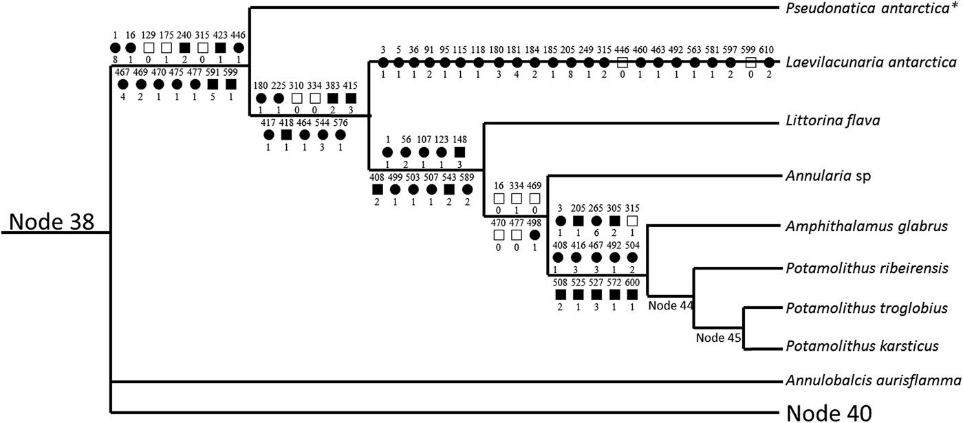
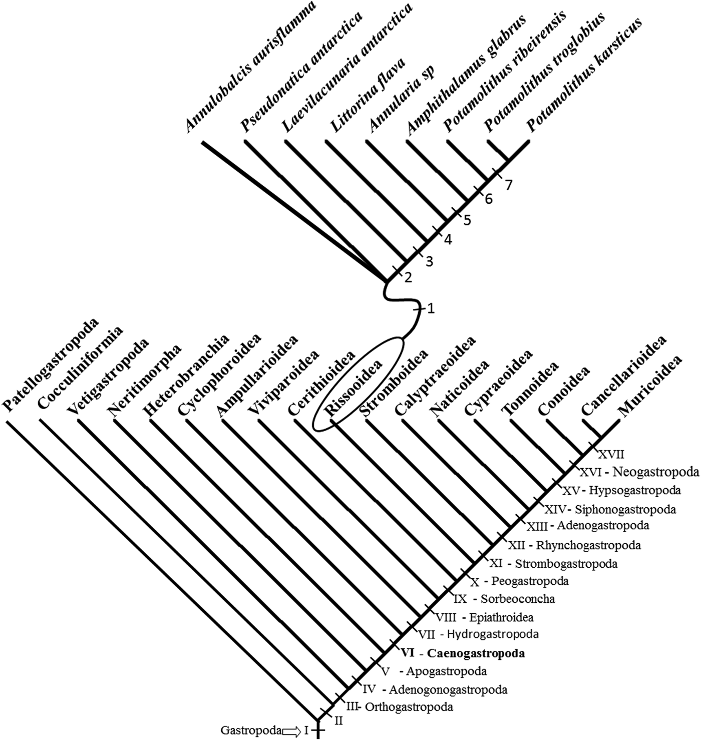
As stated above, the phenotypic characters of both species anatomically described here were coded and inserted in a previous large phylogenetic analysis (Simone, Reference Simone2011), designed to raise the basis of this discussion. The main concern is to interpret the convergence of characters of the studied taxon with the Naticidae, and to ascertain the taxonomic and phylogenetic position of them. The methodology is the same as stated in Simone (Reference Simone2011), and this discussion is performed under the light of that study. All available characters of both studied species are in Table 1 (criteria of Simone, Reference Simone2011), with this analysis resulting in the cladogram shown in Figures 62 & 63. This cladogram is the strict consensus of 80 trees, with a length of 3101, consistency index of 50 and retention index of 94. No additional character was inserted in those already presented in the previous paper (Simone, Reference Simone2011). Pseudonatica antarctica was already present in that paper with the codename ‘Amauropsis’ rossiana, while Laevilacunaria antarctica was simply inserted in the matrix. Both species were introduced in the same region of the cladogram shown in that paper (Simone, Reference Simone2011: figure 20), where ‘A.’ rossiana (now Pseudonatica antarctica) was placed, between the nodes 39 and 42. The new arrangement is shown in Figures 62 & 63, focusing only on that portion of the cladogram, as the remaining remained unaltered. A rearrangement of the synapomorphies of each node is shown in Figure 62, in which 15 synapomorphies support a branch with Pseudonatica antarctica and its sister taxon (Figure 63: node 1), as part of a trichotomy with Annulobalcis aurisflamma Simone & Martins Reference Simone and Martins1995 (an Eulimidae – Ctenoglossa), and node 40. The node preceding P. antarctica can be regarded as Rissooidea–Littorinoidea clade (sensu Simone, Reference Simone2006) and, from the synapomorphies, the more interesting are a globose shell (character 1), the reduction of the ommatophore (character 129), the exposition of pericardium in pallial cavity (240), the reduction of jaw plates (315), the radular lateral tooth with few cusps (423), the oesophageal gland as wide ventral diverticulum (467) possessing transverse septa (469), and a looped capsule gland (591).
Portion of cladogram resulting from insertion of phenotypic data of species studied herein in the cladogram of Simone (Reference Simone2011), with synapomorphies of each node shown. Number of nodes and codes of characters as in Simone (Reference Simone2011) (symbols: black squares = non-homoplastic synapomorphy; white squares = reversion; black circles = ingroup convergences), characters = number above, states = number below each symbol. Number of some nodes as in Simone (Reference Simone2011) (strict consensus of 80 trees, L = 3101, CI = 50, RI = 94). * coded as ‘Amauropsis’ rossiana in Simone (Reference Simone2011).

Abstracting cladogram of Simone (Reference Simone2011) with the inclusion of species studied herein and some neighbouring species in above tree, showing the arrangement of the cladogram with some nodes numbered.

Laevilacunaria antarctica was allocated in a following branch, as sister taxon of remaining Rissooidea–Littorinoidea taxa (Figure 63: node 2), being supported by 11 synapomorphies. From them, the more interesting are the osphradium with ~2/3 of gill length (character 180), the reduction of hypobranchial gland (character 225), the loss of the pair of retractor muscles of the buccal mass (m2) (334) and of the pair of ventral tensor muscles (m11) (383), radular rachidian with basal cusps (417), and the closure of the pallial vas deferens (544) including penis (576).
The remaining arrangement of the cladogram (Figure 62), preceding Littorina flava (King & Broderip, Reference King and Broderip1832) (Figure 63: node 3) is similar to that reported previously (Simone, Reference Simone2011), with the set of synapomorphies of each node very similar, which will not be discussed here.
Taking into consideration the argument by Simone (Reference Simone2006, Reference Simone2011), that the formal taxon Littorinoidea is a paraphyletic arrangement of basal Rissooidea, it is possible to consider the entire taxon simply as ‘Rissooidea’. The same arrangement was obtained herein (Figures 62 & 63), in that littorinoideans Pseudonatica antarctica (zerotulid), Laevilacunaria antarctica, Littorina flava (both littorinids), and Annularia sp (annulariid/pomatiid) resulted in a grade preceding traditional Rissooidea, here represented by the node preceding Amphithalamus glabrus Simone 1996 (Figure 63: node 5), here supported by 15 synapomorphies. In addition, it seems appropriate to note here that what was called Annularia sp. in Simone (Reference Simone2011), appears now to belong to the genus Chondropoma Pfeiffer, 1847.
The similarities of presently studied species with naticids are noteworthy, and they are beyond the external appearance of shell and operculum. The similarity with naticids was already pointed out in the original description of Laevilacunaria antarctica by Martens (Reference Martens1885: 92). The more basal taxa of Naticidae have a globose shell, with the aperture entirely occluded by a corneous, paucispiral operculum (Simone, Reference Simone2011); these attributes are also found in the two species here studied anatomically. Besides the shell and operculum, both species herein studied also share a series of convergent characters with basal naticids, such as a wide-sized foot (Figures 14, 37–41), with a well-developed propodium (despite lacking a clear digger propodium like those of naticids), a complex foot attachment with the operculum (at least in L. antarctica – Figure 16), a ridge-like osphradium surrounded by a satellite fold (Figures 15 & 43) (present in some basal naticids, such as Amauropsis Mörch 1857 – Simone, Reference Simone2011: 182), and, more remarkably, a higher concentrated pallial oviduct (Figures 23, 24, 57 & 59) possessing the albumen and capsule glands mixed with each other in a somewhat coiled way. As discussed below, according to the present cladistic study, those characters are interesting convergent features. The reasons possibly can be attributed to a convergent globose shell associated to a locomotion beyond rocky substrates, i.e. in unconsolidated ones. The pallial oviduct, in particular, has some coiling posterior region in some littorinoideans (Reid, Reference Reid1989, Figure 9), which can produce a benthic gelatinous spawn (Picken, Reference Picken1979b).
Regarding a general phenotypic discussion, and based on the data published in Simone (Reference Simone2011 and references therein), the following interesting aspects can be raised. The lack of a clear digging propodium like that of the naticids indicates that possibly these Antarctic rissooideans can crawl on soft substrates, but with weak capacity for digging through it; on the other hand, a large foot seems useful to crawl on ice or through snow that eventually falls on the rock. Naticids mostly lack anterior pedal glands, which is found in the present species; anterior pedal glands produces mucus to help the animals crawl, it looks more useful in snails that move on hard substrates than on soft ones, justifying its reduction in naticids. The head-foot of the naticids is somewhat mischaracterized, with a true head difficult to find and mostly lacking eyes; this does not happen in the presently studied species, which possess the ordinary, plesiomorphic head (Figures 14 & 37). The naticids possess a well-developed pleurembolic proboscis, the tip bearing the ABO (accessory boring organ), used for making holes in the shell of their prey; this is naturally absent in the presently studied species, which have an ordinary snout (Figures 18 & 42). The complexity of the attachment of the operculum with the pedal opercular pad (Figures 16 & 39) of the present rissooideans is noteworthy, and is also found in most naticids and is absent in remaining gastropods, in which the attachment produces a single, elliptical scar. The reason for this complexity is still obscure, but possibly is related to the firm grip in opercular closure; as the globular shell, common in both groups, has a relatively wide aperture, the operculum is primordial to protect the retracted snail, and so the need for a strong grip. The naticids are part of a branch called Rhynchogastropoda Simone, Reference Simone2011, in such main characters is the bipectinate osphradium. However, some basal naticoideans, belonging to the genus Amauropsis, reverted to a ridge-like osphradium (node 28 of Simone, Reference Simone2011). In that genus, the osphradium is also surrounded by a glandular, ciliated ridge, called a satellite fold. A remarkably similar situation is found in the presently studied species (Figures 15, 17 & 43–45: of). Possibly the satellite fold is developed in order to provide extra mucous flow to a necessary osphradium, which lacks the filaments of a bipectinate organ; as no satellite fold is found in any bipectinate-bearing species. The singular naticoidean pallial oviduct is related to the production of a famous spawn in a form of a coiled, sandy, flattened ribbon (Murray, Reference Murray1962); despite having a similar-shaped pallial oviduct, the species studied here apparently do not build similar structured spawns (Picken, Reference Picken1979a). All these similar features between both presently studied rissooideans and the naticoideans are certainly convergences, and still matter for further investigations. Even though no experiment on comparative ecology has been done, it is possible to speculate that the globose shell on a wide foot, which the naticoideans and Antarctic species studied here have in common, looks well-adapted to crawl through unconsolidated substrates, such as sand (naticoideans) and snow (Antarctic species). This possibly explains the convergences pointed out herein.
It is important to emphasize that the definition of Zerotiludae within Littorinoidea–Rissooidea branch is not totally clear, and even the authors that introduced it reported that it can be a para- and even polyphyletic arrangement (Warén & Hain, Reference Warén and Hain1996: 281). However, the validity of Zerotulidae has not so far been challenged (e.g. Aldea & Troncoso, Reference Aldea and Troncoso2008), and the inclusion of Pseudonatica appears plausible based on its characters discussed above.
Based on the revised cladogram after the inclusion of both studied species (Figures 62 & 63) in the cladogram of Simone (Reference Simone2011), it is clear that they belong to the basal Rissooidea, mostly called Littorinoidea. They in fact have the general morpho-anatomical aspect of the littorinoideans (Reid, Reference Reid1989, Reference Reid2002; Simone, Reference Simone1998), such as the snout, the ridge-like osphradium, the oesophageal gland and the elongated radular sac. However, and as stated before (Simone, Reference Simone2006, Reference Simone2011), there is no true synapomorphy supporting a formal taxon Littorinoidea, a reason for the result as a paraphyletic arrangement. However, the presence of the large oesophageal gland is noteworthy, as it apparently is convergent with higher caenogastropods called Adenogastropoda (Simone, Reference Simone2011: node 96). Such a gland is present in the Naticoidea, Cypraeoidea, Tonnoidea and occurs as the gland of Leiblein and the venom gland in Muricoidea and Conoidea respectively (Simone, Reference Simone2011). Its presence in the littorinoideans is a remarkable convergence with the adenogastropods. When analysing the oesophageal glands of both groups no clear difference is distinguished, as in both groups the gland is ventral, diverticulum-like, fulfilled by transverse septa. Although the convergent oesophageal gland of so far known littorinoideans could support a monophyletic taxon, this character was not sufficient for such a conclusion in the present cladistic analysis (Figures 62 & 63).
Both species studied here possess some similarities in their anatomy, and they differ in the following main features: Pseudonatica antarctica has ommatophore (Figure 14: om), which is absent in Laevilacunaria antarctica (Figure 38) and most rissooideans. Pseudonatica antarctica lacks as well a developed epipodium as that of L. antarctica (Figures 37–41: ep). The arrangement of the scar of opercular pad attachment is also different (Figures 16 & 39). The salivary glands of P. antarctica are somewhat deformed (Figure 22: sg), while those of L. antarctica is ring-like, bearing their distal end weakly attached with each other (Figures 46 & 50). Laevilacunaria antarctica have a pair of oesophageal crops in the anterior oesophagus (Figures 46 & 47: ec), and the oesophageal gland is sac-like (eg); such a crop is absent in P. antarctica, which also have a bulged oesophageal gland (Figure 22). In the odontophore, both species remarkably possess the pair of accessory protractor muscles of buccal mass (Figures 20 & 51: m14), but differ in the length of the horizontal muscle (shorter in L. antarctica – Figure 53: m6, compare with Figure 21), and by pair m7 fused with each other in P. antarctica (Figure 21) and separated in L. antarctica (Figure. 53). The nerve ring of L. antarctica is more compact (Figures 60 & 61) than that of P. antarctica (Figure 25), and the salivary gland of that species does not pass through it, a feature shared with the Neogastropoda.
With the present level of knowledge, it is difficult to interpret the meaning of these differences. They can be indicative of any level of taxonomical separation, from simply species level up to the family level. Certainly molecular tools could bring new insights, and help in the resolution of a better-resolved taxonomy and phylogeny. Antarctic mollusc species sometimes present unexpected outcomes when investigated from a molecular point of view (see, e.g. Barco et al., Reference Barco, Schiaparelli, Houart and Oliverio2012), in which the new subfamily Pagodulinae of muricids was introduced.
ACKNOWLEDGEMENTS
Special thanks to Flavio Dias Passos, Unicamp, for the donation of the Antarctic samples studied herein. To Philippe Bouchet, MNHN, for inviting us to examine the MD55 material housed at MNHN and allowing us to study and publish the specimens treated herein. To José Coltro Jr for providing the trip for Paris.
FINANCIAL SUPPORT
This project is part supported by FAPESP (Fundação de amparo a Pesquisa do Estado de São Paulo), proc. # 2010/11253-9.