Insect consumption of plant-based food sources occurs in many forms, ranging from the wood-feeding termites (Blattodea) to the discretely targeted phloem or xylem feeders. When consuming wood or leaf materials, various enzymatic strategies are required to extract nutrients (Calderon-Cortes et al. Reference Calderon-Cortes, Quesada, Watanabe, Cano-Camacho and Oyama2012). Glycosyl hydrolases involved in hydrolysis of cellulose and xyloglucans are some of the enzymes deployed to deconstruct the plant cell wall, allowing access to both intracellular and cell wall components. Leaf harvesting ants such as Acromyrmex echinatior Forel (Hymenoptera: Formicidae) transfer decomposing leaf material, rich in a fungal-produced xyloglucanase, into fresh leaf material, enhancing the release of cell wall and intracellular substrates used by the Fungi being cultivated by the ants (Kooij et al. Reference Kooij, Pullens, Boomsma and Schiott2016). While the insect-associated cellulolytic activities were originally thought to be produced only by the insect gut microflora, discovery of insect-encoded glycosyl hydrolases began with evidence of a family 9 beta-1,4 endoglucanase from the termite Reticulitermes speratus (Kolbe) (Blattodea: Rhinotermitidae) (Watanabe et al. Reference Watanabe, Noda, Tokuda and Lo1998). Additional cellulolytic glycosyl-hydrolase-encoding genes were subsequently identified, comprised primarily of glycosyl hydrolase 9 in insects and glycosyl hydrolase 5 in nematodes (Watanabe and Tokuda Reference Watanabe and Tokuda2010; Willis et al. Reference Willis, Oppert, Oppert, Klingeman and Jurat-Fuentes2011; Eyun et al. Reference Eyun, Wang, Pauchet, ffrench-Constant, Benson and Valencia-Jiménez2014; Pauchet et al. Reference Pauchet, Saski, Feltus, Luyten, Quesneville and Heckel2014; Shelomi et al. Reference Shelomi, Watanabe and Arakawa2014, Reference Shelomi, Heckel and Pauchet2016).
The glycosyl hydrolase 12 enzymes, which hydrolyse cellulose and/or xyloglucans, have been missed from the list of published insect-encoded glucanases. This may be due to the structural differences from other glycosyl hydrolase members. When the first endoglucanase genes were cloned from a phytopathogenic fungus (Wang and Jones Reference Wang and Jones1995), they were shown to lack the linker region and cellulose-binding domain found in endoglucanases encoded by saprophytic fungi. This paradigm remains to date, wherein the preponderance of phytopathogen-encoded endoglucanases lacks a cellulose-binding domain. The few phytopathogen-encoded endoglucanases that have a cellulose-binding domain are expressed only during necrotrophic/saprophytic stages of plant colonisation, suggesting recognition of the cellulose-binding domain by plant surveillance mechanisms. The glycosyl hydrolase family 12 enzymes are currently the only exception to the paradigm. In the case of glycosyl hydrolase 12, the majority of encoded proteins, whether of phytopathogen or saprophyte origin, lack the cellulose-binding domain. Lacking a highly conserved cellulose-binding domain can allow the sequences to be overlooked when searching for glycosyl hydrolases.
The family 12 glycosyl hydrolases have internal conserved sequences that can be used when trying to identify the gene products, which was done when identifying the first glycosyl hydrolase 12 members present in a plant pathogen (Costanzo et al. Reference Costanzo, Ospina-Giraldo, Deahl, Baker and Jones2006). During our ongoing studies on the presence and role of plant-pathogen-produced glycosyl hydrolase family 12 proteins that have xyloglucanase and/or endoglucanase activity, we have used internal conserved amino acid sequences to find glycosyl-hydrolase-12-encoding genes in the genomes of insects and nematodes. Evidence is presented for glycosyl hydrolase 12 genes in three different phloem feeding psyllids and a plant parasitic nematode. The glycosyl hydrolase 12 enzymes have thus far been absent from the list of published insect- and nematode-encoded glycosyl hydrolases.
Initial searches for glycosyl-hydrolase-12-encoding genes were made using National Center for Biotechnology Information TBLASTN (https://blast.ncbi.nlm.nih.gov) with the three conserved amino acid regions of glycosyl hydrolase 12 (Costanzo et al. Reference Costanzo, Ospina-Giraldo, Deahl, Baker and Jones2006). Searches covered all available nucleotide sequences including whole genome sequencing and transcriptomes and found the following: one transcript from Diaphorina citri Kuwayama (Hemiptera: Psyllidae) (DQ673432), one transcript from Pachypsylla venusta (Osten-Sacken) (Hemiptera: Psyllidae) (GAOP01107961), one transcript from Acanthocasuarina muellerianae Taylor (Hemiptera: Psyllidae) (GAYY01136395), and two transcripts from Xiphinema index Thorne and Allen (Dorylaimida: Longidoridae) (CV508600 and CV127906).
Whole samples of D. citri were obtained as ethanol-preserved samples (D. Hall, United States Department of Agriculture-Agricultural Research Service, Fort Pierce, Florida, United States of America). The D. citri, originally collected from Citrus Linnaeus (Rutaceae), was air dried and then homogenised in 0.5 mL of water with a micropestle in a 1.5-mL centrifuge tube. After brief centrifugation, the supernatant was collected and used for subsequent assays. Purified DNA samples of P. venusta (D. Sloan, Colorado State University, Fort Collins, Colorado, United States of America), X. index, and X. americanum Cobb (L. Carta, United States Department of Agriculture-Agricultural Research Service, Beltsville, Maryland, United States of America) were also used.
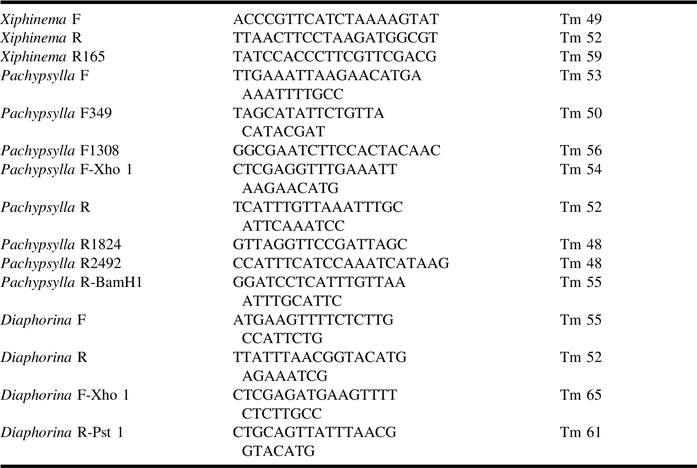
Primers (Table 1) to each end of the coding region, or partial region, were used to amplify target regions from DNA samples of D. citri, P. venusta, and X. index. Genomic regions were amplified with GoTaq polymerase (Promega) using the programme of: initial denaturing cycle at 95 °C for three minutes, followed by 30 cycles of using the lower melting point for each primer pair for 15 seconds, 72 °C extension for 1.5 minutes, and denaturation at 95 °C for 30 seconds. A final extension at 68 °C for 10 minutes completed each programme. Polymerase chain reaction samples were run in 1% agarose gels and bands excised and purified for cloning. Polymerase chain reaction products were cloned into TOPO TA (Invitrogen, Rockville, Maryland, United States of America), followed by transformation into Escherichia coli (Migula) Castellani and Chalmers (Enterobacteriaceae). Positive colonies were used to purify plasmid, followed by sequencing (Macrogen, Rockville, Maryland, United States of America). Due to the length of the P. venusta intron, additional internal primers were used to extend the sequence reads in both directions until a fully overlapping sequence was achieved. Primers to the 5′ region of X. index were used to determine if X. americanum encodes a glycosyl hydrolase 12 gene.
Primer names, sequences, and melting points (Tm).

Glycosyl-hydrolase-12-encoding clones from D. citri were also amplified with primers containing Xho 1 and Pst 1 sites, while the P. venusta clone was amplified with primers containing Xho1 and BamH1. The cloned products were digested and ligated to the binary vector pGD. Plasmid was generated in E. coli, purified, and used to electroporate Agrobacterium tumefaciens Smith and Townsend LB 4404 (Invitrogen). Cells were cultured and used for succulent plant leaf infiltration as previously described (Jones Reference Jones2016). Samples from infiltrated succulent leaves were tested using azure blue cellulose and xyloglucan at 0.1 mg/mL in water and incubated at 37 °C overnight (Jones Reference Jones2016). Two infiltration events and extractions were performed. Enzymatic assays and absorbance readings at A600 were carried out after overnight incubation.
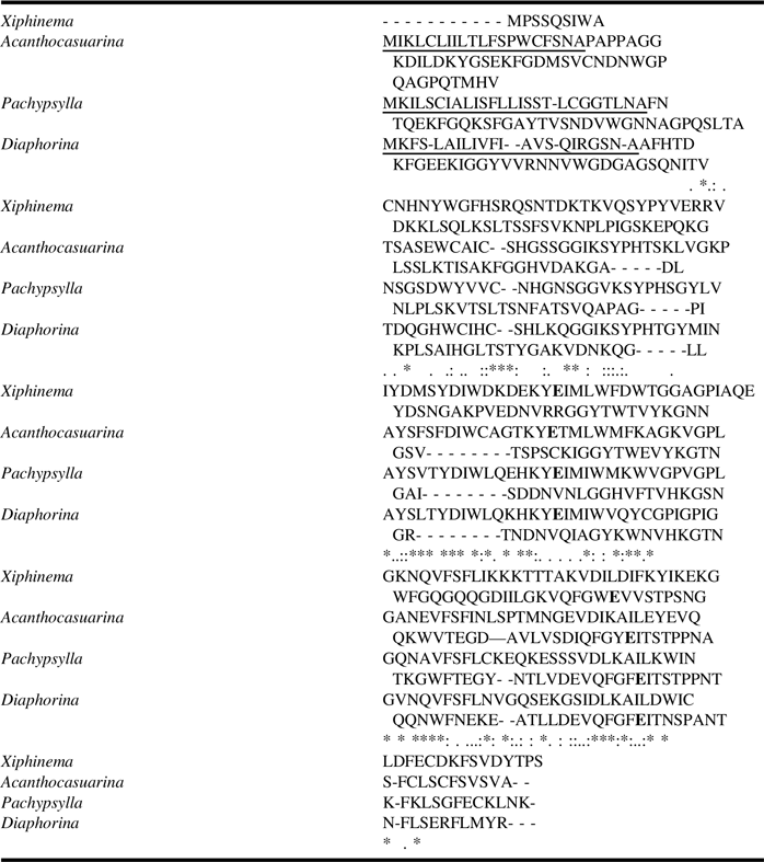
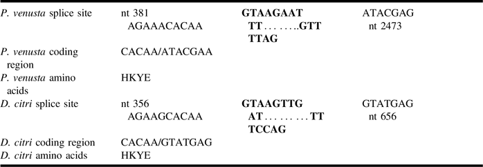
The three psyllid glycosyl-hydrolase-12-encoding genes and the nematode gene differ somewhat in amino acid sequence, while retaining the glutamic acid residues (in bold letters) is required for enzymatic activity (Table 2). The genomic structure is markedly different with P. venusta (submitted as GenBank MF962878) harbouring an intron that is nearly 10-fold larger (2 kilobases versus 283 base pairs) than the intron harboured by D. citri (submitted as GenBank MF962879). Interestingly, the large and the small introns have the same donor and acceptor splice sites (Table 3). Only in silico analysis of A. muellerianea (Taylor et al. Reference Taylor, Jennings, Purcell and Austin2011) was possible due to an inability to access insect specimens or DNA samples.
Alignment of amino acids.

Sequences are based on translation of coding regions of genomic sequences from this study, except for Acanthocasuarina, where the sequence is based on a single mRNA transcript (GAYY01136395). The Xiphinema index sequence information is lacking at the 5′ region of the genomic sequence. Catalytic glutamates are in bold letters. Predicted signal peptides are underlined. Alignments were prepared using Clustal Omega (www.ebi.ac.uk).
Symbols represent – (*) fully conserved, (:) strongly similar, and (.) weakly similar.
Predicted splice regions correlate with transcript sequence.

Start and end of intron regions are shown in bold. The Pachypsylla venusta intron is almost 10-fold larger than the Diaphorina citri intron, while no intron is present in this region of the Xiphinema index genome. Predicted splice sites based on SplicePort (http://spliceport.cbcb.umd.edu).
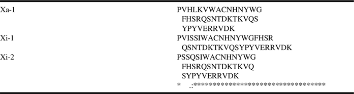
The nematode X. index genomic region encoding a glycosyl hydrolase 12 lacks an intron. Previous information on this nematode was limited to a partial mRNA (CV508600). Along with our genomic sequencing, a 3′ mRNA sequence from a basal bulb sample is polyadenylated (CV127906), supporting eukaryotic nematode origin. The 5′ region of this gene is also present in X. americanum (Table 4). Differences in amino acid sequence in this small region may be useful as an additional tool in identifying differences among similar nematodes. Testing of D. citri protein produced by Agro-infiltration of a genomic construct (allowing intron splicing) demonstrated endoglucanase activity (A600 = 0.155 ± 0.051) but negligible xyloglucanase activity, while the P. venusta had little endoglucanase activity (A600 = 0.091 ± 0.028) and negligible xyloglucanase activity.
Comparison of a 5′ region of Xiphinema using Xiphinema forward and Xiphinema reverse 2 primers.

Amino acid sequence variations at the amino terminus suggest that they might be used to distinguish Xiphinema isolates. (Xa-1) Xiphinema americanum from this study, (Xi-1) X. index from this study, and (Xi-2) X. index from GenBank (CV508600). Alignments constructed with Clustal Omega.
The specific temporal and spatial expression of these genes in the individual insects have not been fully assessed due to a lack of biological material; however, the presence of RNA transcripts in the GenBank database indicates that they are expressed in D. citri, P. venusta, and X. index.
Identification of glycosyl-hydrolase-12-encoding genes in the genomes of psyllids and a root-feeding plant parasitic nematode expands the range of known families of glycosyl hydrolases encoded by insects and nematodes. The variation in size and the presence or absence of an intron in the glycosyl-hydrolase-12-encoding genes studied in this paper would provide an interesting area for study of gene transfer. The origin of glycosyl hydrolases and other genes in insect genomes has, in some cases, been attributed to horizontal gene transfer (Pauchet and Heckel Reference Pauchet and Heckel2013; Sloan et al. Reference Sloan, Nakabachi, Richards, Qu, Murali, Gibbs and Moran2014). The genomes harbouring a gene with an intron would need to originate from a eukaryote, which is a less common source of gene transfer than a prokaryote. The presence of an intron has the potential to expand the possible transcripts generated, due to alternate splicing. This has been demonstrated in a glycosyl hydrolase 5 endoglucanase gene encoded by the plant pathogen Phytophthora de Bary (Peronosporaceae) (Costanzo et al. Reference Costanzo, Ospina-Giraldo, Deahl, Baker and Jones2007). If the genes were acquired through horizontal transfer, the differences in intron size suggest differing genetic origins of each transfer.
The role of glycosyl hydrolase 12 enzymes in psyllid biology remains to be determined. As phloem feeders, they share similarities with Aphididae (Hemiptera), but aphids appear to have glycosyl hydrolase 9 enzymes (GenBank XP_022167267 from Myzus persicae (Sulzer) and XM_001944739 from Acyrthosiphon pisum Harris) like many other phytophagous insects. The possibility that aphids encode glycosyl hydrolase family 12 needs further investigation. Certain family 12 enzymes have discreet activities that are overlooked using standard endoglucanase and xyloglucanase substrates. For example, only one glycosyl hydrolase 12 among a number tested was capable of inducing plant cell wall loosening and expansion (Park and Cosgrove Reference Park and Cosgrove2012). Genetic screening, followed by cloning and enzymatic assays, would allow for sufficient enzyme to detect activity.
Insect-encoded glycosyl hydrolases can be blocked by plant-encoded defensive proteins. The glycosyl hydrolase 10 endoxylanase from the coffee borer, Hypothenemus hampei (Ferrari) (Coleoptera: Curculionidae), was shown to be inhibited by XIP-1 (Padilla-Hurtado et al. Reference Padilla-Hurtado, Flórez-Ramos, Aguilera-Gálvez, Medina-Olaya, Ramírez-Sanjuan, Rubio-Gómez and Acuña-Zornosa2012), a bifunctional protein that binds to and inactivates glycosyl hydrolase family 10 and glycosyl hydrolase family 11 (Payan et al. Reference Payan, Leone, Porciero, Furniss, Tahir and Williamson2004). A similar glycosyl hydrolase inhibitor protein, TAX-I, inhibits only glycosyl hydrolase 11 xylanases (Gebruers et al. Reference Gebruers, Debyser, Goesaert, Proost, Van Damme and Delcour2001). Both XIP and TAX are found in monocots, while dicots encode a type of inhibitor protein that primarily targets the glycosyl hydrolase 12 family of endoglucanase/xyloglucanase enzymes. These inhibitor proteins, referred to as xyloglucan-specific endoglucanase inhibitor proteins, are generally represented by two or three copies in the dicot genome, and up to 12 copies in potato and tomato (Jones Reference Jones2012). They are produced in response to both abiotic and biotic stresses and can be found in the apoplast and xylem fluids (Houterman et al. Reference Houterman, Speijer, Dekker, De Koster, Cornelissen and Rep2007), and have been suggested to be associated with resistance to the root lesion nematode Pratylenchus neglectus (Rensch) Filipjev and Schuurmans Stekhoven (Rhabditida: Pratylenchidae) (Oldach et al. Reference Oldach, Peck, Nair, Sokolova, Harris, Bogacki and Ballard2014). Screening of xyloglucan-specific endoglucanase inhibitor proteins for interaction with insect glycosyl hydrolase 12 enzymes may provide a novel method for interfering with psyllid or nematode feeding and/or pathogen transmission.
Acknowledgements
Research funding was provided through United States Department of Agriculture, Agriculture Research Service project 8042-21220-256-00D.