Introduction
Eating can be a great pleasure but its failure can lead to undernutrition. Further, eating disorder can also decrease quality of life (QOL), particularly among older adults (Leow et al., Reference Leow, Huckabee, Anderson and Beckert2010). Appetite depends on the taste of food, its visual impression is also important. Appetite is affected by past empirical memory of food and depends greatly on sensation through the eyes, nose, and ears. Therefore, the color and shape of food that is cooked in consideration of its visual impression can have great effects on appetite. Dietary intake decreases gradually with age and progression of dementia is considered to be a factor contributing to decreased dietary intake. We often experience problems with eating behavior when caring for dementia patients. However, taste in food can often change and dietary intake decreases, even in the absence of dysphagia.
Taste signals are first received at the taste buds in the tongue, soft palate, and surrounding areas controlled by the cranial nerves (VII, IX, X, XI) via the thalamus (Nakamura et al., Reference Nakamura, Tokumori, Tanabe, Yoshiura, Kobayashi and Nakamura2013). Thus, it is not surprising that the sight of food is sufficient to activate the insula (Simons et al., Reference Simons, Martin and Barsalou2005). In addition to food appearance activating gustatory processing areas including the insula, insular responses to the sight and thought of food may be modulated by the internal state of the body (Malik et al., Reference Malik, McGlone, Bedrossian and Dagher2008). Dementia patients exhibit taste disorder. Steinbach et al. (Reference Steinbach, Hund, Vaitl, Heinrich, Foster and Burger2010) used “taste slip” and an odor test using “Sniffin Sticks,” and observed decreased olfaction and taste in Alzheimer's disease (AD) patients compared with healthy volunteers.
Common approaches for patients with eating disorders use “easy-to-swallow food,” which is a gel containing water, protein, and fat, similar to the normal biological constituents of food, and has been produced to prevent aspiration and eliminate pharyngeal residues by providing a homogeneous and appropriate viscosity. Although easy-to-swallow food has been established to provide food in patients with swallowing disorders, “soft food” has been invented with a focus on maintaining food form while improving dietary intake. It was developed as a new food type with improved shape and appearance (Kuroda, Reference Kuroda2001). Food shape provides pleasure and satisfaction to patients by bringing color and shape to the meal. For example, most “soft fish food” are prepared in the shape of a fish in conditions that are close to the original state of the food ingredients. Soft food is effective to not only improve QOL but also to increase dietary intake for dementia patients (Kuroda, Reference Kuroda2001). “Soft food” has been developed by focusing on form perception and vision-related semantic memory. We hypothesized that cognitive function related to eating may be damaged and exhibit effects on eating behavior in dementia.
Regarding taste memory, there have been no reports whether dementia patients have difficulty in remembering taste of food, although transgenic mice model showed impaired implicit taste memory as well as explicit spatial memory (Janus et al., Reference Janus2004). AD patients have semantic memory disorders. According to the “hub-and-spoke” theory of conceptual knowledge (Rogers et al., Reference Rogers, Lambon Ralph, Garrard, Bozeat, McClelland and Hodges2004), we considered the tests assessing the name (“hub”) and the attributes or property (“taste spoke”) of foods. The “hub-and-spoke” model posits the existence of a pan-modal integrative “hub” region, the anterior temporal lobe (Pobric et al., Reference Pobric, Jefferies and Lambon Ralph2010), as well as the modality-specific knowledge region (“spoke”; Hoffman et al., Reference Hoffman, Jones, Matthew and Ralph2012). Patients can name something they can see but cannot recognize the attributes. Regarding food, these patients may name the food when eating a meal but may not accurately recognize what kind of food it is. Food attributes or properties include the ingredients that were used as well as their taste. Errors of food perception may cause the experience of the food to be different from what is expected, which may cause discomfort in patients and affect eating behavior. Therefore, we focused on the cognitive function of eating a meal and hypothesized that dementia patients have a disorder of taste-related semantic memory. The modality-specific knowledge region associated with taste or gustatory function has been investigated (Binder and Desai, Reference Binder and Desai2011). However, there have been no studies of taste-related semantic memory, i.e. “taste cognition” for AD and vascular dementia (VaD) patients; although several previous studies have conducted taste perception tests (Steinbach et al., Reference Steinbach, Hund, Vaitl, Heinrich, Foster and Burger2010).
In this study, we examined whether AD or VaD patients accurately recognized both the name of food and its taste property. Specifically, patients who could accurately name the food might not necessarily recognize its taste property. For example, if a patient is able to name the “curry and rice” but cannot imagine the appropriate food ingredients, he/she would find it difficult to accurately recognize “curry and rice.” Furthermore, if a patient is suspected to have a disorder of taste perception, he/she may not recognize a lemon to be “acid.” To examine this state, we developed questions concerning matching of food ingredients of the meal with taste perception. Using the question concerning matching with food ingredients of the meal, we further confirmed whether patients accurately responded with respect to common foods, such as curry and rice. In the questions related to taste cognition, a test of basic taste, i.e. sweetness, saltiness, vinegar, and bitterness was conducted. A taste perception test was also administered to evaluate perception. To confirm the hypothesis that the insula is associated with taste cognition, vascular lesions were evaluated extensively in VaD patients using magnetic resonance imaging (MRI), and positron emission tomography (PET) was used to assess AD patients.
Materials and methods 1
Institutional facility
The study was performed in a long-term care facility in Tome, Miyagi prefecture, northern Japan. It is a 150-bed institution including a 40-bed special care unit for dementia. A dietician and a speech therapist work there. All of the patients in the study were institutionalized. Data from healthy adults were obtained at a memory clinic in Osaki, Miyagi prefecture, northern Japan. Tome and Osaki are typical agricultural areas neighboring each other, and people have similar eating habits.
Participants 1
Thirty AD patients (4 men and 26 women) and 20 VaD patients (8 men and 12 women) were studied. The inclusion criteria were: (1) staying at the institute for more than three months with no changes in drug treatments, (2) AD was diagnosed according to the NINCDS-ADRDA (National Institute of Neurological and Communicative Disorders and Stroke–Alzheimer's Disease and Related Disorders Association) criteria (McKhann et al., Reference McKhann, Drachman, Folstein, Katzman, Price and Stadian1984) and VaD according to the NINDS-AIREN (National Institute of Neurological Disorders and Stroke–Association Internationale pour la Recherché et l'Enseignement en Neurosciences) criteria (Roman et al., Reference Roman, Tatemichi, Erkinjuntti, Cummings, Masdeu and Garcia1993), and (3) AD patients did not exhibit any cerebrovascular diseases (CVDs), as shown by MRI (see below), and VaD patients revealed CVDs in the responsible areas causing cognitive decline.
The exclusion criteria were: (1) poor visual acuity even when corrected to less than 10/20 for each eye, or color blindness, as assessed by a ophthalmologist independent of this study, (2) other diseases, such as malignant tumor or infection, or systemic diseases causing cognitive impairment, i.e. vitamin B6/12 deficiency or hypothyroidism, (3) history of schizophrenia or visual hallucination, (4) dysphagia, as assessed by a speech pathologist, (5) treatment with drugs that could affect appetite, (6) aphasia or communication disorders, (7) Mini-Mental State Examination (MMSE; Folstein et al., Reference Folstein, Folstein and Mchugh1975) scores of 5 or less that could affect understanding the test instruction, (8) disliking the consumption of any of the “test-meals” (see below) to exclude the possibility of a “favor effect,” and (9) showing behavioral abnormalities, such as delusion. Using the Behavioral Pathology in Alzheimer's Disease Frequency-Weighted Severity scale (BEHAVE-AD-FW; Monteiro et al., Reference Monteiro, Boksay, Auer, Torossian, Ferris and Reisberg2001), we extensively assessed delusional ideas to exclude those manifesting “delusion of poisoning,” which could affect the results (Nakatsuka et al., Reference Nakatsuka, Meguro, Tsuboi, Nakamura, Akanuma and Yamaguchi2013).
Since we investigate the semantic memory on taste in AD, patients with semantic dementia should be excluded. The patients did not meet the criteria of semantic dementia based on family interview, neuropsychological assessment nor evidence of temporal lobe atrophy in MRI, as confirmed by the current diagnostic criteria (Gorno-Timpi et al., Reference Gorno-Timpi, Hillis, Weintraub, Kertesz, Mendez and Cappa2011).
As for controls, 15 healthy controls (HC; 5 men and 10 women) were also studied. They were cognitively healthy, based on the MMSE scores over 26 and observations by their families. They all had no CVDs on MRI images and met the exclusion criteria.
The mean (SD) ages in the HC, AD, and VaD groups were 83.8 (7.7), 84.5 (6.6), and 84.9 (5.6) years, respectively, showing no group differences. The mean (SD) MMSE scores were 26.6 (2.5), 14.5 (6.0), and 14.2 (3.6) in the HC, AD, and VaD groups, respectively, showing a significant group difference (F = 25.478, p < 0.01). Post hoc tests showed significant (p < 0.01) differences between the HC and AD groups, and between the HC and VaD groups. No AD–VaD differences were observed. According to the questionnaire answered by the family, the participants exhibited no remarkable differences in their daily lifestyle related to cooking; i.e. they were all able to prepare “test-meals.”
Written informed consent was obtained from each of the participants and from the family of those with dementia according to the Declaration of Helsinki (BMJ 1991;302:1194). The study was approved by the ethical committee of Tohoku University Graduate School of Medicine, as well as those of the respective institutes.
Magnetic resonance imaging
All participants underwent MRI (Achieva 1.5T, Philips Electronics, Japan) at the hospital. Combined axial T1-weighted, T2-weighted, and FLAIR (fluid attenuated inversion recovery) images were used to evaluate atrophy and CVD. Lesions were considered to be CVD when they exhibited low intensity on the T1-weighted or FLAIR images and high intensity on the T2-weighted image at the same location, with diameters ≥ 4 mm (Ishii et al., Reference Ishii, Meguro, Yamaguchi, Ishikawa and Yamadori2007). The thalamus and insula, included in the taste pathway, together with the frontal, temporal, parietal, occipital lobes, basal ganglia were visually assessed for the presence of CVDs. A board-certified neurologist (M.N.) and a geriatrician (S.Y.) assessed the findings independently and were in agreement. In the event of a disagreement, a board-certified senior neurologist (K.M.) made the final decision.
Food Cognition Test
Naming (foodnaming) and matching with food ingredients (foodmatching)
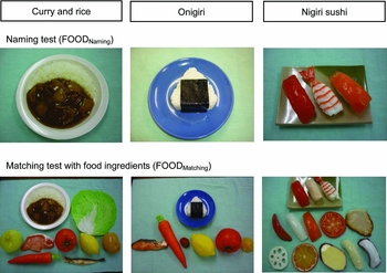
We developed an original Food Cognition Test, to confirm whether three relatively common kinds of food models, “nigiri sushi,” “onigiri,” and “curry and rice” were understood (Figure 1). To exclude the effect of odor, replicas were used. They were realistic replicas prepared by a Japanese company that provides the replicas for restaurants, and were all matched with real foods by the participants. The frequency and familiarity were “high class” according to the “Guidelines for nutrition care and management in geriatric healthcare facilities” (Kato et al., Reference Kato, Shimizu and Sugiyama2005).
The upper line shows the three food models, “curry and rice,” “onigiri,” and “nigiri sushi,” whereas lower line represents the food item models of “potato,” “lemon,” “tomato,” “pork,” “onion,” “shishamo (fish),” “cabbage,” and “carrot.” Upper models were used for FOODNaming, and lower models were used for FOODMatching.

First, the naming (FOODNaming) was tested showing a replica model of “curry and rice.” A tester asked “What is this?” to a participant after showing a food model. The participant was then asked to confirm whether the food item shown matched with the meal, i.e. was included in or arranged as part of the meal (FOODMatching). The food item models of “potato,” “lemon,” “tomato,” “pork,” “onion,” “shishamo (fish),” “cabbage,” and “carrot” were presented separately to the participant, the name was enquired and the participant was asked whether the item was included in the “curry and rice.” Similarly, the tester presented the second meal model “onigiri (rice ball)” and enquired about the name “onigiri,” followed by the separate presentation of food item models of “tomato,” “salmon,” “lemon,” “pickled Japanese plum,” “carrot,” and “takuan,” enquiry about the name and whether the item was included in the “onigiri.” Finally, the tester presented the third meal model “nigiri sushi” and enquired about the name “nigiri sushi,” followed by the separate presentation of food item models of “tuna,” “roast pork,” “salmon,” “tomato,” “squid,” “sweet potato,” “bonito,” “orange,” “shrimp,” “octopus,” “lotus,” “scallop,” and “sea bream,” enquiry about the name and whether the item was included in the “nigiri sushi.” The number of correct answers was scored. In FOODNaming, correct responses for the total three kinds of meals and food earned 30 points, and in FOODMatching, correct answers for all earned 27 points.
Matching with taste perception (tastematching)
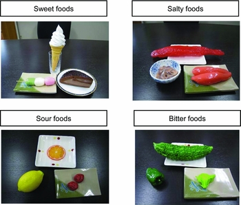
Three respective items of food models showing “sweet,” “salty,” “sour,” and “bitter” were presented and participants responded with the name and taste of 12 items in total (Figure 2).
The food models for “sweet” were “rice dumpling,” “cake,” and “soft cream,” those for “salty” were “salted salmon roe,” “salted cod roe,” and “salted squid,” those for “sour” were “lemon,” “pickled Japanese plum,” and “orange,” and those for “bitter” were “nigauri,” “butterbur sprout,” and “green pepper,” respectively.

The food models for “sweet” were “rice dumpling,” “cake,” and “soft cream,” those for “salty” were “salted salmon roe,” “salted cod roe,” and “salted squid,” those for “sour” were “lemon,” “pickled Japanese plum,” and “orange,” and those for “bitter” were “nigauri,” “butterbur sprout,” and “green pepper,” respectively. After it was confirmed that the participant could identify the food, the participant selected one of the tastes: “sweet,” “salty,” “sour,” and “bitter,” described on a piece of paper (TASTEMatching). The order of presentation of food was random in each test. The maximum score was 12, as the total number of correct answers for 12 items.
Perfect test–retest reliability was observed for the Food Cognition Tests for the HC groups and the dementia patients who agreed to retest (n = 38). The remaining 12 dementia patients did not agree to perform the retest. The Cronbach's α values were 0.84, thus, confirming the validity of the evaluations.
Taste Perception Test
A clinical taste test was conducted using the filter disc method established by otolaryngologists (Tomita, Reference Tomita1986). The filter disc method is quantitative, easily available, requires only a short time and requires no specific training. The measurement site is an area that is accurately controlled by the cranial nerves VII, IX, X, and XI (Tomita, Reference Tomita1986). We administered this Taste Perception Test to eight healthy young adults to examine four test sites on the tongue and confirmed that there were no differences from the tongue tip to the chorda tympani nerve field (data not shown). Therefore, for the participants, the tests were conducted at one site on the tip of the tongue, in consideration of the burden and safety of these tests.
Since it was preferable to choose substances with four kinds of taste from the ones used previously, to compare the test results with the previous report by von Skramlik et al. (Reference Von Skramlik Schwarz1956), sugar was used for “sweetness,” salt for “saltiness,” tartrate for “vinegar,” and quinine hydrochloride for “bitterness,” respectively. Test procedures involved placing a reagent of “sweet,” “salty,” “sour,” and “bitter” on the tongue tip in sequence. The participant identified the tastes by presenting a card describing the taste as “sweet,” “salty,” “sour,” or “bitter.” Before the next taste, the participant rinsed his/her mouth with water to eliminate any residual taste. The instructions for the test order were that sweetness, saltiness, or vinegar could be selected as the first taste, but that bitterness must be tested last. The intensity of the taste was increased from Nos. 1 to 5 in order and the number of correct answers was examined. The average taste of participants with normal taste was Nos. 2 to 3. The results are presented as the mean number of correct answers.
Dietary intake
Dietary intake was determined in accordance with the “Guidelines for nutrition care and management in geriatric healthcare facilities” (Kato et al., Reference Kato, Shimizu and Sugiyama2005). The percentage of dietary intake was estimated by calculating the mean for three days before the test; summing the total energies of the main and side dishes and between meal food intake. The scores were null (0%) to complete intake as 10 (100%). According to the guideline, scores less than 7 (70%) were defined as “decreased dietary intake.” For example, when a patient took a complete intake (100%) of a rice bowl, 40% intake of a side dish, and a full intake (100%) of a snack, the total intake was 70%, scored as 7.
Materials and methods 2
Participants 2
To test the hypothesis that the insula is associated with taste cognition, 20 AD patients were selected from among Participants 1 to undergo PET. They were classified into two MMSE-matched subgroups, i.e. the normal TASTEMatching AD subgroup (10 women, mean MMSE = 11.5), and the impaired TASTEMatching AD subgroup (10 women, mean MMSE = 11.4), with the groups showing normal and impaired scores on TASTEMatching, respectively. There were no significant group differences for MMSE scores. Other semantic memory rather than taste cognition was assessed with the naming of daily objects included in the Western Aphasia Battery; two subgroups did not show any differences (data not shown). Using 18F-fluorodeoxyglucose (FDG)-PET, cerebral metabolic rate for glucose (CMRglc) was calculated.
Positron emission tomography
The PET study was performed using a model SET 2400W scanner (Shimadzu, Japan) at the Cyclotron Radioisotope Center, Tohoku University, at rest with the eyes open. The PET room was kept well-lighted and quiet. A short cannula was placed in the median cubital vein for blood sampling. A transmission scan (six minutes) was performed with a 68Ge/68Ga external rotating line source approximately 30 minutes after a bolus intravenous injection of 185 mBq of FDG. Forty minutes after the injection, a two-dimensional (2D) emission scan (ten minutes) was performed for quantitative analysis, followed by a three-dimensional (3D) emission scan (five minutes). Blood samples were collected 40 minutes after the injection and 18F radioactivity was measured using a well-type scintillation counter. The plasma glucose concentration was also measured 40 minutes after the injection. A time activity curve was estimated from the plasma radioactivity using a simplified method (Wakita et al., Reference Wakita, Imahori and Ido2000).
CMRglc parametric images were obtained from each 2D image using an autoradiographic method (Phelps et al., Reference Phelps, Huang, Hoffman, Selin, Sokoloff and Kuhl1979). Basic image processing and voxel-based data analysis were performed using SPM2 (Statistical Parametric Mapping, 2002). PET data were subjected to an affine and non-linear spatial normalization to the standard Montréal Neurological Institute PET template of SPM2 and to reslicing of 2 × 2 × 2 mm, followed by smoothing using an isotropic Gaussian filter of 12 mm in diameter. The anatomically standardized images were then normalized by analysis of covariance (ANCOVA) to a mean voxel count of 50. Subgroup image differences were compared by paired t-test, with significance accepted if the voxels survived a corrected threshold of p < 0.001 for multiple comparisons.
Results
Food Cognition Test
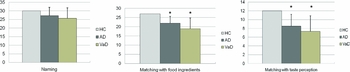
All healthy participants responded correctly for FOODNaming, FOODMatching, and TASTEMatching. Figure 3 presents the findings of the Food Cognition Test. No significant group differences were noted for FOODNaming; however, significant group differences were observed for FOODMatching and TASTEMatching. The AD and VaD groups showed lower scores than the HC groups; no AD–VaD difference was observed.
Shown are the means with SDs. No significant group differences were noted for FOODNaming. Significant group differences were observed for FOODMatching (ANOVA, F = 9.648, p < 0.01) and TASTEMatching (ANOVA, F = 8.746, p < 0.01). For both tests, significant differences were observed between the HC and AD groups (post hoc test, p < 0.01), and the HC and VaD groups (post hoc test, p < 0.01); no AD–VaD difference was observed. HC = healthy control; AD = Alzheimer's disease; VaD = vascular dementia. *: different from HC.

In the AD group, the correlation coefficient between MMSE and FOODNaming was 0.630 (Pearson, p < 0.01), and that between MMSE and FOODMatching was 0.666 (Pearson, p < 0.01). The partial correlation coefficient between MMSE and FOODMatching, using FOODNaming as a control variable, was 0.379 (p = 0.056). In the VaD group, the correlation coefficient between MMSE and FOODNaming was –0.112 (p = 0.775) and that between MMSE and FOODMatching was 0.078 (p = 0.842).
In the AD group, the correlation coefficient between MMSE and TASTEMatching was 0.731 (Pearson, p < 0.01). The partial correlation coefficient between MMSE and TASTEMatching, using FOODNaming, and “Taste Perception Test” as control variables, was 0.455 (Pearson, p < 0.05), which suggested that taste cognition correlated with the MMSE independent of these variables. No such correlations were observed in the VaD group.
Taste Perception Test
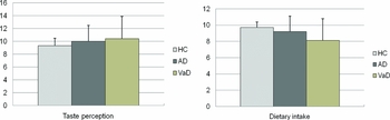
The Taste Perception Test results are shown in Figure 4 (left part). There were no significant group differences. In the AD group, a significant correlation was observed between MMSE and Taste Perception Test scores (Pearson, r = –0.581, p < 0.01); showing a decreasing tendency of taste with disease severity, consistent with the previous study (Steinbach et al., Reference Steinbach, Hund, Vaitl, Heinrich, Foster and Burger2010). No remarkable taste differences (sweet, salty, sour, and bitter) were noted for the correlations with MMSE scores, or between the Taste Perception Test scores and any scores on the Food Cognition Tests.
Shown are the means with SDs. No group differences were noted for the Taste Perception Test and the dietary intake scores. HC = healthy control; AD = Alzheimer's disease; VaD = vascular dementia.

In the VaD group, there was no significant correlation between MMSE and Taste Perception Test scores (Pearson, r = –0.177, p = 0.648), which may be influenced by the food properties independent of the disease severity.
Dietary intake
The mean dietary intake scores are shown in Figure 4 (right part). There were no significant group differences. Twelve dementia patients among the 50 patients exhibited decreased dietary intake, with scores 7 (70%) or less. Eight of the 12 patients exhibited decreased TASTEMatching scores (six of them also exhibited decreased FOODMatching scores). For the remaining 38 dementia patients with normal dietary intake, eight of them exhibited decreased TASTEMatching scores (six of them also exhibited decreased FOODMatching scores). There was a significant difference (at 0.01 level) for the impaired TASTEMatching between the decreased dietary intake group (8/12) and the normal dietary intake group (8/38; χ 2-test, χ 2 = 6.75 > χ 1 2 (0.01) = 6.64).
Food with a different taste that was provided as a normal meal (e.g. sweet salted salmon roe, sour rice dumpling) was prepared and given to the patients to test whether they could be convinced of such taste for the possible improvement of dietary intake. One AD patient (Case #1) and one VaD patient (Case #2) exhibited improved dietary intake after such intervention.
Case #1: a 94-year-old woman, diagnosed as AD with an MMSE score of 10, showed a 40% decrease in dietary intake. She provided good responses to the FOODNaming and FOODMatching, but poor responses to TASTEMatching. The Taste Perception Test score was low. She responded that the salted salmon roe was “sweet” in TASTEMatching. She described her experience as “delicious,” after actually eating the sweet salted salmon roe prepared by the dietician. PET image of the bilateral insula showed decreased CMRglc.
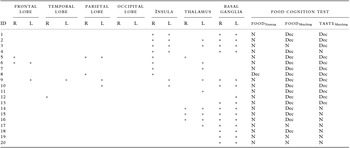
Case #2: an 81-year-old woman, diagnosed as VaD with an MMSE score of 10, showed a 60% decrease in dietary intake (ID 3 in Table 1, see below). She showed the same test pattern as Case #1, but the Taste Perception Test score was average. MRI revealed CVDs in the bilateral basal ganglia and the insula, together with the left thalamus. She responded that rice dumpling was “sour” in TASTEMatching. She described her experience as “delicious,” after actually eating the sour rice dumpling prepared by the dietician.
Vascular lesions and Food Cognition Test in VaD

Dashed line between ID 10 and ID 11 separates the participants with and without insular lesions. The participants with insular lesions (right or left) showed impaired TASTEMatching (9/10), compared with those without insular lesions (3/10) (χ 2-test, χ 2 = 5.21 > χ 1 2 (0.025) = 5.02).
VaD = vascular dementia; R = right; L = left; N = normal score; Dec = decreased score.
Basal ganglia include the putamen and the globus pallidus.
*Presence of vascular lesion.
Neural basis: PET and MRI findings
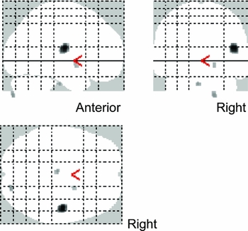
A voxel-based analysis revealed that CMRglc in the right insula was lower in the lower taste cognition AD subgroup, compared with the higher test score AD subgroup, as presented in Figure 5.
Glucose utilization of the right posterior insula was significantly lower (p < 0.001, corrected for multiple comparisons) in the AD subgroup, which exhibited impaired Food Cognition Test (matching with taste perception). The illustrated voxels show the right insula: coordinates (x, y, z) = (46, –12, 18), cluster size (voxels) = 154, z score = 4.88.

Table 1 presents the distribution of the vascular lesions in the VaD patients. While there was heterogeneity in the vascular lesions in the VaD group, the participants with insular lesions (right or left) showed impaired TASTEMatching, compared with those without insular lesions. For both the AD and VaD groups, there were no remarkable findings for the anterior temporal lobes, which was associated with the “hub” of semantic memory.
Discussion
Food Cognition Tests
In the HC group, the responses to FOODNaming, FOODMatching, and TASTEMatching were extremely consistent. This suggested that participants had a disorder if their responses were different from that of the HC group.
In the AD group, responses to FOODNaming and FOODMatching were less accurate than the HC group, although the former was insignificant. The correlation between MMSE and FOODNaming suggested that correct naming could become disturbed as disease severity increased. Furthermore, the correlation between MMSE and FOODMatching suggested that the attributes or property of the meal decreased with the increase in severity. While the probability of a partial correlation between MMSE and FOODMatching, using FOODNaming as a control variable, did not reach significance (p = 0.056), the possibility cannot be ruled out that MMSE and FOODMatching exhibit a slight correlation, independent of FOODNaming. However, for the Taste Cognition Test, a significant partial correlation was noted between the MMSE and TASTEMatching, using FOODNaming, and “Taste Perception Test” as control variables, showing that “taste cognition” may be disturbed in the AD group. This suggested that attributes or “taste spoke” tended to decrease with disease severity, despite the continued ability to respond using the correct names (“hub”).
In the VaD group, responses to FOODNaming, FOODMatching, and TASTEMatching were also less accurate than the HC group, similar to the AD group. However, none of the correlations with MMSE were significant. Disorder of cognitive function varied in the VaD patients because the lesions and symptoms were dependent on each patient.
Taste Perception Test
In the AD group, a significant correlation was observed between the MMSE and Taste Perception Test scores. Taken together with the results above, the severity of the AD neurodegenerative processes could affect the bottom-up sensory process, as well as the top-down cognitive process. Based on such global dysfunction, specific dysfunction of the specific region (e.g. the insula) may add and modify the disorders of taste cognition in AD. The lack of remarkable correlations between the Taste Perception Test scores and any scores on Food Cognition Tests indicated that the impaired cognitive process could not be simply attributed to the decreased bottom-up sensory process.
In the VaD group, however, there were no correlations between the MMSE and any of the tests scores, including Taste Perception. The results suggested that for VaD, the regional role (e.g. the insula) was important for taste cognition.
Semantic memory
AD patients have semantic memory disorders. As described above, based on the “hub-and-spoke” theory of conceptual knowledge (Rogers et al., Reference Rogers, Lambon Ralph, Garrard, Bozeat, McClelland and Hodges2004), we developed original tests regarding the name (“hub”) (FOODNaming) and the attributes or property (taste “spoke”) of foods (TASTEMatching). Contrary to the report that semantic dementia patients showed “hub” damage first followed by visual “spoke” damage (Hoffman et al., Reference Hoffman, Jones, Matthew and Ralph2012), we revealed that the “hub” was relatively spared in AD or VaD, whereas the “taste hub” (TASTEMatching) was impaired. Negative findings of the anterior temporal lobe associated with the “hub” (Pobric et al., Reference Pobric, Jefferies and Lambon Ralph2010) supported this discussion. FOODMatching might be associated with the “action” semantics, since the knowledge of food ingredients was easily retained for people who usually prepared such meals. Since the participants disclosed no remarkable difference on daily lifestyle according to the questionnaire answered by their families, further investigation is needed to clarify the point.
Several studies have reported a category-specific naming disorder (living vs. non-living) in AD (Martinaud et al., Reference Martinaud, Opolczynski, Gaillard and Hannequin2009). Although it remains controversial, non-living things tended to be better named than living things. Foods are made of “living” things. Our patients did not exhibit any differences in the naming scores between FOODNaming and the daily objects (non-living things) by the Western Aphasia Battery (WAB); this category difference did not explain the results. Furthermore, all of the “test meals” were familiar to the participants and those who disliked eating any of the “test-meals” were excluded. Thus, the results were not attributed to the preference effect.
We first showed that AD or VaD patients exhibited impaired taste-related semantic memory (taste cognition), despite the fact that they could name the meal and their taste perception was intact.
Neural basis
Impaired self-awareness of action or anosognosia for hemiplegia may be associated with the right insula lesion (Baier and Karnath, Reference Baier and Karnath2008). We previously reported that VaD patients improved their functional status by a comprehensive physical and psychosocial approach via right insula activation. Patients with insular lesions cannot integrate external and internal (physical) information well. Taste cognition and taste perception were similar. The insula is associated with taste, and its dysfunction is linked with cognitive impairment. In addition, “taste cognition,” a property of semantic memory, has been shown to be damaged independently, which suggests that “taste cognition” exists independent of the entire semantic memory and may be damaged specifically. We demonstrated that “taste cognition” was specifically damaged in patients with both AD and VaD, and that the strongest candidate culprit lesion of the disorder of “taste cognition” was the insula.
Decreased dietary intake was observed in Cases #1 and #2, and both of them exhibited poor responses to TASTEMatching. While this finding does not immediately allow us to conclude that taste cognition is involved in dietary intake, further studies are necessary to investigate this possibility. The importance of the orbitofrontal-insular-striatal circuit in high-level regulation of feeding was reported by the indication of binge eating rather than a decrease in dietary intake (Woolley et al., Reference Woolley, Gorno-Tempini, Seeley, Rankin, Lee and Matthews2007). Voxel-based MRI analyses suggested that there was damage in the right orbitofrontal-insular-striatal circuit. While our AD or VaD patients did not exhibit binge eating, the suspected similar neuronal basis should be investigated further.
A previous acute stroke study reported that taste perception disorders occur frequently after stroke, especially with the anterior circulation subtype (Heckmann et al., Reference Heckmann, Strossel, Lang, Neundorfer, Tomandi and Hummel2005). For the right-left difference, a recent stroke study (Stevenson et al., Reference Stevenson, Miller and McGrillen2013) reported similar effects for the tongue, as well the insula, regarding the taste; the left insula group responded poorer on tasks relative to the right insula group. Our VaD participants were all in a chronic state in the care institute, and no such laterality was noted. Further investigation is needed to clarify these points.
Dietary intake care based on cognitive aspects
Patients with good dietary intake were included among the patients with decreased Taste Perception Test scores and most of them ate normal food with shape. Therefore, vision-related semantic memory was considered important for each meal. Dietary intake did not correlate significantly with MMSE, naming, or taste cognition. A small number of patients exhibited decreased dietary intake, which prevented us from drawing appropriate conclusions. Various approaches have been undertaken for patients with reduced dietary intake in care facilities in consideration of the meal patterns, swallowing and food preference. However, dietary intake is rarely improved. We suggest that in future studies, disorder of “taste cognition” should be assessed in dementia patients in actual care facilities.
The findings on Cases #1 and #2 provide support to the “as if” somatic marker hypothesis based on the deteriorated cognitive framework. As these were observed in only two patients, further investigation should be conducted to test this hypothesis.
In general, the quality of experience is integrated by bottom-up processes, reflecting characteristics of the stimulus of the sensory organs, and top-down processes, associated with the individual beliefs, desires, and expectations. Visual perception is affected by prior conceptual structures, as well as by characteristics of the visual stimulus itself, and assessments of an individual's ability are influenced by the expectations of his/her ability, as well as by objective performance measures. The taste studies provided sufficient foundation for investigating the influence of conceptual information on subjective experience (Lee et al., Reference Lee, Frederick and Ariely2006). Prior intellectual information and subjective expectations can bias objective informational search. Emotional experience is not only based on the effect of physical internal organs, like taste perception, but is also derived from explicit recall of the experience (Damasio et al., Reference Damasio, Grabowski, Bechara, Damasio, Ponto and Parvizi2000), like the emotional experience of memory of taste (taste cognition). This is supported by what we call the “as if” somatic marker hypothesis (Damasio, Reference Damasio1994), which could also explain the effect of the insula on smoking relapse vulnerability in addicted smokers (Janes et al., Reference Janes, Pizzagalli, Richardt, deB Frederick and Chuzi2010). We can discuss the possibility that the cognitive framework of the dementia patient was deteriorated as Cases #1 and #2.
Limitations and future issues related to this study
The Taste Perception Test using the filter disc method is usually conducted based upon the tongue site. However, in this study the test was conducted at only one site on the tip of the tongue. This method must be evaluated further, to confirm the uniformity of the taste response. Furthermore, the taste cognition test should be conducted concomitant with olfactory tests. However, while the test required a long time to administer, the taste perception was focused and no olfactory tests were conducted in this study, in consideration of the burden to the patients. In future investigations, the contents of the tests should be redesigned to improve these aspects.
Conclusions
We administered an original Food Cognition Test and Taste Cognition Test using food models. The latter examined how the participants perceived the “taste” after seeing the food. Some of the dementia patients exhibited disorder of “taste cognition” in the absence of any taste perception disorder. Furthermore, this disorder might occur independent of disorders of naming. It was suggested that that “taste cognition,” the most important property of food that is seen, was specifically damaged. PET findings of the AD group, taken together with the MRI results of the VaD group, suggested that disorder of “taste cognition” was highly related to damage of the insular cortex.
Conflict of interest
None.
Description of author's roles
K. Meguro designed the study, supervised the data collection, and wrote the paper. T. Suto, Y. Kato, K. Tezuka, S. Yamaguchi collected the data and assisted with writing the paper. M. Nakatsuka was responsible for the statistical design of the study and for carrying out the statistical analysis.