Introduction
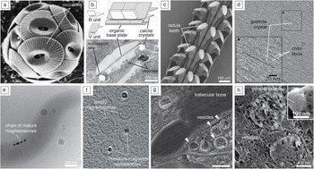
Mineralized tissues support locomotion (e.g., bones), predation (teeth), and defense (shells), and are used for sensing acceleration (e.g., vertebrate otoliths and otoconia), gravity (statoliths), light (lenses), and magnetic fields (magnetosomes), often doubling as nutrient/waste storage.Reference Weiner and Lowenstam1 Many of their remarkable properties emerge from the hierarchical organization of organic and inorganic building blocks (Figure 1)Reference Marsh2–Reference Mahamid, Sharir, Gur, Zelzer, Addadi and Weiner7 (also see the June 2015 MRS Bulletin issue: Biomineralization). Despite new experimental techniquesReference Hendley, Tao, Kunitake, De Yoreo and Estroff8–Reference Ross12 and continuous improvements in computational approaches,Reference Wallace, Hedges, Fernandez-Martinez, Raiteri, Gale, Waychunas, Whitelam, Banfield and De Yoreo13,Reference Demichelis, Raiteri, Gale, Quigley and Gebauer14 we are still far from understanding how the organisms direct mineral nucleation and subsequent processes that lead to assembly.Reference Baumgartner, Dey, Bomans, Le Coadou, Fratzl, Sommerdijk and Faivre15–Reference Gal, Kahil, Vidavsky, DeVol, Gilbert, Fratzl, Weiner and Addadi17 This article focuses on our current understanding of two critical aspects of biomineralization—(1) how self-assembled surfaces direct location, orientation, and timing through heterogeneous nucleation; and (2) the effect of confinement within small volumes on the rate of phase transformations.
Heterogeneous nucleation and confinement in biomineralization. (a) Coccoliths are elements of the extracellular coccosphere in coccolithophores, a class of unicellular marine algae. Heterococcoliths (labeled coccoliths) are composed of a ring of elaborately shaped calcite (CaCO3) single crystals attached to an organic base plate. (b) Coccoliths are composed of alternating, interdigitated R and V units that differ in shape and crystallographic orientation. Precipitation is confined within vesicles inside the cell and involves heterogeneous, oriented nucleation in specific locations along the periphery of the baseplate.Reference Marsh2,Reference Marsh, Ridall, Azadi and Duke3 (c–d) Limpets are intertidal mollusks that graze on rocks using (c) exceptionally hard, iron oxide-based radula teeth. (d) During mineralization, goethite crystals nucleate on a preformed matrix of chitin fibers.Reference Sone, Weiner and Addadi4 Inset is a high-magnification image of the black box. (e–f) In magnetotactic bacteria, (e) magnetite nanoparticles are confined within magnetosome vesicles, (f) allowing for alignment and navigation in magnetic fields.Reference Scheffel, Gruska, Faivre, Linaroudis, Plitzko and Schüler5,Reference Abreu, Sousa, Aronova, Kim, Cox, Leapman, Andrade, Kachar, Bazylinski and Lins6 (g–h) Calcium phosphate mineral precipitates (labeled mineral globules) in intracellular vesicles observed in neonatal mouse calvaria, a model for human bone mineralization.Reference Mahamid, Sharir, Gur, Zelzer, Addadi and Weiner7 Inset is a high-magnification image of a mineral globule.

A brief review of nucleation in biomineralization
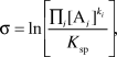
Biomineralization typically starts with precipitation from a supersaturated aqueous solution.Reference De Yoreo, Gilbert, Sommerdijk, Penn, Whitelam, Joester, Zhang, Rimer, Navrotsky, Banfield, Wallace, Michel, Meldrum, Colfen and Dove16,Reference Navrotsky18 The driving force for the formation of a new phase with the chemical formula  $\left[ {{{\left( {{A_1}} \right)}_{{k_1}}}{{\left( {{A_2}} \right)}_{{k_2}}} \ldots {{\left( {{A_i}} \right)}_{{k_i}}}} \right]$ can be expressed in terms of the supersaturation:
 $${\rm{\sigma }} = {\rm{ln}}\left[ {{{\mathop \prod \nolimits_i {{[{{\rm{A}}_i}]}^{{k_i}}}} \over {{K_{sp}}}}} \right]$$,
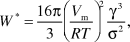
where K sp is the solubility product of the new phase, [Ai] refers to the activity of the aquo-complex of the i th species (Ai) in solution, and k i is the associated stoichiometric coefficient. Clusters of the new phase arise from concentration fluctuations. According to classical nucleation theory (CNT), a cluster that is exactly of critical size, the nucleus, is in unstable equilibrium, and either loss or gain of additional building blocks will reduce its free energy. Smaller clusters will on average shrink, and larger ones will grow. The reversible work of nucleation W* is the free-energy cost (barrier) associated with the formation of a spherical, incompressible nucleus:
 $${W^*} = {{16{\rm{\pi }}} \over 3}{\left( {{V_m} {\over {RT}}} \right)^2}{{{{\rm{\gamma }}^3}} \over {{{\rm{\sigma }}^2}}}$$,
where γ is the (average) surface free energy of the newly formed interface, R is the universal gas constant, T is temperature, and V M is the molar volume of the new phase.Reference Debenedetti19 As discussed in the article by Sear in this issue, the intrinsic nucleation rate J V, the number of nuclei formed per unit time and unit volume, then takes the form:Reference De Yoreo and Vekilov20
 $${J_{\rm{V}}} \propto {e^{ - {{{W^*}} \over {{k_{\rm{B}}}T}}}}$$,
where k B is the Boltzmann constant.
In the presence of any heterogeneities in the parent phase (heterogeneous nucleators), the height of the barrier is reduced by a structure factor, S, which depends upon the specific geometry of the nucleus and interface between the nucleus and the nucleator.Reference Sear21 Alternatively, the impact of heterogeneous nucleators can be expressed by assigning a net effective surface free energy, γnet:Reference Hu, Nielsen, Freeman, Hamm, Tao, Lee, Han, Becker, Harding, Dove and De Yoreo22,Reference Hamm, Giuffre, Han, Tao, Wang, De Yoreo and Dove23
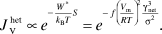 $$J_{\rm{V}}^{het} \propto {e^{ - {{{W^*}} \over {{k_{\rm{B}}}T}}S}} = {e^{ - f{{\left( {{{V_m} \over {RT}}} \right)}^2}{{{\rm{\gamma }}_{net}^3} \over {{{\rm{\sigma }}^2}}}}}$$.
The nucleation rate is extremely sensitive to the presence of heterogeneous nucleators, about which little is known in bulk experiments. Consequently, determination of γ from nucleation rate data is difficult, and a wide range of values have been reported (i.e., calcite [64–1480 mJ/m2]).Reference Forbes, Radha and Navrotsky24–Reference Sohnel and Mullin27 Similar to other crystalline solids, nucleation at moderate supersaturation is expected to proceed extremely slowly when heterogeneous nucleators are absent.Reference Forbes, Radha and Navrotsky24,Reference Bruno, Massaro, Pastero, Costa, Rubbo, Prencipe and Aquilano25 Metastable amorphous calcium carbonate (ACC), however, forms readily under similar conditions. While ACC is frequently observed in vivo and in vitro, the mechanism of its formation and subsequent transformation into one of the crystalline polymorphs of calcium carbonate remains the topic of much debate.Reference Wallace, Hedges, Fernandez-Martinez, Raiteri, Gale, Waychunas, Whitelam, Banfield and De Yoreo13,Reference Gebauer and Cölfen28
Nucleation on surfaces
Irrespective of whether a metastable phase such as ACC precedes the formation of a biomineral, the onset of mineralization likely involves heterogeneous nucleators. In biomineralization, these heterogeneous nucleators include particles, fibers, and membranes constructed from (macro)molecules of the organic matrix, phospholipid bylayers with associated macromolecules that delimit the mineralization site, and the surface of any precursors present.Reference Sommerdijk and de With29,Reference Meldrum and Cölfen30 A good example of the intricate control over crystal orientation by heterogeneous nucleation is the assembly of alternating V and R units on the organic baseplate of the coccolith (see Figure 1b).Reference Marsh2–Reference Mahamid, Sharir, Gur, Zelzer, Addadi and Weiner7,Reference Nudelman, Lausch, Sommerdijk and Sone31,Reference Nudelman32
The impact of the interface on nucleation density, polymorph, and crystallographic orientation is apparent in studies of self-assembled monolayers (SAMs).Reference Aizenberg33,Reference Love, Estroff, Kriebel, Nuzzo and Whitesides34 These early reports attribute rapid nucleation of calcite on carboxylate-terminated alkanethiol-based SAMs to a close match in lattice parameters, but ignore the effect of strain at coherent interfaces.Reference Turnbull and Vonnegut35 Strain has subsequently been shown to modulate calcite crystal shape during growth.Reference Pokroy and Aizenberg36 Recently, it has become clear that during oriented nucleation, both sides of the interface cooperatively reorganize.Reference Quigley, Rodger, Freeman, Harding and Duffy37 A fundamental limitation of the early experiments is that conclusions were based on observations made fairly far into the growth stage. Much detail regarding relative rates of nucleation and subsequent growth rates of nuclei, for instance, in different orientations, is therefore lost, and unambiguous interpretation is challenging.
Microfluidic devices have been used to continuously generate solutions of fixed concentrations and pass them over a functionalized interface where crystals nucleate and grow at a constant supersaturation, while allowing in situ observations.Reference Forbes, Radha and Navrotsky24 Heterogeneous nucleation rates of calcite on SAMs were found to be consistent with the predictions of CNT,Reference Hu, Nielsen, Freeman, Hamm, Tao, Lee, Han, Becker, Harding, Dove and De Yoreo22,Reference Hamm, Giuffre, Han, Tao, Wang, De Yoreo and Dove23,Reference Giuffre, Hamm, Han, De Yoreo and Dove38 and provide estimates of γnet in the range of ∼70–100 mJ/m2 for polysaccharide- and alkanethiol-functionalized surfaces, thereby establishing a lower bound for the surface energy of calcite. Furthermore, there is a direct relationship between γnet and the free energy of binding between calcite and a series of functional groups, consistent with the conventional wisdom that “good binders are good nucleators.”Reference Hamm, Giuffre, Han, Tao, Wang, De Yoreo and Dove23
Functional patterning of substrates is thought to be integral to controlling the location of nucleation events, and by extension, hierarchical assembly in biomineralization.Reference Aizenberg39,Reference Gong, Wang, Ihli, Kim, Li, Walshaw, Chen and Meldrum40 Looking ahead, such patterning will be essential to improving our understanding of cooperative effects, such as the synergy gained from the presence of both carboxylate and amine-terminated SAMs in the formation of amorphous silica.Reference Turnbull41
Nucleation in confinement
We refer to a system as confined if (a) the phase transformation occurs in one or more small volumes of a supersaturated solution; and (b) transport of at least one building block of the new phase is prevented between these volumes. After division of a bulk system into a large number of small volumes, only a subset will contain suspended nucleators,Reference Turnbull41 and phase transformation cannot “spread” from one volume to the next. Ideally, the boundary is a poor heterogeneous nucleator, and the rate converges to that of homogenous nucleation.Reference Turnbull41–Reference Sear43
Phase transformation in confinement will appear to slow down unless very large numbers of droplets are observed. This is because in any system consisting of N identical volumes V d, with a uniform steady-state intrinsic nucleation rate J V, the fraction of droplets in which nucleation has occurred is (see the Sear article in this issue):Reference Sear44
 $$f = 1 - {e^{ - {J_{\rm{V}}}N{V_d}t}}$$,
where t is the time. The time until nucleation has occurred in 50% of the droplets (τ1/2) is inversely proportional to the number and volume of the droplets (Figure 2a–b). In other words, even if the barrier is unchanged, the extrinsic nucleation rate, J = J VNV d, decreases with decreasing system size.
The effect of confinement on nucleation. (a) Plot of the fraction f of N volumes in which nucleation has occurred against log time (J V = 108 m–3 s–1; V d = 65 pL). (b) Log–log plot of  ${\rm{\tau }}_{1/2}^{ - 1}$ versus N, showing the increase in conversion rate with system size. (c–e) Effect of confinement in spherical containers with r c = 30 nm on supersaturation for calcite particles (K sp = 10–8.48 M2, V m = 36.9 cm3/mol, γ = 0.1 J/m2). (c) Fractional drop in supersaturation as a function of particle radius r for initial supersaturation values ranging from 1 to 5. (d) Contour plot of the left-hand side of Equation 6 as a function of the initial supersaturation and the particle radius. The solution of Equation 6 is indicated by the red contour line. (e) Contour plot of the critical supersaturation
 ${\rm{\sigma }}_i^*$ for nucleation of calcite as a function of the surface free energy and the confining radius. Substantially higher initial supersaturations are required for nucleation as the degree of confinement and the interfacial energy of the nucleus increase. Note: J V, number of nuclei formed per unit time and unit volume; τ1/2, time until nucleation that has occurred in 50% of the droplets; K sp, solubility product of the new phase; V m, molar volume of the new phase; γ, surface free energy; σi, initial supersaturation; σf, final supersaturation; V d, volume of drop; r c, confining radius.

When confinement approaches the nanoscale, the assumption that nucleation does not affect the composition of the aqueous phase breaks down. For example, if nucleation of an AB solid at some initial σi consumes an amount of A and B such that their activities each drop by one-half, the supersaturation is reduced by Δσ = σi − σf ≅ 1.4, where σf is the final supersaturation. Therefore, the particle that just formed is subcritical in size and cannot be the nucleus. This effect is more pronounced in smaller confining volumes for less soluble precipitates and at lower initial supersaturation (Figure 2c).
One can show that at a given σi in a confined system of radius r c, only a particle with radius r << r c that meets the following equality is in equilibrium after formation:
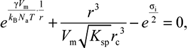 $${e^{{{{\rm{\gamma }}(V_m}} \over {{k_{\rm{B}}}{N_{\rm{a}}}T}} \cdot {1 \over r}}} + {{{r^3}} \over {{V_m} \sqrt {{K_{sp}}} {r_c}^3}} - {e^{{{{{\rm{\sigma }}_i}} \over 2}}} = 0$$,
where N a is Avogadro’s number. Below a critical initial supersaturation of  ${\rm{\sigma }}_i^*$, this equation does not have a solution, and nucleation cannot occur. Above
 ${\rm{\sigma }}_i^*$, the smaller of the two real solutions corresponds to the nucleus and the larger to the particle in final, stable equilibrium (i.e., the end point of the phase transformation; see Figure 2d–e).
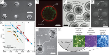
Closely related to the biological systems that inspired them, synthetic liposomes (vesicles bounded by one or more lipid bilayers) have been used to study phase transformations of iron oxides, calcium phosphates, and earth alkaline carbonates in confinement (Figure 3a).Reference Mann, Hannington and Williams51,Reference Collier and Messersmith52 When CaCO3 is precipitated in liposomes between 50 nm and 50 µm in diameter (∼130 aL to 130 nL, Figure 3c), ACC forms rapidly, and the supersaturation with respect to calcite is reset to  ${\rm{\sigma }} = \ln \left[ {K_{sp}^{ACC}/K_{sp}^{calcite}} \right] \approx 4.8$, where
 $K_{sp}^{ACC}$ is the solubility product of ACC, and
 $K_{sp}^{calcite}$ is the solubility product of calcite. This increases the nucleation barrier. In sufficiently small liposomes, the supersaturation may even fall below
 ${\rm{\sigma }}_i^{\rm{*}}$, such that calcite formation becomes impossible. Where nucleation is possible, the time until it is observed in 50 out of 100 liposomes further increases by a factor of 104 for the largest liposomes, and a factor of 1013 for the smallest liposomes when compared to bulk reaction in 130 mL because of the effect of small volumes on the extrinsic nucleation rate. Taken together, confinement effects dramatically delay the formation of calcite and increase the lifetime of ACC (Figure 3c).Reference Tester, Whittaker and Joester46
(a) Calcite crystals nucleated on the (001) plane on a self-assembled monolayer (SAM) of HS(CH2)11SO3– supported on Pd.Reference Aizenberg, Whitesides and Black45 (b) Nucleation rate as a function of (constant) supersaturation for calcite nucleated on SAMs with a range of functional head groups follows predictions of classical nucleation theory, with constant slope proportional to the interfacial energy, γ. Reprinted with permission from Reference Reference Hamm, Giuffre, Han, Tao, Wang, De Yoreo and Dove23. © 2014 National Academy of Sciences. (c) Amorphous calcium carbonate (ACC), rather than calcite, precipitates in confinement in giant liposomes, and is stable for long periods of time.Reference Tester, Whittaker and Joester46 (d) Calcium phosphate precipitates formed in double emulsion droplets eventually transform into hydroxylapatite.Reference Shum, Bandyopadhyay, Bose and Weitz47 (e) Calcium carbonate precipitation in aqueous droplets trapped on a patterned SAM results in extended stability of ACC, but eventually, calcite forms; this is likely due to heterogeneous nucleation at the triple line or on the SAM.Reference Stephens, Ladden, Meldrum and Christenson48 (f) ACC formed in semiconfining pores (diameter d = 800 nm) in a track-etch membrane.Reference Loste, Park, Warren and Meldrum49 (g) Partial confinement between a cylinder with radius ℓ and a flat substrate resulted in differential stabilization of several metastable intermediates (ACP, OCP, and OHAp) that are typically observed sequentially in hydroxylapatite synthesis.Reference Wang, Christenson and Meldrum50 Note: J 0, nucleation rate; σ, supersaturation.

The versatility of confinement effects was demonstrated by the precipitation and transient stabilization (for hundreds of seconds) of a range of amorphous metal carbonates (MCO3, M = Ca, Sr, Ba, Mn, Cd, Pb) in individually levitated drops with a volume of ∼4 µL.Reference Wolf, Müller, Barrea, Kampf, Leiterer, Panne, Hoffman, Emmerling and Tremel53 Higher throughput was achieved by spray drying from a microfluidic nebulator, where confinement effects, coupled with high supersaturation rates and rapid quenching (drying), resulted in long-term stabilization.Reference Amstad, Gopinadhan, Holtze, Osuji, Brenner, Spaepen and Weitz A54 Microfluidic devices have also been used to generate surfactant-stabilized microemulsions of aqueous droplets in oil, which offer low polydispersity and improved efficiency of encapsulation compared to liposomes (Figure 3d).Reference Shum, Bandyopadhyay, Bose and Weitz47,Reference Romanowsky, Abate, Rotem, Holtze and Weitz55 Confinement further allowed investigating the impact of reagent concentrations on CaCO3 polymorph selectivity and of solubility on nucleation of KNO3.Reference Laval, Crombez and Salmon56 In the CaCO3 system, confinement in droplets trapped on a patterned SAM (V d ∼ 1 pL, Figure 3e) resulted in a rapid ACC formation, but delayed nucleation of crystalline polymorphs, despite the presence of a relatively powerful nucleating surface.Reference Stephens, Kim, Evans, Meldrum and Christenson57
A range of effects consistent with confinement have also been observed in systems where, unlike in liposomes or droplets, transport of building blocks is possible in at least one direction in space. For example, it has been inferred that the nucleation rate of calcite decreases more rapidly than that of ACC as the diameter of pores in track-etch membranes is reduced from 3 μm to 200 nm (Figure 3f).Reference Loste, Park, Warren and Meldrum49,Reference Schenk, Albarracin, Kim, Ihli and Meldrum58 Similar observations were made in the calcium phosphate system.Reference Cantaert, Beniash and Meldrum59 The wedge between a solid half-cylinder resting on a flat substrate constitutes a more complex system (Figure 3g), where the degree of confinement can be thought of as rapidly increasing with decreasing distance to the contact line.Reference Stephens, Ladden, Meldrum and Christenson48,Reference Wang, Christenson and Meldrum50,Reference Wang, Christenson and Meldrum60 Consequently, metastable precursors of crystalline calcium carbonates, sulfates, and phosphates persist for much longer times closer to the contact line. As a result, the “Ostwald–Lussac” sequence of increasingly stable phases observed over time in the bulk system is instead patterned in space. While quantitative treatment of these partially confined systems is more difficult because much depends on geometrical parameters, diffusivities, and mixing, they can be used to rapidly screen for metastable precursors that can be challenging to observe in bulk reactions.
Conclusions
Heterogeneous nucleation and confinement are integral factors to biological control over location, timing, and polymorph selection in biomineralization, but remain challenging to probe quantitatively. This article highlights recent in vitro experiments that show how the influence of heterogeneous nucleators can be understood in the context of CNT without needing to invoke “nonclassical” effects. Based on recent experiments and simple thermodynamic considerations, we suggest three ways in which confinement reduces nucleation rates of crystalline biominerals. Amorphous intermediates appear to be less affected. As a result, the lifetime of amorphous precursors can be dramatically longer, even in the absence of any stabilizing additives. This effect may open a window for an organism to manipulate the amorphous material with a low risk of spurious nucleation. Controlled transformation into a crystalline polymorph could then occur by introducing a heterogeneous nucleator into the confined volume, before or after the amorphous intermediate has formed.