Introduction
Schistosomes are parasitic flatworms that constitute an important public health problem. Adult worms live in their host’s bloodstream where they can reside for many years by importing nutrients across their tegument (Kusel et al., Reference Kusel, Al-Adhami and Doenhoff2007). However, the Schistosoma mansoni lifespan in permissive hosts under laboratory conditions still remains unclear, since experiments are concluded before the experimental host's longevity is reached. According to data in the literature, the worm's lifespan ranges from approximately 1.5 to 10 years (Fulford et al., Reference Fulford, Butterworth, Ouma and Sturrock1995) and may reach beyond three decades (Harris et al., Reference Harris, Russell and Charters1984).
The free-living stages of schistosomes are self-supporting and they are dependent on the energy reserves they acquired in their previous host. Once inside the host, substrates are plentiful, and adult worms live inside the mesenteric vasculature, where the female flukes lay eggs in the capillary venules. Some of the eggs penetrate through into the intestine lumen and are excreted in the faeces (Farah et al., Reference Farah, Mola, Kariuki, Nyindo, Blanton and King2000) and others become lodged in the tissues. Both in vivo and in vitro kinetics of oviposition are characterized by initial low egg production followed by a maximum production and progressive reduction in the number of eggs (Rocha et al., Reference Rocha, Rocha, Pedroso, Colosimo and Coelho1995; Barth et al., Reference Barth, Fernandes and Rodrigues1996).
Studies of reproductive ageing have been carried out in a variety of organisms. In animals, ageing is characterized by widespread degenerative changes in tissue morphology and function that result in a progressive decrease in the ability to reproduce (Hughes et al., Reference Hughes, Evason, Xiong and Kornfeld2007). Why study ageing of Schistosoma mansoni?
It is known that the faecal egg count in human schistosomiasis mansoni infection progressively decreases during the chronic phase of the disease (Barth et al., Reference Barth, Fernandes and Rodrigues1996). Although the mechanisms involved in such diminished egg output are not well understood, some studies have correlated them with both structural and functional changes in the host intestine (Moreels et al., Reference Moreels, De Man, Bogers, De Winter, Vrolix, Herman, Van Marck and Pelckmans2001). In addition, infertile worm pairs were observed during chronic S. mansoni infection in mice (Cheever et al., Reference Cheever, Mosimann, Deb, Cheever and Duvall1994). Nevertheless, long-term studies describing morphological effects of ageing on adult schistosomes are lacking. Previous studies in our laboratory have shown that confocal images enable detailed morphological analysis of the adult S. mansoni reproductive system (Neves et al., Reference Neves, Pereira, de Oliveira, Gomes and Machado-Silva1998, Reference Neves, Costa-Silva, Martinez, Branquinho, de Oliveira, Lenzi, Gomes and Machado-Silva2004, Reference Neves, de Lamare Biolchini, Machado-Silva, Carvalho, Branquinho, Lenzi, Hulstijn and Gomes2005), and further investigations have provided new data on genital anomalies in adult schistosomes (Neves et al., Reference Neves, Costa-Silva, Martinez, Biolchini Cde, Lenzi, Gomes and Machado-Silva2003).
The present paper is aimed at gaining greater insight into the effects of ageing on adult schistosomes recovered from the water rat, Nectomys squamipes, using confocal laser scanning microscopy. One animal in our investigation ceased to pass schistosome eggs in its faeces and died after 70 weeks under laboratory conditions. Reported as a natural host of S. mansoni in Brazil (Machado-Silva et al., Reference Machado-Silva, Galvao, Presgrave, Rey and Gomes1994), infected water rats have a maximum life expectancy of 2 years under natural conditions (Modena et al., Reference Modena, dos Santos Lima and Coelho2008).
Materials and methods
Animal procedures
All procedures were carried out in accordance with the conventional guidelines for experimentation with animals (Ellery, Reference Ellery1985). Nectomys squamipes specimens were captured in the municipality of Sumidouro (22°02′46″S; 42°41′21″W), which is in an area of S. mansoni transmission in the state of Rio de Janeiro (Brazil), and then transferred to polypropylene rodent cages covered with stainless-steel screens. Under laboratory conditions (Rodrigues e Silva et al., Reference Rodrigues e Silva, Machado e Silva, Faerstein, Lenzi and Rey1992), these animals were fed on maize and cucumber and had free access to water.
Parasitological procedures
To determine whether the captured water rats were infected, faecal samples were taken from each animal and examined by both the Kato–Katz thick smear technique (Katz et al., Reference Katz, Chaves and Pellegrino1972) and the spontaneous sedimentation method (Lutz, Reference Lutz1919). Because one rat died after 70 weeks of captivity, after excreting viable S. mansoni eggs in its faeces for 65 weeks and only unviable eggs thereafter, hepatic perfusion was immediately performed (Smithers & Terry, Reference Smithers and Terry1965).
Recovered S. mansoni worms (six males and one female) were fixed in AFA (alcohol, formalin and glacial acetic acid) at room temperature, stained with 2.5% hydrochloric carmine, dehydrated in a series of alcohols (70%, 90% and absolute), clarified in methyl salicylate with Canadian balsam (1:2) and preserved as whole-mounts on glass slides. Confocal images were captured with a LSM 510–ZETA confocal laser microscope (Zeiss, Germany), under reflected mode with a 543 nm He/Ne laser and LP 570 filter. Morphological changes in the tegument, suckers and reproductive organs of adult worms were the focus of this study.
Results
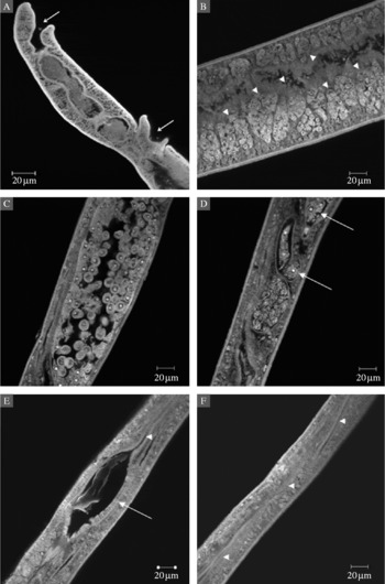
A view of the female S. mansoni anterior end showed that oral and ventral suckers were apparently normal (fig. 1A). Tomographic images revealed follicular vitelline glands distributed in two lateral bands (fig. 1B) and numerous vitelline cells. At the posterior end of the ovary (fig. 1C), oocytes leave the ovary and proceed down the short oviduct, the walls of which dilate in the presence of spermatozoa to form the seminal receptacle (fig. 1D). Towards the anterior end, the common vitelline duct joins the oviduct, which expands slightly to form the ootype, comprising flattened cells and unclear nuclei, where a developing embryo enclosed by its shell was observed (fig. 1E). The ootype opens into a long uterus with no evidence of eggs (fig. 1F).
Images of female S. mansoni stained with hydrochloric carmine. (A) Oral and ventral suckers (arrows); (B) vitelline glands (arrowheads); (C) ovary with oocytes; (D) seminal receptacle and oocytes (arrows); (E) ootype (arrow) and uterus (arrowhead); (F) uterus (arrowheads).

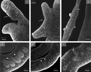
At the anterior end of males, small spines covered both suckers and the gynaecophoric canal (fig. 2A–C). As for the reproductive system, testicular lobes ranged from 1 to 7 and germinative cells were absent (fig. 2D). Spermatozoa were apparent in the seminal vesicle (fig. 2F).
Images of male S. mansoni stained with hydrochloric carmine. (A) Spiny outer surface of the oral sucker (arrow); (B) ventral sucker with spines (arrow); (C) spines (arrowhead) located in one of the margins of the gynaecophoric canal (arrow); (D) testicular lobes filled with few germinative cells (arrowheads); (E) normal testicular lobes filled with cells (arrowheads); (F) seminal vesicle and ejaculatory duct (arrow).

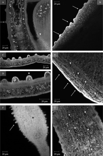
The dorsal surface of male worms bore unevenly distributed tegumental tubercles, extensive areas without any or with some rudimentary tubercles (fig. 3B, C and E). The developed tubercles were covered in spines (fig. 3D), while at the subtegumental level, vacuolated areas were detected (fig. 3E and G). Male adult worms showed a feeble degree of muscularization on the dorsal surface (fig. 3G) and in suckers.
Images of the tegument of male S. mansoni stained with hydrochloric carmine. (A) Lateral view of normal tegumental tubercles (arrow) and gynaecophoric canal with spines (arrowhead); (B) lateral view of tegumental tubercles (arrows); (C) normal tegument with large tubercles; (D) tubercles with spines; (E) tegument without tubercles (arrows) and subtegumental vacuolated areas (arrowheads); (F) dorsal surface with tubercles (arrow) and normal muscle bundles organized in circular, longitudinal and diagonal muscle fibres (arrowheads); (G) dorsal surface with flacid musculature and vacuolated areas (arrowheads).

Testicular lobes filled with germinal cells (fig. 2E) and dorsal tubercles (fig. 3A) were apparent in normally developed tissues. Muscle fibres were arranged in bundles in the dorsal surface and muscle, with muscle bundles comprising circular, longitudinal and diagonal muscle fibres (fig. 3F).
Discussion
Ageing is characterized by widespread degenerative changes in tissue morphology and function that result in a progressive decrease in the ability to reproduce (Hughes et al., Reference Hughes, Evason, Xiong and Kornfeld2007). The extension of lifespan appears to be affected by a shift from reproductive to non-reproductive maintenance (Longo & Fabrizio, Reference Longo and Fabrizio2002). Data in the literature have shown that S. mansoni faecal egg counts in infected humans progressively decrease through the chronic phase of the disease (Rocha et al., Reference Rocha, Rocha, Pedroso, Colosimo and Coelho1995). Possibly, older worms in a long-term infection could present infertility. The assumption that reproductive ageing of human-derived schistosomes is responsible for decreased egg counts in chronic schistosomiasis stages requires further morphological studies (Kastner et al., Reference Kastner, Kohn, Teixeira and Pitanga1975). However, because of limitations involved in human host research, it is difficult to characterize mechanisms that influence reproductive ageing of worms in human models (Wilson et al., Reference Wilson, Langermans, van Dam, Vervenne, Hall, Borges, Dillon, Thomas and Coulson2008).
Nevertheless, schistosomes grown in laboratory animals may provide an attractive model for the study of worm infertility, even though most experimental studies are terminated before reaching the longevity of the experimental host, in which schistosomes have a shorter lifespan (Cheever et al., Reference Cheever, Mosimann, Deb, Cheever and Duvall1994). These issues were overcome here by the use of permissive host-derived schistosomes, which were recovered from one rat that died after 70 weeks in captivity. After shedding viable eggs for a long time in faeces, this rat eliminated only unviable eggs, suggesting that older parasites may have age-related degenerative changes in the reproductive system. Previous studies have shown that ovarian dysfunction is the primary reason for the reproductive failure of schistosome pairs grown in vitro (Irie et al., Reference Irie, Basch and Beach1983). It is also possible that atrophy of the ovary and vitellaria leads to lower egg production and thus decreased faecal egg output (Wilson et al., Reference Wilson, Langermans, van Dam, Vervenne, Hall, Borges, Dillon, Thomas and Coulson2008).
In this study, ovaries, vitellaria and oocytes were shown to be apparently normal. However, the ootype was not composed of cylindrical cells with plaited bases and nuclei with flabby chromatin, in contrast to its previously described typical architecture (Neves et al., Reference Neves, de Lamare Biolchini, Machado-Silva, Carvalho, Branquinho, Lenzi, Hulstijn and Gomes2005). This finding suggests that the mechanism for eggshell production by the S. mansoni ootype during organogenesis may be impaired. A previous study in vitro showed that within the ootype and uterus the oocytes were not embedded within egg-like material (Irie et al., Reference Irie, Basch and Beach1983). It is noteworthy that eggshells were found with marked alterations on the intestinal wall of the naturally infected rat that ceased shedding viable eggs (Rodrigues e Silva et al., Reference Rodrigues e Silva, Machado e Silva, Faerstein, Lenzi and Rey1992).
The morphological changes evidenced in the reproductive system were not limited to females. Despite the fact that testicular lobes ranged from 1 to 7, their content showed areas with no reproductive function in spermatocytes. A recent investigation reported that schistosomes derived from chronically infected rhesus macaques also showed a reduced number of spermatocytes (Wilson et al., Reference Wilson, Langermans, van Dam, Vervenne, Hall, Borges, Dillon, Thomas and Coulson2008), which might account for decreased spermatozoa observed in the seminal receptacle.
In the present study, confocal images showed that at the anterior end of the male worms, oral and ventral suckers and the gynaecophoric canal had spines. This is consistent with our previous report based on scanning electron microscopy (Machado-Silva et al., Reference Machado-Silva, Lanfredi and Gomes1997). Within the oral sucker and on the dorsal surface, male worms showed a flabby musculature, which is likely to be related to decreased muscular tone, affecting both adhesion capacity and dislodgment within the mesenteric vessels.
The tegument of schistosomes is an important interface between the parasite and its intravascular environment in the host (Abath & Werkhauser, Reference Abath and Werkhauser1996). The capacity to acquire nutrients across the tegument surface and to escape from the host immune response depends on its structural integrity (Braschi et al., Reference Braschi, Curwen, Ashton, Verjovski-Almeida and Wilson2006). Earlier studies have shown that schistosomes derived from permissive hosts (mice and water rat) bore tubercles with spines which were mainly concentrated in the middle region of the tegument (Machado-Silva et al., Reference Machado-Silva, Lanfredi and Gomes1997). In the present investigation, unevenly distributed tubercles or extensive areas without tubercles were found on the worm dorsal surface. The presence of spines on the tegument of adult schistosomes is known to be related to their age and physiological condition (Southgate & Agrawal, Reference Southgate and Agrawal1990). In addition, vacuolization of the subtegumental area might lead to impaired migration and supply of host nutrients. Although the exact aetiologies of the alterations reported herein are unknown, the involvement of free radicals in ageing has been described (Harman, Reference Harman2006). Taken together, the present results raise issues on possible causes of infertility in older male and female schistosomes.