Introduction
The order Mycocaliciales Tibell & Wedin (Mycocaliciomycetidae Tibell) is a group of small, non-lichenized ascomycetes with pin-like ascomata. In past years, these species were often treated as part of Caliciales s. lat. and, as such, usually called ‘pin-lichens’. However, several molecular phylogenetic studies have shown that mycocalicioid fungi are related to the Eurotiomycetidae and Chaetothyriomycetidae (Eurotiomycetes) (Geiser et al. Reference Geiser, Gueidan, Miadlikowska, Lutzoni, Hofstetter, Fraker, Schoch, Tibell, Untereiner and Gueidan2006; Schoch et al. Reference Schoch, Sung, López-Giráldez, Townsend, Miadlikowska, Hofstetter, Robbertse, Matheny, Kauff and Wang2009; Prieto et al. Reference Prieto, Baloch, Tehler and Wedin2013). The superficial similarities in habitat ecology and ascomata shape with species of Caliciales s. str. and Coniocybaceae have apparently resulted from convergence. The taxonomy of Mycocaliciales has been reviewed several times since the description of Mycocalicium subtile (Pers.) Szatala (Szatala Reference Szatala1925), especially by Tibell (e.g. Tibell Reference Tibell1984, Reference Tibell1987, Reference Tibell1991). Titov's monograph (Titov Reference Titov2006) presents a good summary of the currently known Holarctic species. For an overview of the history, taxonomy, genera and species, see also Tuovila (Reference Tuovila2013).
The generic delimitation of mycocalicioid fungi is unsatisfactory, as pointed out by, for example, Hawksworth (Reference Hawksworth1980), Samuels & Buchanan (Reference Samuels and Buchanan1983), Titov (Reference Titov1994) and Tibell & Vinuesa (Reference Tibell and Vinuesa2005). Tuovila (Reference Tuovila2013) tried to solve the ambiguity but was unsuccessful due to the complexity of the problem and the unexpectedly high diversity within the group. Species circumscription has been vague and many carry several synonyms. Examples include Chaenothecopsis pusilla (Ach.) A. F. W. Schmidt and Mycocalicium subtile (see Titov Reference Titov2006), both of which have become dumping grounds for species that do not have some distinctive, clearly discernible characters.
The reputed lack of sufficient, stable morphological characters (Schmidt Reference Schmidt1970; Tibell Reference Tibell1984; Vinuesa Navarro Reference Vinuesa Navarro2002) has generated some attempts to establish new characters for species delimitation in the Mycocaliciales. The analysis of spore ontogeny with transmission and electron scanning microscopy (Tibell Reference Tibell1984), the study of cultured anamorphs (Tibell Reference Tibell1997), and uncharacterized chemical compounds in ascomata and anamorphs (Vinuesa Navarro Reference Vinuesa Navarro2002) have offered some informative, although somewhat labour intensive characters. So far, the delimitation and identification of mycocalicioid species has been based mainly on the morphology and anatomy of the ascomata and ascospores (Tibell Reference Tibell and Ahti1999; Titov Reference Titov2006). In addition to the spore septation and size, and the presence of an ornamentation on the spore wall, the pigmentation of the spore septum has also been used as a distinctive character (Tibell Reference Tibell1982). In the ascoma, importance has been ascribed to the number and width of the hyphal layers in the excipulum and to the organization and pigmentation of hyphae in the excipulum and stipe. These hyphal characters are visible best or only in thin microtome sections, which were introduced for species delimitation by Schmidt (Reference Schmidt1970). The emphasis on using thin sections in species delimitation has confused the interpretation of the anatomy of mycocalicioid species. As a consequence, several distinctive morphological characters, especially the textura type of excipulum and stipe, have been overlooked. While thin sections can reveal some features in the internal structure of the ascoma, the method is also laborious and often difficult to achieve for those identifying mycocalicioid species.
Chemical characters currently used for delimiting mycocalicioid species are rather few. The use of potassium hydroxide (KOH, usually 5–10%) for identification was first mentioned by Vainio (Reference Vainio1890), but the distinctive colour reactions of certain Chaenothecopsis species to KOH were first described by Nádvornik (Reference Nádvornik1940). Unspecified blue iodine reactions of mycocalicioid fungi have been observed by many lichenologists, seemingly first by Tuckerman (Reference Tuckerman1872). Keissler (Reference Keissler1938) and Lettau (Reference Lettau1940) routinely mention blue iodine reactions of the hymenium. However, Schmidt (Reference Schmidt1970) did not recognize the value of iodine, probably because Lettau's otherwise confusing species concepts obscured the specificity of these reactions. Given the impact of Schmidt's taxonomic treatment (Reference Schmidt1970) on the prevailing practice of species delimitation, it is understandable why iodine (either in Lugol's or Melzer's reagent) has not been routinely used.
Our studies (e.g. Tuovila Reference Tuovila2013; Tuovila et al. Reference Tuovila, Schmidt, Beimforde, Dörfelt, Grabenhorst and Rikkinen2013, Reference Tuovila, Davey, Yan, Huhtinen and Rikkinen2014; Rikkinen et al. Reference Rikkinen, Tuovila, Beimforde, Seyfullah, Perrichot and Schmidt2014; Selva & Tuovila Reference Selva and Tuovila2016) have revealed an unexpectedly high diversity of mycocalicioid species, based on DNA sequences as well as on morphology and anatomy. While the true number of mycocalicioid species is unknown, and our molecular data are far from sufficient (see Tuovila Reference Tuovila2013) to formulate an evolutionarily solid hypothesis of the phylogeny of Mycocaliciales, we concentrate in the current investigation on practical methods for studying and identifying specimens. The search for new anatomical, morphological and chemical characteristics will also help clarify species and generic concepts within the group.
Here we present an overview of all the characters for mycocalicioid fungi and discuss the variation and potential use of these for species delimitation and identification. We also present several previously unused or misinterpreted morphological, anatomical and chemical characters of mycocalicioid species, many of which have already been incorporated into descriptions of new mycocalicioid species (Tuovila et al. Reference Tuovila, Cobbinah and Rikkinen2011a, Reference Tuovila, Larsson and Rikkinenb, Reference Tuovila, Schmidt, Beimforde, Dörfelt, Grabenhorst and Rikkinen2013, Reference Tuovila, Davey, Yan, Huhtinen and Rikkinen2014; Rikkinen et al. Reference Rikkinen, Tuovila, Beimforde, Seyfullah, Perrichot and Schmidt2014; Selva & Tuovila Reference Selva and Tuovila2016). Our emphasis is on anatomical characters that can be seen in properly prepared squash mounts.
Material and Methods
Biological material
This study was based mainly on material collected and studied in Tuovila (Reference Tuovila2013), additional new material acquired in 2013–2015, and on selected specimens from herbaria H, TUR, O and UPS. The material contains specimens of several species, from several regions and locations. Lists of specimens used for representative drawings are given in Supplementary Material Table S1 (available online). Some of these drawings have already been published as part of species descriptions (e.g. Tuovila et al. Reference Tuovila, Cobbinah and Rikkinen2011a, Reference Tuovila, Schmidt, Beimforde, Dörfelt, Grabenhorst and Rikkinen2013, Reference Tuovila, Davey, Yan, Huhtinen and Rikkinen2014). In addition to these specimens, hundreds of additional specimens (not listed) were studied in order to distinguish and delimit the characters presented in the Results. Species of Sphinctrina Fr. were not studied as we could not find them in our field studies and the material in herbaria is scarce. However, these species have a well-supported position within Mycocaliciales (Rikkinen et al. Reference Rikkinen, Tuovila, Beimforde, Seyfullah, Perrichot and Schmidt2014; Tuovila et al. Reference Tuovila, Davey, Yan, Huhtinen and Rikkinen2014) and they probably share at least some of the characters presented here.
Microscopy
Macroscopic features of the specimens were observed using a stereomicroscope. Microscopic characters were observed and measured from squash preparations in water and water-based solutions using a compound microscope (Leica DMLS) with a ×100 oil-immersion objective, ×10 oculars, and additional ×1.5 or ×2.0 magnifications. Between 10 and 20 mature, released spores were measured, randomly selected from each ascoma. Both length and width were measured in the same spore to derive the Q-value (length/width). Potassium hydroxide (10%, KOH), Lugol's reagent (IKI), Melzer's reagent (MLZ), ammonical Congo red (CR), Cresyl blue (CRB) and nitric acid (N) were used to observe specific chemical reactions and for better contrast in some diagnostic morphological structures (see Supplementary Material Table S2, available online). Drawings were made by hand, with a drawing tube (Leica), and digitally refined and arranged for publication.
Squash preparations
Specimens were studied as fresh as possible to observe the characters in living condition (Baral Reference Baral1992). The textura and pigmentation in stipe and excipulum vary considerably and offer multiple morphological characters (see below). Since these structures need to be observed in surface view, carefully prepared squash mounts are necessary. The outer layers of the ascomata of mycocalicioid species usually consist of heavily pigmented, thick-walled hyphae. This, in addition to the often complex structure of the epithecium, poses some challenges when viewed in squash preparations. First and foremost, if not done properly, the squashing of material will break the cover glass. The following methods help avoid breakage: (1) transfer one (or more) ascomata into the medium (water or other suitable solvent), (2) let the ascomata soak in the medium for at least 5 min, (3) carefully put the cover glass on top of the medium and ascoma, (4) gently press with finger (soft paper between finger and glass to avoid greasy staining), and (5) finish squashing by tapping carefully, for example with the eraser end of a pencil. Using a thicker (#2) instead of a thinner (#1) cover glass can also help prevent breakage. Some species are easier to squash, and the hardness/softness of the ascoma is another species-specific character worth noting.
Results
Macroscopic characters
Ascoma size and colour
The size of the ascoma is an important, albeit imprecise diagnostic feature. Populations of mycocalicioid fungi typically include several age cohorts of mature ascomata, developed during consecutive growing seasons. In addition, the size of mature ascomata can vary both between and within populations, sometimes differing by up to twofold. While the size differences between and within populations are more likely to reflect microvariation in environmental conditions during ascoma development, the size of mature ascomata usually varies only within certain limits and can be used for identification in combination with other characters. The smallest ascomata encountered were less than 0.5 mm high and the largest were anywhere from 1.5 to 5.0 mm high.
The ascomata of most mycocalicioid species are dark in colour under incident light. Although black is by far the most common colour, shades of green (olive to moss green) and brown (from dark to pale yellowish brown) are also regularly encountered. Some species have a distinct reddish, greenish or bluish tinge or hue to the otherwise dark ascomata. The colour of immature versus mature ascomata can also be different; the immature ascomata are often black without the hues of mature ascomata.
The surface of the stipe and/or capitulum may be shiny or matt. In addition, some species have coloured pruina on the stipe, and sometimes also on the capitulum. Pruina may originate from senescent, fracturing hyphae (Tibell & Titov Reference Tibell and Titov1995), or from extracellular pigmented crystals (Bonar Reference Bonar1971; Selva & Tibell Reference Selva and Tibell1999). In many species, mature ascospores accumulate on the top of the epithecium and this may have a significant effect on the colour and overall appearance of the capitulum.
Branching
Branching of the ascoma, resulting in two or multiple capitula in one ascoma, is a common feature in mycocalicioid species. While a pronounced tendency to produce branched ascomata, as well as the type of branching, are species-specific features, all branching species can also produce non-branched ascomata. Similarly, most predominately non-branching species occasionally produce one or several branched ascomata. In some types of branching, the capitulum may only be divided into distinct lobes (Fig. 1F & M), while in others there may be fully separated capitula but without individual stipes (Fig. 1Q–S). Examples of common branching types are illustrated in Fig. 1. The frequent branching and proliferation of certain resinicolous species was discussed in Tuovila (Reference Tuovila2013) and Tuovila et al. (Reference Tuovila, Schmidt, Beimforde, Dörfelt, Grabenhorst and Rikkinen2013).
Diagrammatic representations of typical capitulum shapes (A–N) and branching patterns (O–U) of mycocalicioid fungi. Capitulum shape: A & C, cupulate. B, conical. D, convex. E, lentiform. F, lobed or multiheaded. G, ellipsoid. H, spherical. I, semiglobate. J, emarginate. K & L, flabelliform (flattened K). L–N, irregular. The shapes of irregular capitula can show great inter- as well as intra-specific variation, but the overall form (e.g. elongated in N, flat in L) is usually constant. Note the variation in the width of the capitulum relative to the stipe (A vs C) and in the shape of the epithecium (e.g. raised in A, flat in C). Note also the difference between long and slender stipes (L, M, P), and thick stipes (C, D, I, J). Branching patterns: O, from the stipe. P, through the capitulum. Q, from whorls. R–T, branching from stipe (S, from upper part; R, from middle & T, from base). U, from (pseudo) stroma. Diagram J is a cross-section; all others are overall profiles. Species and voucher information can be found in Supplementary Material Table S1 (available online).

Stipe
The length and width of the stipe vary between species, and both features can have value in species delimitation. However, as the overall size of ascomata of mycocalicioid species varies between species (see above), these variables often have to be assessed in relation to each other. Some examples of the overall variation in stipe length and width can be seen in Fig. 1. It is noteworthy that slender stipes tend to be longer (Fig. 1L & M), and shorter stipes are commonly thicker (Fig. 1J). In most species the stipe is of more or less even width, from top to bottom (e.g. Fig. 1A–C). More rarely, in some species, the stipe widens at the base (Fig. 1I) or just below the capitulum (Fig. 1D). According to Rikkinen (Reference Rikkinen1999), the length of the stipe seems to be dependent on ecological conditions. Known species with very short stipes were reviewed by Tibell & Ryman (Reference Tibell and Ryman1995).
Capitulum
The young, emerging ascomata of mycocalicioid fungi tend to have very small capitula or, at the very least, no capitula at all. Before the development of the capitulum, the developing ascoma often has a sharp apex, and the capitulum is not distinguishable macroscopically from the stipe (see also Rikkinen Reference Rikkinen1999). The size and shape of the capitulum typically change notably during the maturation process. Thus, when using the shape of the capitulum as a morphological character, one should always compare ascomata of the same age. Some typical capitulum shapes of mycocalicioid fungi are illustrated in Fig. 1. The capitulum is composed of excipulum and epithecium, and their relative proportions should also be assessed when using the capitulum as a morphological character. Excluding Sphinctrina, mycocalicioid species do not form a mazaedium like the ‘true’ pin-lichens. However, the excipular margin is usually distinguishable and the border of excipulum and epithecium is easy to observe, often with a stereomicroscope.
Microscopic characters
Vital taxonomy
Vital taxonomy has revealed new characters in several groups of ascomycetes (Baral Reference Baral1992). However, in mycocalicioid species, there seems to be very little variation between the living and dead state of the ascomata. The only difference in the morphology of living and dead ascomata is the structure of ascus apices. Mycocalicioid species are xeromorphic and there is no need to dry the specimens, although the substratum may need to dry. It is often hard to tell from fresh collections whether the ascomata are living or dead. However, green and brown colours, in particular, tend to fade after many years in the herbarium and distinctively green-coloured species might be only pale yellow after 10 years in the herbarium. Also, Tibell & Titov (Reference Tibell and Titov1995) noticed that the distinct KOH+ (red) reaction of Chaenothecopsis dolicocephala Titov could not be observed in specimens that were 2–3 years old or older. This kind of fading or disappearance of pigments with time may be common in other mycocalicioid species as well.
Colours of the hyphae
The pigments of mycocalicioid fungi are in the cell walls of the outermost hyphal layers of the stipe and excipulum (Fig. 2). In all specimens that were macroscopically black, the colour of the hyphae was not black but of some lighter shade when viewed under transmitted light. The hypothecium and the inner part of the stipe may be, and often are, more lightly coloured or even hyaline, but completely hyaline species have not been described and were not encountered in our studies. Different shades of green and brown were the most commonly observed colours, but different shades of grey, yellow and brownish red to wine red have also been recorded. The colour of the outermost layers of stipe and excipulum (and epithecium, if one exists) is species-specific. However, it must be noted that old ascomata in many species tend to have much darker colours than young ones. Commonly, the green shades of young ascomata turn brown or even dark brown at maturity or in over-aged ascomata. In addition, the stipe and excipulum may share the same colour or have different combinations of colours, brown and green being the most common combination. In some species the colours are very pale and this is usually (but not always) reflected in the colour observable under incident light.
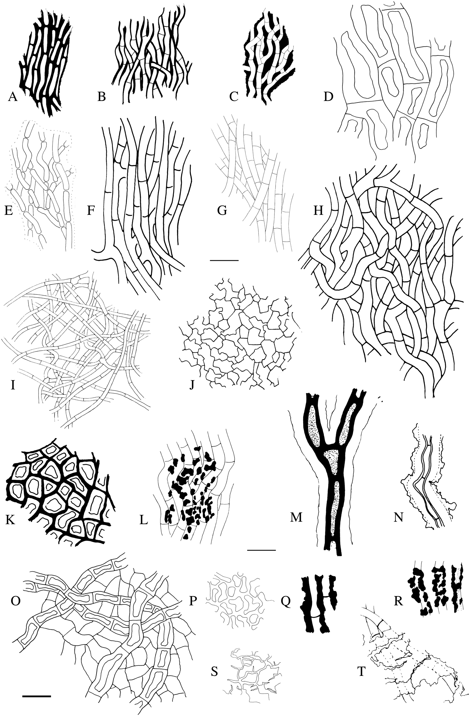
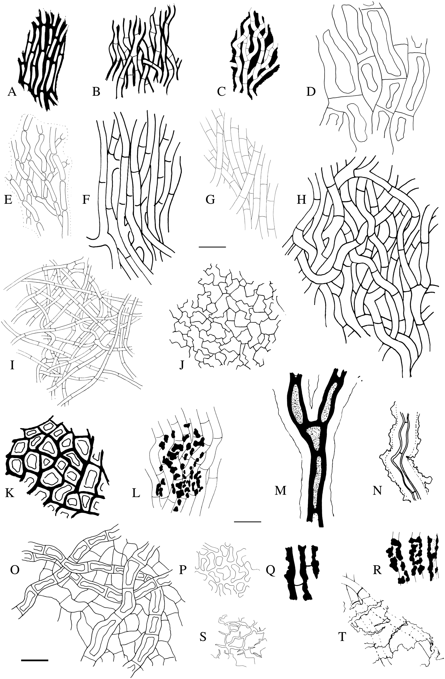
Diagrammatic representations of textura types and pigmentation in the excipulum and stipe of mycocalicioid fungi. Textura types: D & L, textura prismatica (elongated cells in hyphae, more or less periclinal), note that strictly prismatic textura seems to be rare in mycocalicioid species, and their hyphae tend to be at least slightly intertwined producing intermediate forms between prismatica and intricata (A–C, E–G). C, H & I, textura intricata (hyphae intertwined). K & O, textura angularis (cells angular or isodiametric). J & P, textura epidermoidea (irregularly or puzzle-like shaped cells). E, I & P, textura oblita (fused cell walls, note that textura oblita also occurs in intricata and epidermoidea textura types). Septa of hyphae may be either thin (e.g. G–J, N) or thick (e.g. A, D, M) and hyphal walls may be thin (e.g. G, L) or thick (D, M, N). A net of branching hyphae may occasionally cover the excipulum (O). Pigmentation: L, N, Q & R show accumulation of pigment as irregular lumps and pieces on the hyphal walls of the outer layer of the excipulum. S & T, glass-like surface material that breaks down in squash mounts. Species and voucher information can be found in Supplementary Material Table S1 (available online). Scales = 10 μm.
Textura types and hyphal characters
In previous treatments of mycocalicioid fungi, the hyphal cells have usually been described only as elongated or isodiametric. However, the diversity of textura types exceeds this rather crude classification. Some of the textura types were common in both the excipulum and the stipe and some were observed in only one or the other. Some typical textura types of mycocalicioid fungi are illustrated in Fig. 2.
The textura type of the excipulum and stipe in a species may be the same or different. If the same, the hyphae are often somewhat wider and cell walls thicker in the stipe. The textura types are species-specific and provide some of the best distinguishing characteristics in mycocalicioid fungi, especially when combined with hyphal measurements. The observed width of the hyphae varied considerably between species from thin (Fig. 2F) to very thick (Fig. 2D). Commonly the hyphae vary between 2.0 and 6.0 μm, but even 8.0 to 12.0 μm were observed. The hyphal walls were typically at least 0.5 μm and usually around 1.5 μm thick, but species with cell walls up to 6.0 μm thick were encountered. In rare cases, the thick-walled hyphae of the stipe are distinctively two-layered: the outer wall of the hyphae is thick and the inner wall thin, and often one or the other is more strongly coloured (Fig. 2M). The textura types and hyphal measurements are not dependent on each other, so two species may have the same textura type but have distinctively different hyphal measurements. The hyphae in the hypothecium and in the inner part of the stipe are usually thin walled, or at least thinner than in the outer layers. The hyphal width is typically the same or close to the measurements of the hyphae in the outer layers.
The surface of the outer excipulum and/or stipe is sometimes covered by a clearly distinguishable, sparse or densely intertwined net of arching to horizontal hyphae (Fig. 2O). This structure is usually clearly distinguishable from the excipulum and stipe. The hyphal net can sometimes cover the whole ascoma but is more commonly restricted to the upper part of the stipe and lower part of the capitulum. All classical textura types of ascomycetes (see e.g. Eckblad Reference Eckblad1968) were encountered during the study. Textura oblita is usually considered to have hyphae arranged parallel to each other, but in mycocalicioid species the fused, thick cell walls typical for textura oblita occur also in intricata and epidermoidea textura types.
Epithecium
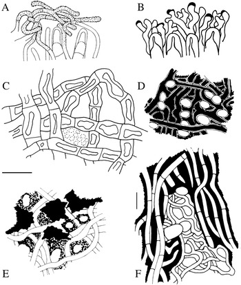
The structure of the epithecium differs from the structural types of the stipe and the excipulum. In our studies we found three different basic structures of the epithecium: formed by paraphyses only (Fig. 3A & B), paraphyses and excipular hyphae (Fig. 3F), or excipular hyphae only (Fig. 3C–E). Some of these epithecium types are illustrated in Fig. 3. One species studied, Mycocalicium calicioides (Nádv) Tibell, did not have any kind of epithecium. Epithecia consisting of both paraphyses and excipular hyphae are usually extremely tight structures and very difficult to break in squash preparations, especially in types where the paraphyses are longer than the asci and form a complex net below the excipular hyphae and/or have amorphous material in the hymenium and/or epithecium.
Diagrammatic representations of epithecium types of mycocalicioid fungi. A & B, paraphyses only, A, paraphyses exceeding and covering asci and branching and B, crown-like, branched tips of paraphyses. C & E, a (loose) net of excipular hyphae. D, excipular hyphae with a very tight, even surface with small holes. F, both paraphyses and excipular hyphae with small holes. Note the amorphous material covering the epithecium in E. Species and voucher information can be found in Supplementary Material Table S1 (available online). Scales = 10 μm; note that the 10 μm scale in F is different.

Paraphyses
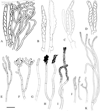
The paraphyses of mycocalicioid fungi are hyaline, and distinct pigments or colours were not observed in any of the species studied. The paraphyses are long and slender, usually 1.0–2.0 μm wide, thin-walled and septate. The septal intervals vary within and between species. The septation was found to be a rather imprecise character for species identification, but it has some consistency in the range of septal intervals. Although in many mycocalicioid species the paraphyses lack differentiating value, some features of paraphyses, when present, are species-specific and can be highly informative. These include the presence and type of branching (Fig. 4E, F & J), surface incrustations and crystals (Fig. 4G–K), and the way the paraphyses contribute to the epithecium (Fig. 3A, B & F). Some characteristic types of paraphyses are illustrated in Fig. 4.
Diagrammatic representations of the asci and paraphyses of mycocalicioid fungi. Asci: A, B & D, with croziers. C, with simple septa (i.e. without croziers). B, covered with amorphous material. Paraphyses: G, unbranched. H & I, branched in the lower part. A, E & J, branched in the upper part. F, branched in the upper part and crown-like. A, E & F, smooth. G & K, covered with different types of crystals, warts or amorphous material. F, apices thickened. Note that the basal cell of a paraphysis is usually indistinct. Species and voucher information can be found in Supplementary Material Table S1 (available online). Scale = 10 μm.

Asci
The asci of all mycocalicioid species are thought to arise from croziers. However, three (sub)tropical species occurring on angiosperm exudates, Chaenothecopsis khayensis Rikkinen & Tuovila, C. pallida Rikkinen & Tuovila and C. resinophila Rikkinen & Tuovila, produce asci without croziers. With careful microscopy and appropriate reagents (we recommend CR) this feature is evident. All known mycocalicioid fungi have cylindrical asci and the ascus form varies little, if at all, between fresh material and dry herbarium specimens. This is in contrast to some other groups of ascomycetes that have a different form and often also a different ascospore orientation in living asci (Baral Reference Baral1992).
Ascus apex
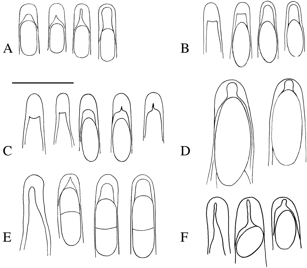
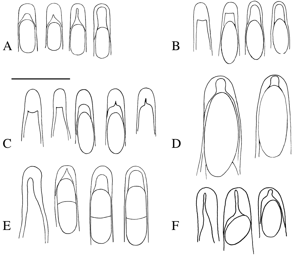
The type of ascus apex in mycocalicioid species varies between species and usually the appearance changes during ascus maturation. In this study several intermediate forms (e.g. Fig. 5C) between the traditional Mycocalicium and Chaenothecopsis types (e.g. Schmidt Reference Schmidt1970) were observed. The distinctive canal reported in most species of Chaenothecopsis (Fig. 5A, second apex from the right) is best seen in dried herbarium specimens. In fresh material, the thickening of the ascus apex is often less pronounced and the characteristic canal appears only after adding a reagent such as KOH or MLZ, which kills the specimen. Whereas the ascus apex type is usually identical in closely related species (e.g. Tuovila et al. Reference Tuovila, Davey, Yan, Huhtinen and Rikkinen2014), in some species the ascus apex has value in species identification. These include the basic structural type, if and how the apex changes during maturation, and the relative size of the ascus apex. Some of the variation in ascus apex anatomy and the canal types are illustrated in Fig. 5.
Diagrammatic representations of the variation in ascus apex in mycocalicioid fungi. Presence of canal: A, D & F, Schmidt's Chaenothecopsis-type with distinct canal in developing ascus tip, narrow (A, F), wide (D), note the canal is present at some stages of development. Thickening of tip: A–C, E, with apex more or less distinctively thickened in young, immature asci. C, Schmidt's Mycocalicium-type with thickened ascus tip, in which the apex is more or less evenly thickened at all stages of ascus development, mature asci sometimes with a faint fracture-like canal. B, vanishing-type ascus apex in which the apex is strongly and evenly thickened in very young to semi-mature stages, but the thickening disappears gradually without the formation of a canal, and mature asci are without thickening. Note that several different intermediate types between the Chaenothecopsis, Mycocalicium and vanishing-types (E, F) can be found. In these a poorly developed canal may be visible, and sometimes observable only with cytoplasm staining (e.g. CRB) reagents and with high magnification. In all examples the asci are arranged by age, youngest on the left, all stages not always presented. Species and voucher information can be found in Supplementary Material Table S1 (available online). Scales = 10 μm.
Ascospores
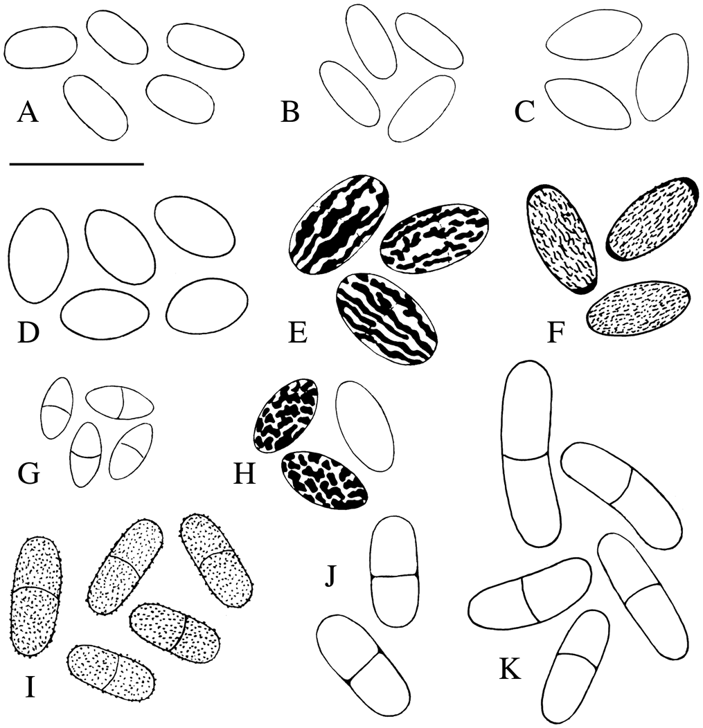
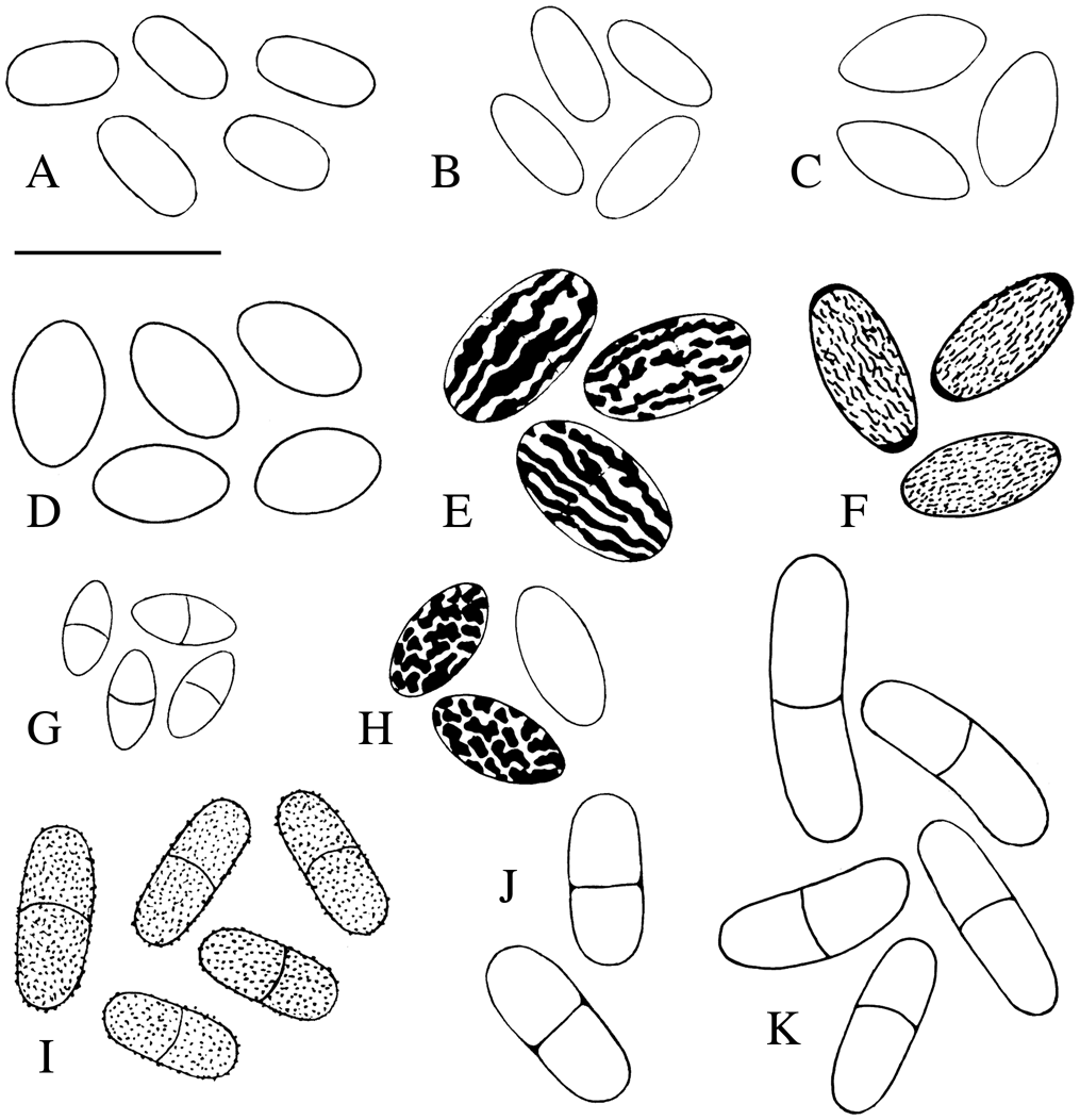
Ascospores have the most important and phylogenetically informative characters in mycocalicioid species. Several spore characters were found to be informative and especially important in species identification: shape, size, ornamentation, and the Q-value (spore length/width, measured from the same spore). Some common ascospore types are illustrated in Fig. 6. The spore length in mycocalicioid species ranges from 3 μm in some Chaenothecopsis up to 75 μm in some Stenocybe species, with spore width between 2–25 μm (Titov Reference Titov2006; Hawksworth et al. Reference Hawksworth, Aguirre-Hudson and Ainsworth2014). Even though the interspecific variation of spore size is quite large, the measured spore lengths of two species can be almost identical. In such cases, the Q-value may differ between species and can help in their identification.
Diagrammatic representations of some spore types of mycocalicioid fungi: A–F & H, aseptate. G & I–K, septate. A, I & J, cylindrical. B, E & F, ellipsoid. C, D, G & H, fusoid. K, phaseoliform. Note the thickened walls at the spore apices (F) and septum thickened at the junction between septum and spore wall (J). Ornamentation, mounted in water: A–D, G & J–K smooth. I, finely spinulose to verrucose. E–F, ridged. H, panther-like. Species and voucher information can be found in Supplementary Material Table S1 (available online). Scale = 10 μm.
Ascospore morphology
Most mycocalicioid species have symmetric, bilateral spores, aseptate to up to 7-septate in some species. Usually the Q-value of the spores is more than 1 and only some species of Sphinctrina have more or less spherical spores (Löfgren & Tibell Reference Löfgren and Tibell1979). Cylindrical and ellipsoid were the most common spore types found in our studies, but others (e.g. fusoid, ovoid and phaseoliform) were also encountered. Tibell (Reference Tibell1982, Reference Tibell1984) was the first to bring attention to the differences in the thickness and appearance of the septum. In some species the septum is thin and lighter than the outer wall (referred to as ‘poorly pigmented’ by Tibell), and this character is also species-specific. The thickness of the septa may also vary during the maturation of the spore. In addition, the septum can be as thick as or thicker than the outer wall, or thickened only at the junction of the septum and outer wall.
Ascospore surfaces
Ascospore surfaces vary from smooth (Fig. 6A–D, J & K) to distinctively ornamented. When present, spore ornamentation is a good character for species identification. Three basic types of ornamentation were observed in the specimens studied: (1) more or less evenly distributed single warts (Fig. 6I), (2) continuous to discontinuous ridges (Fig. 6E & F), and (3) irregularly shaped plates that almost entirely cover the spore surface (Fig. 6H). In some species the developing ornamentation is already visible in immature spores, although fully developed only in mature spores. In other species the ornamentation develops very late and is visible only in mature spores. Although the ornamentation is a good and species-specific character, there seems to be, at least in some species, varying ratios of smooth and ornamented spores between ascomata of the same population, most likely depending on the maturity of the ascoma.
Crystals
Many mycocalicioid species have coloured or hyaline crystals in their ascomata. Some species have crystals only in certain places (e.g. only in the stipe or covering the apices of the paraphyses), while in other species they are scattered throughout the ascoma. The presence, type and position of the crystals are species-specific. The occurrence of different crystals seems to depend on the maturity of the ascoma, for example, very young ascomata often lack crystals typical of the species. Most of the coloured crystals (usually yellow or red) have a distinct colour reaction to some reagents, mainly KOH. The hyaline crystals may be difficult to observe in KOH or water, and they may even dissolve in KOH. However, hyaline crystals are easy to observe in CR, since they usually turn bright red in the reagent (congophilous, CR+). Some species have an almost glass-like, amorphous material that covers the stipe, or sometimes even the whole ascoma, especially when young. In squash preparations the material breaks like ice (Fig. 2S & T) and makes the hyphal structure below difficult to observe. This material usually fully or partly dissolves in KOH and CR.
Chemical reactions and reagents
Colour reactions to potassium hydroxide (KOH) and nitric acid (NO₃) have traditionally been used in the identification of mycocalicioid species (e.g. Titov Reference Titov2006). Well-known reactions to KOH include the dramatic colour reactions of some species with unidentified, crystalline pigments. So far, the following reactions have been described (see e.g. Schmidt Reference Schmidt1970; Tibell Reference Tibell and Ahti1999): red pigment turning green, yellow/red pigment turning red, and yellow turning aniline/violet-red. KOH is also a bleaching reagent, and most of the colour reactions reported for different mycocalicioid species are actually pigments being removed from the cell walls, sometimes leaking into the medium, and eventually fading. Additionally, the hyphae of many mycocalicioid species swell in KOH and the swelling is especially pronounced in species with textura oblita. For this reason, spores and other structures should not be measured in KOH. A reddish colour reaction to nitric acid can be seen in some Chaenothecopsis species, but in most species the colours are not affected or are only slightly intensified. A brown and/or green colour leaking into the medium is a relatively common KOH reaction in mycocalicioid species, and as such is relatively uninformative. However, whether the colour of the hyphal walls leaks or not is a species-specific reaction, and for this reason important to note and report.
Different staining reagents, mainly CRB and CR help to distinguish thin and hyaline structures (e.g. paraphyses, ascus apex characters and croziers). CR usually dissolves the glass-like layer (see above), making the textura type below easier to observe. Some types of hyaline crystals and lightly coloured or hyaline amorphous material of the epithecium and hymenium are also often congophilous and easier to observe.
Iodine reagents and reactions
Lugol's and Melzer's reagents, the two commonly used iodine solutions, yield different reactions and should not be confused with each other (Baral Reference Baral1987). The euamyloid (blue) and dextrinoid (red, sometimes referred to as pseudoamyloid) reactions induced by MLZ are independent of KOH and can be seen with or without KOH pretreatment. These reactions can also be observed by adding MLZ to a squash preparation after IKI has first been applied. The hemiamyloid reaction is usually a red to brownish red reaction but it can also be blue, depending on the iodine concentration of the IKI solution (Baral Reference Baral1987). The hemiamyloid reaction can be observed only with IKI and, by definition, disappears if MLZ is added. Also, by definition, the hemiamyloid reaction cannot be seen if the preparation is first pretreated with KOH. The pretreatment with KOH yields the same (blue) reaction with IKI and MLZ. The euamyloid blue reaction can be induced with both MLZ and IKI, and it is important to test with both reagents to know whether the blue in IKI is hemiamyloid or euamyloid. For a more detailed analysis of these reactions, see Baral (Reference Baral1987).
In the species studied, the euamyloid (blue with MLZ and IKI) reaction was rarer than IKI-specific reactions, and it was observed only in the hymenium. The euamyloid reaction is quite persistent and the observed colour varies from light to dark inky blue. The dextrinoid reaction in MLZ (Baral Reference Baral1987) was not observed in any of the species studied.
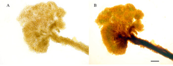
In our studies, two different colour reactions were observed with Lugol's reagent. These reactions seem to be highly sensitive to the freshness of IKI and are usually best seen with freshly made reagent (diluted from stock solution). Firstly, a persistent red reaction may be present in hyphae of the stipe, excipulum and epithecium. The red IKI reaction appears to be most common in the stipe, either in the whole stipe or just at the base. If present in the excipulum, it is usually also present in the stipe, and if present in the epithecium, it is also present in the excipulum and stipe. The observed colour varies from faint greyish pink to strong crimson red, but the shade depends also on the freshness of IKI. The red reaction is often surprisingly clear in IKI (Fig. 7), even though the excipulum and stipe are predominantly very dark in this group. The red colour reaction of excipular tissue was present in the majority of specimens examined. In some species the reaction is absent and in others it is almost impossible to observe, mainly in species with very dark colours. In such species, a faint reddish tint in the extreme basal cells could sometimes be seen.
Pseudohemiamyloid reaction. A, Chaenothecopsis nana Tibell (Alava 21983, TUR 55759) in KOH. B, the same ascoma of C. nana in Lugol's reagent (IKI) after repeated changes between KOH and IKI. The pseudohemiamyloid reaction visible in the excipulum and stipe (red) and hymenium (blue). Scales = 0.1 mm.
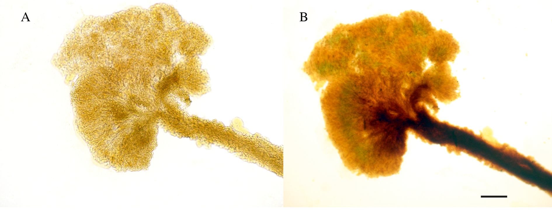
A second reaction, disappearing blue, is present in the hymenium and often also in the hypothecium. In the specimens examined during our studies, this colour reaction was never observed in the excipulum, stipe or epithecium. The observed colour is usually light blue but can vary from faint bluish grey to intense inky blue. The speed of the reaction varies from very fast and transient to nearly persisting, and in some cases the reaction seems to persist almost indefinitely. This reaction is easily missed but can best be observed by first mounting the specimen in water, and after the preparation has partially dried (air has penetrated under the coverslip), a drop of freshly made 1% Lugol's reagent is added to the edge of the coverslip while simultaneously observing the specimen at ×40 magnification. The reaction occurs when the reagent penetrates the squashed ascomata. Sometimes it appears only as a rapidly moving blue ring and may last just a few seconds or even less.
We first considered these red and blue reactions as hemiamyloidity, as described by Baral (Reference Baral1987), and the following articles have described them as such: Tuovila et al. (Reference Tuovila, Cobbinah and Rikkinen2011a, Reference Tuovila, Larsson and Rikkinenb, Reference Tuovila, Schmidt, Beimforde, Dörfelt, Grabenhorst and Rikkinen2013, Reference Tuovila, Davey, Yan, Huhtinen and Rikkinen2014), Rikkinen et al. (Reference Rikkinen, Tuovila, Beimforde, Seyfullah, Perrichot and Schmidt2014), Selva & Tuovila (Reference Selva and Tuovila2016) and Beimforde et al. (Reference Beimforde, Tuovila, Schmidt, Lee, Gube and Rikkinen2017). However, we have recently discovered that these reactions differ from Baral's definition of hemiamyloidity. For this reason, we describe here a new iodine reaction, the pseudohemiamyloid reaction. This is a reversible, KOH resistant/enhanced IKI reaction. It can easily be mistaken for a hemiamyloid reaction, since it disappears with MLZ and can be seen only with IKI. The main and most important difference between this and a hemiamyloid reaction is the reaction to KOH pretreatment.
According to Baral (Reference Baral1987), KOH pretreatment turns all hemiamyloid reactions to euamyloid (the structures that are red in IKI are blue with KOH + IKI or KOH + MLZ). However, our observations revealed that this rule does not apply to mycocalicioid species where the IKI reactions are reversible and KOH resistant, and usually even enhanced with KOH. The blue and/or red colours disappear if IKI is washed away with excess water or MLZ. However, the colours reappear when KOH and IKI (in this order) are added. In our studies we tested different combinations of adding water, KOH, IKI and MLZ and the results were always the same: the red and blue reactions were always visible after adding KOH and IKI (Fig. 7) and they disappeared/were not visible in MLZ. In addition, the red reaction never turned blue with KOH pretreatment as in hemiamyloidity. Since this is in contrast to Baral's description of a hemiamyloid reaction, we consider the blue and red reactions found in our studies to be something different. Although these blue and red reactions are independent of each other, we still consider them to be the same reaction because they react identically to IKI, MLZ and KOH. In species descriptions it should be noted whether it is a blue or red pseudohemiamyloid reaction. To our knowledge, these reactions have not been previously described in any other group of fungi.
Both the red and blue pseudohemiamyloid reactions present in specimens with a water + IKI treatment were present and usually enhanced with KOH pretreatment. One possible reason is that pseudohemiamyloid reactions are easier to observe in structures bleached with KOH. In a few cases, the pseudohemiamyloid reaction was not observed with the water + IKI treatment, but was strong with the KOH pretreatment. In all specimens observed, the disappearing blue reaction to IKI (see above) was more stable (usually not disappearing) with KOH pretreatment. We recommend testing this reaction in mycocalicioid fungi first with IKI only (as described above) and then with KOH pretreatment (KOH + IKI), and after observing the reaction, to add MLZ to test the amyloid reaction.
Discussion
There is a real need for user-friendly practical methodologies to identify fungi, in a range of settings which do not have access to more advanced technologies. Taxonomic studies based on DNA are often at odds with the morphological differences previously found between species. Oftentimes, detailed comparative analyses of the morphology, chemistry and ecology of proposed cryptic species have revealed previously overlooked characteristics (Del-Prado et al. Reference Del-Prado, Ferencová, Armas-Crespo, Amo de Paz, Cubas and Crespo2007; Garrido-Benavent & Pérez-Ortega Reference Garrido-Benavent and Pérez-Ortega2015). This together with the re-examination of previously misunderstood characters of fungi, as was suggested, for example, by Schmitt (Reference Schmitt, Pöggeler and Wöstermeyer2011), would aid in species delimitation without recourse to DNA analyses.
The phylogenetic relationships of Mycocaliciales with other groups of ascomycetes are relatively well known (Geiser et al. Reference Geiser, Gueidan, Miadlikowska, Lutzoni, Hofstetter, Fraker, Schoch, Tibell, Untereiner and Gueidan2006; Prieto et al. Reference Prieto, Baloch, Tehler and Wedin2013). The order seems to have diverged early and is fairly distinct from other members of the Eurotiomycetes (Prieto & Wedin Reference Prieto and Wedin2013; Prieto et al. Reference Prieto, Baloch, Tehler and Wedin2013). Although mycocalicioid fungi share some morphological and ecological characters with their closest relatives in the Eurotiomycetidae and Dothideomycetidae, the distinctiveness of several characters supports their suggested position on a supraordinal (Mycocaliciomycetidae) level (Hibbett et al. Reference Hibbett, Binder, Bischoff, Blackwell, Cannon, Eriksson, Huhndorf, James, Kirk and Lücking2007).
The traditional classification of the Mycocalicium-type and Chaenothecopsis-type morphology (Schmidt Reference Schmidt1970) is oversimplified because a wide range of intermediate forms have been observed in recent decades (Titov & Tibell Reference Titov and Tibell1993; Titov Reference Titov1994; Tibell Reference Tibell1998; Rikkinen et al. Reference Rikkinen, Tuovila, Beimforde, Seyfullah, Perrichot and Schmidt2014). Schmidt differentiated species into four genera of Mycocaliciaceae (excluding Sphinctrina) on the basis of ascus measurements, ascus apex characters, spore septation and hyphal structures of the stipe. These generic delimitations were based on a rather limited number of species and specimens, which apparently skewed some of his conclusions about the morphological variation within the group (Hawksworth Reference Hawksworth1980). As noted already by Samuels & Buchanan (Reference Samuels and Buchanan1983), the four genera cannot be differentiated by these, or any single character, and the current system has clearly become outdated.
Long held tradition and well-established, widely used practices have sometimes created bias in taxonomists who have been reluctant to change. One good example in mycocalicioid fungi is the treatment of Phaeocalicium tibellii Kalb. The genus Phaeocalicium has been described as having ‘periclinally arranged or isodiametric hyphae’ in the excipulum (e.g. Tibell Reference Tibell1996). However, a very different type of structure, textura epidermoidea, was beautifully illustrated and described in P. tibellii by Kalb (Kalb & Hafellner Reference Kalb and Hafellner1992) but was later rejected by Titov as an artefact (Titov Reference Titov2006).
The structure of the ascus and ascus apex of mycocalicioid fungi lack the typical iodine reactions of ascus apices of classical inoperculate and operculate ascomycetes. While the anatomy of the ascus apex cannot be used for currently used generic delimitation, there is consistency in the structure between closely related species (e.g. Tuovila et al. Reference Tuovila, Davey, Yan, Huhtinen and Rikkinen2014). In addition, there seems to be more variation in the structure than previously thought (Fig. 5) and it may yet prove to be a valuable character at the generic level, as soon as more accurate phylogenies are available.
Different iodine reactions are usually species-specific and are widely used for identifying and delimiting species in many fungal groups. However, they have not been systematically used in the characterization of mycocalicioid fungi. These results demonstrate that the different iodine reactions (MLZ/IKI) are an important addition to the chemical characters of mycocalicioid fungi. Although Harder et al. (Reference Harder, Lodge, Petersen, Hughes, Blanco, Frøslev and Læssøe2011) question the reliability of a strictly dichotomic use of amyloid reactions for delimitating species and genera, as long as there is variation in these reactions they should not be overlooked. In this study, the pseudohemiamyloid reaction was somewhat more common than the non-amyloid (inamyloid) or euamyloid reaction, but it remains to be seen how common these iodine reactions are in mycocalicioid fungi.
All the characteristics discussed here can be seen using a regular compound microscope. However, it is essential that the ×100 oil-immersion objective be used. One of our main conclusions is that no single morphological, anatomical or chemical characteristic is enough to distinguish between species (see also Tuovila et al. Reference Tuovila, Davey, Yan, Huhtinen and Rikkinen2014) and that a combination of several characteristics is usually required for identification.
As Heller et al. (Reference Heller, Frandsen, Lorenzen and Siegismund2014) point out, morphology, and evolutionary and ecological processes are all likely to be linked together, although the mechanism is as yet unknown. While our data are insufficient to resolve the generic delimitations of mycocalicioid fungi, some trends in our phylogenetic analyses are recurring. Ascospore form, septation and, to some degree, the textura type of the excipulum, all seem to have strong phylogenetic signals (Tuovila Reference Tuovila2013). Also, on a smaller scale, the red and yellow crystals with strong KOH reactions seem to have some phylogenetic value. These reactions have been described mainly from species with lichen/algal connections. Interestingly, these species seem to belong to two evolutionarily different lineages, one with septate spores and one with aseptate spores. Although it will take time to find a satisfactory phylogeny, we have some promising preliminary results of smaller subclades that show reliable relationships and consistency in morphological characters. For instance, the ‘Mycocalicium sp.’ in Vinuesa et al. (Reference Vinuesa, Sanches-Puelles and Tibell2001) grouped strongly in our analyses (results not shown) with Chaenothecopsis nana and other closely related, undescribed species, all of which had a classic Chaenothecopsis-type ascus apex (also noted in Vinuesa et al. (Reference Vinuesa, Sanches-Puelles and Tibell2001)) and textura epidermoidea in the excipulum.
Finding a reliable phylogeny of any group of organisms can be time-consuming, and linking morphological characters with a molecular phylogeny can be even more difficult and may be contentious, especially if the suggested changes are radical (Heller et al. Reference Heller, Frandsen, Lorenzen and Siegismund2013; Zachos & Lovari Reference Zachos and Lovari2013; Cotterill et al. Reference Cotterill, Taylor, Gippoliti, Bishop and Groves2014). One may argue over whether a population or a clade characterized by a single shared character is worth being taxonomically delimited, or even over how to define a species. However, few question the notion that more data are needed. De Queiroz (Reference De Queiroz2007) emphasizes the need to accumulate evidence from multiple, diagnosable traits. As the number of traits increases, it becomes statistically more likely that the species recognized are in fact distinct. Our main objective in this article is to identify additional morphological characters that can be used to help interpret interspecific variation. We acknowledge that this is not an exhaustive study of the morphological variation in mycocalicioid species but, nevertheless, our results show several potential morphological and histochemical features which could be used for delimiting and identifying mycocalicioid species. These results could be a baseline upon which colleagues could suggest further contributions to the elucidation of a reliable phylogeny of mycocalicioid fungi.
Acknowledgements
This study was partly conducted during the Ph.D. studies of HT at the University of Helsinki and partly in the project ‘Conservation of wood-inhabiting Ascomycetes in changing forest landscapes’ at the University of Jyväskylä. These projects were funded by the Jenny and Antti Wihuri Foundation, the Ella and Georg Ehrnrooth Foundation and the Oskar Öflund Foundation, and by the Ministry of the Environment (PUTTE grant), respectively. Håkon Holien, Steven Selva and Jouko Rikkinen kindly loaned their specimens. We thank the curators of O and UPS for their help with the specimens, and the editors and two referees for valuable comments and suggestions that helped to greatly improve our paper. We are grateful for the opportunity to take an interesting plunge into an unknown area of poorly known fungi.
Author ORCID
Hanna Tuovila, 0000-0001-8875-6977.
Supplementary Material
To view Supplementary Material for this article, please visit https://doi.org/10.1017/S0024282920000481.