INTRODUCTION
Whether conducting behavioural research or establishing population parameters, photo-identification is generally regarded as the most effective, non-invasive method available to researchers for gathering information about cetacean societies in the wild (Evans & Hammond, Reference Evans and Hammond2004). In many dolphin species, individual animals can be reliably distinguished by the unique position and shape of presenting nicks or dorsal edge marks (DEMs) (Würsig & Würsig, Reference Würsig and Würsig1977; Hammond et al., Reference Hammond, Mizroch and Donovan1990), facilitating their recapture over time. Such mark capture-recapture studies have been instrumental to our present understanding of the biology, behaviour, health and ranging patterns of studied populations (e.g. Thompson & Hammond, Reference Thompson and Hammond1992; Baird & Whitehead, Reference Baird and Whitehead2000; Kreb, Reference Kreb2004; Auger-Méthé & Whitehead, Reference Auger-Méthé and Whitehead2007; Robinson et al., Reference Robinson, O'Brien, Berrow, Cheney, Costa, Eisfeld, Haberlin, Mandleberg, O'Donovan, Oudejans, Ryan, Stevick, Thompson and Whooley2012). However, not all delphinids are well-suited to photo-identification, due to their practical inaccessibility in deep, offshore waters, their highly gregarious nature (with some species occurring in group sizes of several hundred to thousands even), or simply because of low numbers of naturally marked individuals in some species.
The short-beaked common dolphin (Delphinus delphis L.) can be notoriously difficult to study for all of the above reasons and, as a result, many of the basic parameters of studied populations, such as the size, distribution, home range and social structure for example, remain poorly understood (Murphy et al., Reference Murphy, Winship, Dabin, Jepson, Deaville, Reid, Spurrier, Rogan, López, González, Read, Addink, Silva, Ridoux, Learmonth, Pierce and Northridge2009). Only a low percentage of individuals are thought to exhibit useful, distinguishing marks for mark-recapture analyses. Nevertheless, photo-identification catalogues exist for the species in several parts of the world (e.g. Neumann et al., Reference Neumann, Leitenberger and Orams2002a; Bearzi et al., Reference Bearzi, Politi, Agazzi, Bruno, Costa and Bonizzoni2005) and DEMs (in the form of tears, notches and indents), fin colouration, pigmentation patterns and physical deformities have all been used for the recognition of individual common dolphins.
Both natural and anthropogenic sources have been identified in the formation of DEMs in delphinids, including social, intra-specific interactions (e.g. Scott et al., Reference Scott, Mann, Watson-Capps, Sargeant and Connor2005), interactions with other cetacean species and predators (e.g. Corkeron et al., Reference Corkeron, Morris and Bryden1987; Jefferson et al., Reference Jefferson, Stacey and Baird1991; Wedekin et al., Reference Wedekin, Daura-Jorge and Simões-Lopes2004), boat strikes and propeller wounds (e.g. Wells & Scott, Reference Wells and Scott1997), non-lethal interactions with fishing gears (e.g. Read, Reference Read2008) and even scarring from biopsies and tagging studies (e.g. Scott et al., Reference Scott, Wells, Irvine, Mate, Leatherwood and Reeves1990; Bearzi, Reference Bearzi2000). In the present study, two common dolphin identification catalogues from the Bay of Gibraltar and the Moray Firth in north-east Scotland were examined to document the relative frequency, distribution and shape of presenting DEMs in selectively ‘marked’ individuals and to investigate the potential causes responsible for their occurrence.
METHODS
Individually recognizable dolphins were examined from two long-term photo archives opportunistically collected during dedicated boat surveys in the Bay of Gibraltar (GIB) from 2001 to 2008 (P Linares, unpublished data) and the Moray Firth (MF) from 2009 to 2013 (Robinson et al., Reference Robinson, Eisfeld, Costa and Simmonds2010). Marked individuals exhibiting at least one or more prominent DEMs were selected from each archive. Thereafter, the best available right or left image for each individual was scored for image quality (after Urain et al., Reference Urain, Hohn and Hansen1999):
 $${\rm Dorsal\; image\; quality}\; = \; \displaystyle{{(C1 + C2 + \; A + PV + \; P)} \over 3}$$
(with each numeral being scored between 1 and 3)
where C1 = clarity, C2 = contrast, A = angle, PV = proportion of fin visible and P = proportion of the frame filled by the fin, with each feature being assigned a value between 1 and 3 (1 representing the lowest and 3 the highest quality in each case). Accordingly, only individuals with a score of 3 or above were used in the following analysis.
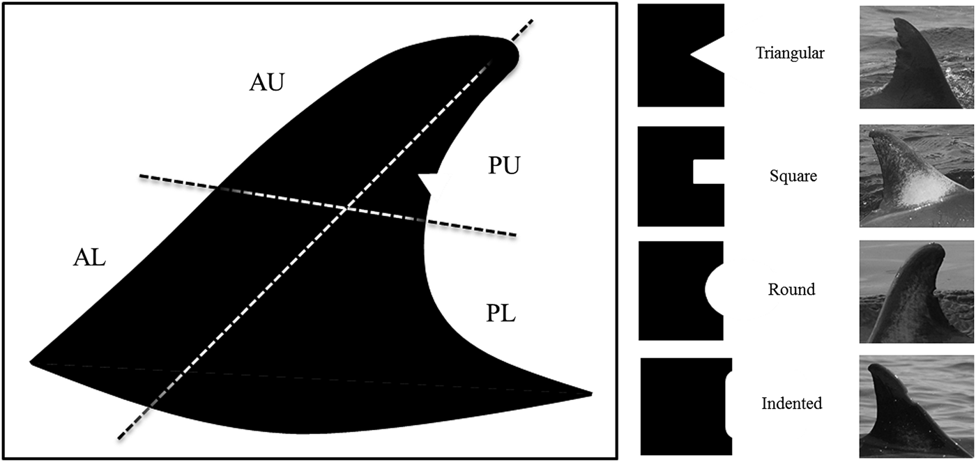
A total of 617 quality images of individually marked animals were subsequently selected – 520 from the larger GIB dataset and 97 from the smaller MF dataset – from which thorough counts of all presenting DEMs were made. A dorsal fin layout system (adapted from Tetley et al., Reference Tetley, Baumgartner, Björgvinsson, Robinson and Wald2007) was used to map the respective positions and shapes of observed DEMs along the anterior/posterior and upper/lower margin of the fin (Figure 1). Differences in the frequency and occurrence of DEMs within and between the respective GIB and MF datasets were evaluated using Chi-squared tests. Individuals were further inspected for markings potentially inflicted by fishing gear and were subsequently categorized as ‘consistent’, ‘likely consistent’ or ‘not consistent’ with fisheries interactions (after Baird et al., Reference Baird, Mahaffy, Gorgone, Cullins, McSweeney, Oleson, Bradford, Barlow and Webster2015). Where information was available on the sex of individuals – from observations of the post-anal hump in males or the presence of young calves in females – evidence for a sex bias in the number and type of presenting nicks was also examined.
Showing the dorsal fin layout system used to document and assign the relative positions (AU, anterior upper (AU) and lower (AL) and posterior upper (PU) and lower (PL)) and shapes (triangular, square, round or indented) of presenting DEMs in the common dolphin.

RESULTS
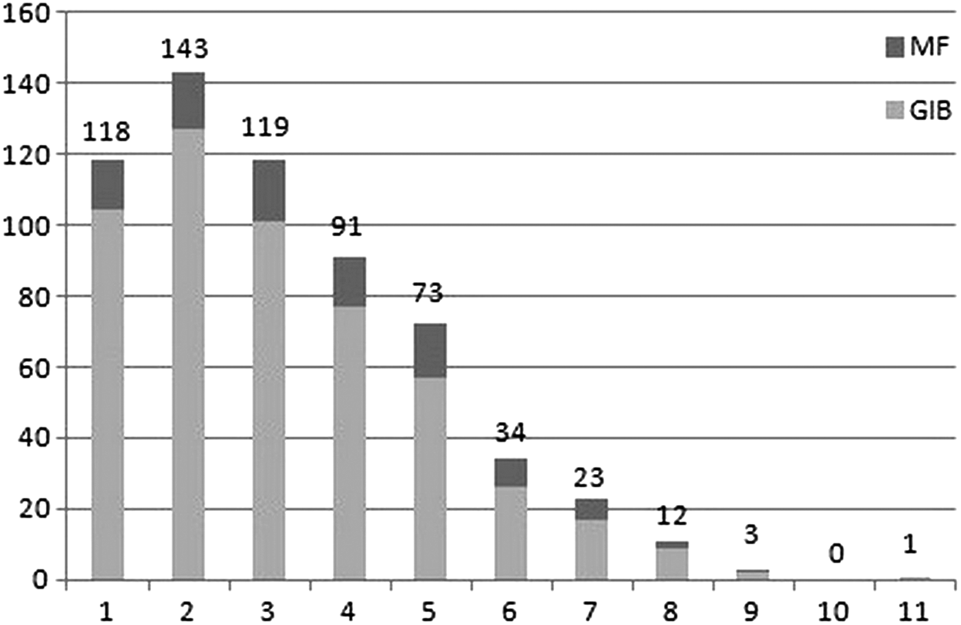
From the 617 common dolphins examined in the present study, a total count of 1989 DEMs was made. The number of DEMs in individual animals ranged from 1 to 11 (median = 3, mean = 3.2 ± 1.9), with almost 70% of all dolphins exhibiting between 2 and 5 nicks (Figure 2). 97.3% of all the dolphins examined exhibited one or more nicks in the posterior dorsal margin, with 87% of animals exclusively displaying DEMs in the posterior margin only. Conversely, 13.1% of animals exhibited DEMs in the anterior dorsal edge, with just 2.4% exhibiting nicks in the anterior fin margin alone.
Histogram showing the range and number of DEMs exhibited by individual ‘marked' common dolphins from the Bay of Gibraltar (GIB) and Moray Firth (MF) (N = 617).
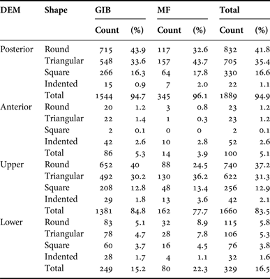
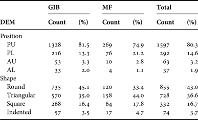
A chi-squared test revealed that DEM positions did not occur by chance in either GIB or MF populations (χ2 = 15.7, df = 3, P = 0.0025). Indeed, almost 95% of all DEMs observed in the pooled dataset occurred along the posterior margin of the dorsal fin, and 83.5% were positioned in the upper region of examined fins (Table 1). The highest frequency of nicks in both MF and GIB datasets were subsequently recorded in the upper posterior (PU) region of the dorsal fin, with the lowest frequency being observed in the lower anterior (AL) region (Table 2).
The occurrence and frequency of dorsal edge mark (DEM) shapes in the posterior, anterior, upper and lower regions of the dorsal fin in ‘marked’ common dolphins from Gibraltar (GIB) and the Moray Firth (MF) respectively (GIB: N = 1630; MF: N = 359).
Summarizing the distribution of DEMs and the presenting shapes observed in common dolphins from Gibraltar and the Moray Firth (N = 1989) (PU posterior upper, PL posterior lower, AU anterior upper and AL anterior lower).
The distribution of DEM shapes also occurred unevenly within populations (χ2 = 17.4, df = 3, P = 0.001). In both GIB and MF datasets, approximately 80% of the DEMs observed were either round or triangular in profile (Table 2), although round nicks were proportionally higher than triangular nicks in the GIB dataset and vice versa in the MF dataset. Round, triangular and, to a lesser extent, square (or notched) DEMs were also recorded in significantly higher proportions along the posterior dorsal edge than the anterior edge (Table 2). In contrast, indented DEMs, though generally seen in much lower numbers than all other nick shapes, were observed in higher numbers along the anterior dorsal than the posterior dorsal edges, and were seen to represent more than half of all the DEMs occurring along the anterior fin.
A high percentage of animals from both the GIB and MF exhibited DEMs, injuries or disfigurements that were ‘consistent’ or ‘likely consistent’ with fisheries interactions. In the MF dataset, 27% of the individuals examined showed evidence of damage from fishing gears, compared with 14% in the larger GIB dataset. Five animals from GIB had missing fins and a single animal from the MF had a collapsed/disfigured dorsal. However, information on the sex of these animals was not available. Gender confirmation was available for just 14% of the animals in the combined GIB and MF dataset. Subsequently, an analysis of sex bias in relation to fisheries interactions was not possible in this investigation. Nevertheless, the number of DEMs in identified adult females (mean 3.9 ± 1.9, range 1–7) (N = 17) was found to be proportionally lower than in confirmed adult males (mean 5.1 ± 2.0, range 1–11) (N = 69), which also exhibited more extensive body scarring than females and more generally ragged-looking fins.
Matching pigmentation patterns, on both the right and left hand sides of the dorsal fin, were further noted in this study, as also documented by Neumann et al. (Reference Neumann, Leitenberger and Orams2002a). This patterning varied widely between individuals (Figure 3) but was nevertheless widely prevalent in both GIB (68%, N = 356) and MF animals (65%, N = 63) and appeared to be consistent in recaptured individuals, during the duration of this study period at least, for photo-identification purposes.
Dorsal pigmentation patterns observed in D. delphis from the Bay of Gibraltar and Moray Firth. Degrees of pigmentation varied from mild to extensive (left to right).
DISCUSSION
As in other delphinids commonly studied using photo-identification methods, the common dolphin evidently exhibits a wide range and diversity of dorsal edge markings (DEMs). In the Bay of Gibraltar (GIB) and Moray Firth (MF) animals examined herein, a remarkable similarity was seen in the frequency, position and shapes of the DEMs observed. Of particular note were the high number of presenting DEMs in a large percentage of individuals from both datasets – the majority of animals exhibiting between two and five DEMs – and the predominant occurrence (~95%) of nicks in the posterior, trailing fin edge. The total counts and location of presenting DEM shapes were also highly comparable in both populations, indicating that the causal processes determining DEM formation are probably very similar.
Intraspecific aggression between rivals and associates is commonplace in odontocete societies (e.g. Kato, Reference Kato1984; Herzing, Reference Herzing1996; MacLeod, Reference MacLeod1998; Connor et al., Reference Connor, Read, Wrangham, Mann, Connor, Tyack and Whitehead2000; Robinson, Reference Robinson2014) and in the common dolphin sexual dimorphism and testes size signify a promiscuous mating system (Murphy et al., Reference Murphy, Collet and Rogan2005). Thus, physical exchanges between animals – in the form of biting, jawing, body slamming and tail hitting, characteristic of delphinids (e.g. Smuts & Smuts, Reference Smuts and Smuts1993; Herzing, Reference Herzing1996) – may account for a large proportion of the DEMs recorded in the species, particularly in adult males competing for female consortships. Indeed, where information on gender was available in the GIB and MF datasets, identified males in the present study not only exhibited more typically ragged fins with a greater number of nicks than adult females, but also showed a greater prevalance of body and dorsal fin scarring, as similarly reported in bottlenose dolphins (Tursiops truncatus Montagu) by Rowe & Dawson (Reference Rowe and Dawson2009). Male–female and female–female interactions in common dolphins are thus assumed to be less prolific than between-male interactions, suggesting female D. delphis are perhaps more tolerant and less competitive for food, territory and mates than rival males.
In addition to interactions between conspecifics, presenting DEMs might also arise from interactions with other species during which time interspecific agonistic exchanges between animals may occur. The common dolphin frequently associates in mixed-species groups with striped dolphins (Stenella coeruleoalba Meyen) for example (Frantzis & Herzing, Reference Frantzis and Herzing2002). However, interactions with Risso's dolphins (Grampus griseus Cuvier) (Frantzis & Herzing, Reference Frantzis and Herzing2002), killer whales (Orcinus orca L.) (Jefferson et al., Reference Jefferson, Stacey and Baird1991), tunafish (e.g. Das et al., Reference Das, Lepoint, Loizeau, Debacker, Dauby and Bouquegneau2000) and several shark species (Long & Jones, Reference Long, Jones, Klimley and Ainley1996) have additionally been reported, which might all be further implicated in this respect.
Since the posterior (trailing) edge of the dorsal fin is conspicuously thinner and more easily torn than the thicker, collagenous tissue forming the anterior (leading) edge, it is hardly surprising that the majority of DEMs observed in the present examination were located here. Interestingly however, over 13% of the animals examined from the GIB and MF datasets exhibited DEMs along the anterior (leading) margin of the fin (N = 100) in contrast to <1% of photo-identified bottlenose dolphins from the same geographic regions (Linares, unpublished data; Robinson et al., Reference Robinson, O'Brien, Berrow, Cheney, Costa, Eisfeld, Haberlin, Mandleberg, O'Donovan, Oudejans, Ryan, Stevick, Thompson and Whooley2012). More than 50% of the anterior edge nicks recorded in this study were also notably indented in shape, as if the dorsal fin had been struck by a solid object. A significant impact to the base of the anterior dorsal region could conceivably result in fin collapse through loss of structural support, and such injuries are commonly associated with fisheries interactions, as explicitly reported by Baird & Gorgone (Reference Baird and Gorgone2005) in false killer whales (Pseudorca crassidens Owen).
Common dolphins are known to be particularly vulnerable to by-catch (Morizur et al., Reference Morizur, Berrow, Tregenza, Couperus and Pouvreau1999; De Boer et al., Reference De Boer, Leaper, Keith and Simmonds2008) and dorsal fin disfigurements and/or mutilations in free-ranging or incidentally stranded animals are a clear sign of non-lethal entanglement in fishing gears (Kirkwood et al., Reference Kirkwood, Bennet, Jepson, Kuiken, Simpson and Baker1997; Kiszka et al., Reference Kiszka, Pelourdeau and Ridoux2008). In the present study, high percentages of the animals examined displayed DEMs that were ‘consistent’ or ‘likely consistent’ with fisheries interactions. Five individuals from the GIB photo-identification catalogue exhibited completely missing dorsal fins, which could have major implications for their survival and/or reproduction – as the dorsal fin is known to be important for thermoregulation, providing cooled blood to the female reproductive system (Rommel et al., Reference Rommel, Pabst and McLellan1993). However, observations in the Moray Firth in 2008 of a nursing common dolphin completely missing her dorsal fin may imply that, even with such major losses, adult females may still be able to adequately thermoregulate and reproduce in some cases. All the same, the number of animals exhibiting anterior DEMs and/or missing fins in the GIB and MF datasets might be representatively low due to high levels of mortality from by-catch in both populations (e.g. De Boer et al., Reference De Boer, Leaper, Keith and Simmonds2008).
Since DEMs are acquired and accumulated with age (Hammond et al., Reference Hammond, Mizroch and Donovan1990) and may also be gender-specific (e.g. Marley et al., Reference Marley, Cheney and Thompson2013), disfigurements aside, those individuals exhibiting the highest frequency of DEMs are predictably mature, adult animals, from which representative population data can be obtained. While adult common dolphin males may be detected from extensive scarring and observations of the post-anal hump (e.g. Neumann et al., Reference Neumann, Russell, Orams, Baker and Duignan2002b), marked females can also be reliably sexed from their visible associations with calves, and thus recapture data for both genders are plausible in the species. In addition to presenting DEMs, scars and deformities (including injuries resulting from boat strikes and propeller wounds, where these occur), pigmentation patterns, anomalies and dorsal fin colour, as observed in the present study and reported by Neumann et al. (Reference Neumann, Leitenberger and Orams2002a) and Stockin & Visser (Reference Stockin and Visser2005), may further be used for individual recognition in these delphinids as they may be consistent over time.
The number of recaptures of individual common dolphins in the GIB and MF datasets ranged from two to 11 sightings. However, only 12.8% of the marked animals identified in this investigation were recaptured in two or more years (Robinson & Linares, unpublished data). That saying, the photographic images used in this study were largely opportunistic in nature, as encounters were for the most part unpredictable, and school sizes of up to 300+ animals were not uncommon, making photo-identification impractical (e.g. Robinson et al., Reference Robinson, Eisfeld, Costa and Simmonds2010). In this respect, information on the percentage of marked individuals proportional to group sizes in the GIB and MF datasets was not available for further investigation, although a record of the fine-scale spatial and temporal occurrence of the individuals recaptured in these datasets is certainly of some considerable interest.
As by-catch likely represents the most common and widescale threat to common dolphin populations, the occurrence, prevalence and type of dorsal fin marks or injuries (e.g. missing fins or disfigurements) detected could provide a functional measure of the intensity of interactions with incidental fisheries. The MF population examined herein, for example, were visibly more marked than the GIB animals, and exhibited a higher prevalence of skin lesions/abrasions on the dorsal fin, body and tail stock, as commonly observed in UK by-caught animals drowned in small-mesh trawl nets used by purse-seine fisheries (Kuiken et al., Reference Kuiken, Simpson, Allchin, Bennett, Codd, Harris, Howes, Kennedy, Kirkwood and Law1994). Conversely, fin amputations typically associated with long-line, hand-line or gill net entanglements (e.g. Baird & Gorgone, Reference Baird and Gorgone2005; Kiszka et al., Reference Kiszka, Pelourdeau and Ridoux2008) may be far more common in the GIB population. That said, lower rates of dorsal fin injury in specific populations could be attributed to elevated mortality rates from the fishing methods and/or gear-types used (Baird et al., Reference Baird, Mahaffy, Gorgone, Cullins, McSweeney, Oleson, Bradford, Barlow and Webster2015), so this information certainly needs to be interpreted with care. Nevertheless, used in conjunction with strandings data, photo-identification records of D. delphis evidently provide a functional monitor of the sources (e.g. gill nets versus long lines) and levels of interactions with local fisheries, as well as a precautionary measure of how different populations may be affected by different fishing practices.
ACKNOWLEDGEMENTS
We would like to thank Paul Linares for his dedicated fieldwork in the Bay of Gibraltar from 2001 to 2008 and for the provision of photographs used in this study. The Moray Firth dataset was collected between 2009 and 2013, with field assistance provided by Marina Costa, Ben Williams, Jamie Vaughan, Gary Haskins and Tom Bean. We thank the many volunteers who assisted with data collection during boat surveys in Scotland and Gibraltar. CCG would also like to thank staff from the Faculty of Engineering and the Environment at the University of Southampton for advice and guidance provided during the early stages of this study.
FINANCIAL SUPPORT
We thank Care for the Wild International (recently merged with the Born Free Foundation) for ongoing financial support.