Aneurysms of the sinus of Valsalva are rare lesions that present either as isolated lesions, or in association with anomalies such as a ventricular septal defect.Reference Goldberg and Krasnow1 The most feared complication of such an anomaly is the sudden catastrophic rupture of the aneurysm. Unfortunately, due to the clinical silence and asymptomatic state of most patients with unruptured aneurysms, they are usually not diagnosed or repaired prior to rupture. Patients with ruptured sinus of Valsalva typically present with rapidly progressive cardiac failure, with complaints of dyspnoea, chest pain, and fatigue.Reference Goldberg and Krasnow1, Reference Azakie, David, Peniston, Rao and Williams2 We present two unique paediatric cases where ruptured sinus of Valsalva was diagnosed based on detection of new murmurs during routine physical examinations in otherwise asymptomatic children.
Patients and methods
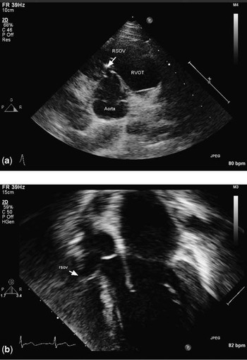
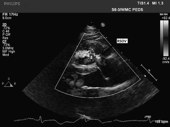
An 11-year-old asymptomatic boy presented to the cardiology clinic for evaluation of a heart murmur. His past medical history was not suggestive of chest trauma or significant infections. On physical examination, his heart sounds were normal; a grade III ejection systolic murmur and an early diastolic murmur were audible at the left sternal border. Electrocardiogram showed right axis deviation with an increase in right ventricular forces suggesting right ventricular hypertrophy. The echocardiogram showed a large aneurysm of the sinus of Valsalva with rupture into the right ventricular outflow tract, a subaortic membrane, and a ventricular septal defect (Figs 1a and 2). During surgical repair, a large aneurysm of the right sinus of Valsalva with a small rupture defect was seen to protrude into the right ventricular outflow tract, and the ventricular septal defect was just below it. Surgical repair involved patch closure of the ventricular septal defect, repair of the aneurysm of the sinus of Valsalva, resection of the subaortic membrane, and closure of a patent foramen ovale. Post-operative echocardiogram showed an intact ventricular septal defect patch, no residual membrane, and no leak from the ruptured sinus of Valsalva. The patient was discharged home on the third day on no medications.
(a) Parasternal short-axis view showing the rupture of sinus of Valsalva into the right ventricular outflow tract. (b) Apical 3 chamber view shows rupture of the right sinus of Valsalva with the aneurysm protruding into the right ventricular outflow tract and the right coronary cusp prolapsing into the ventricular septal defect.

Parasternal long-axis view with colour flow Doppler revealing flow across the ruptured sinus of Valsalva (arrow), superior to the aortic annulus.

A 9-year-old girl with a diagnosis of a small ventricular septal defect in infancy was noted to have a new diastolic murmur on a clinic follow-up visit. She had not been seen since initial presentation for 8 years, and had been completely asymptomatic. Parents denied any history suggestive of trauma to the chest or infective endocarditis. On examination, she had a loud to-and-fro systolic and diastolic murmur over the left sternal border and an active precordium. Diagnostic workup included an echocardiogram that revealed ruptured sinus of Valsalva with continuous left-to-right shunting of blood into the right ventricular outflow tract from the aorta (Fig 1b). The right coronary cusp prolapsed into the perimembranous ventricular septal defect, restricting it and causing mild aortic regurgitation. This was further confirmed on transoesophageal echocardiogram at the time of surgery. Surgical correction of the ventricular septal defect and ruptured sinus of Valsalva was performed. She remained healthy on follow-up examination. Echocardiogram showed normal cardiac function with no residual lesions.
Comment
Aneurysms of the sinus of Valsalva are generally uncommon; the incidence among Caucasians is 0.14–0.23%, while among Asians it is as high as 3.5%.Reference Goldberg and Krasnow1 Aneurysms of the sinus of Valsalva can originate in the right, left, or non-coronary sinus. Ruptured sinus of Valsalva of the right sinus is the most common, occurring in 75–90%, and non-coronary sinus in 10–25% cases. Males between 11 and 67 years are predominantly affected.Reference Goldberg and Krasnow1
The aetiology of sinus of Valsalva aneurysms lies in the anatomy and embryology of the structures. The structural defect involves a lack of continuity between the aortic annulus and aortic media. Embryologically, there may be an incomplete fusion of the aortopulmonary and interventricular septum. As a result of the weakened tissues and continued high-pressure exposure, a finger-like aneurismal sac develops.Reference Goldberg and Krasnow1 This ruptures, usually secondary to either chest trauma or infective endocarditis, further weakening the thinned out wall, leading to a sudden left-to-right shunt and cardiac failure.
Surgery forms the mainstay of treatment for ruptured sinus of Valsalva. Ruptured and symptomatic unruptured aneurysms are clear indications for surgery. If left alone, the shunt through the defect increases over time leading to failure and damage to the aortic valve structure. Treatment for unruptured asymptomatic aneurysms is not entirely straightforward; some experts believe that close monitoring is the best approach, while others recommend early surgical repair. The primary goals for surgery include closure of the aneurysm of the sinus of Valsalva, removal of the aneurysmal sac, and repair of any associated defects. The operative mortality is low (1%) in those with uncomplicated aneurysm of the sinus of Valsalva repair. Surgical intervention is very successful; long-term survival rate has been shown to be between 82% and 97%.Reference Ott3
The usual clinical presentations of ruptured sinus of Valsalva have been well described. Rupture usually occurs in young adulthood. Detection of a continuous to-and-fro murmur along the left sternal border should suggest the possibility of ruptured sinus of Valsalva. Symptoms include abrupt onset of dyspnoea, fatigue, chest pain, and paroxysmal nocturnal dyspnoea with orthopnoea. Occasionally, patients can present with atrioventricular conduction defects, arrhythmias, or even sudden death.Reference Goldberg and Krasnow1 The rupture may start small and progressively enlarge, and thus the symptoms can progress from mild-to-severe with time.
In 2001, Vural et alReference Vural, Sener, Tasdemir and Bayazit4 reviewed cases over a 15-year time period and showed that of 34 ruptured cases there were no asymptomatic cases reported. The age range of these patients was 11–25 years, indicating the predominance of symptomatic presentation in this age range. In a larger study by Au et alReference Au, Chiu, Mok, Lee, Cheung and He5, of 53 patients, both adults and children, with ruptured sinus of Valsalva, only three patients (3.8%) presented with asymptomatic murmurs. Kakos et alReference Kakos, Kilman, Williams and Hosier6 reviewed the diagnosis and management of aneurysm of the sinus of Valsalva in children, emphasising the typical presentations in children. On the basis of these previous studies, it is clear that an asymptomatic ruptured sinus of Valsalva aneurysm is an uncommon presentation in adulthood, and occurs even more rarely in children.Reference Burakovsky, Podsolkov, Sabirow, Nasedkina, Alekian and Dvinyaninova7 In a large study by Nowicki et al involving 125 patients with ruptured sinus of Valsalva, asymptomatic patients were found to be significantly younger, which is shown to be statistically significant with a p-value of less than 0.005.Reference Nowicki, Aberdeen, Friedman and Rashkind8
This report highlights the need for a high index of suspicion while evaluating new continuous murmurs in children, regardless of the symptoms. Early surgical repair of this condition is warranted to prevent the rupture from widening, leading to cardiac failure.
Acknowledgements
The authors are grateful to Mr Michael Scotton, RDMS, Echocardiography technician at Westchester Medical Center for his technical assistance in procuring the images. The authors have no disclosures or financial support for this paper.