Introduction
Pleistocene osseous deposits in Central Europe offer valuable insights into past faunal communities and palaeoenvironment. However, due to depositional and taphonomic factors, the sites are usually biased toward the representation of selected taxa, being just narrow windows into past biocoenoses. For example, archaeological sites preserve mainly remains of taxa that were the choice of Palaeolithic hunters, such as mammoth and reindeer (Pfeifer Reference Pfeifer2022; Piskorska et al. Reference Piskorska, Stefaniak, Krajcarz and Krajcarz2015). Caves may preserve long stratigraphic sequences, but the assemblages are commonly dominated by cave dwellers, such as cave bear that used caves as hibernacula e.g., Stajnia and Biśnik caves (Baca et al. Reference Baca, Popović, Stefaniak, Marciszak, Urbanowski, Nadachowski and Mackiewicz2016; Krajcarz et al. Reference Krajcarz, Krajcarz and Marciszak2014). Types of sites that have a potential for recording broader mammalian community are trap caves—that accumulate random victims that accidentally fell down the vertical abyss; or caves that served as carnivores dens—where bone collectors have accumulated remains of their prey. Graniczna Cave presents a rare opportunity to investigate a diverse and well-preserved assemblage from such a context.
Graniczna Cave, discovered in the 1990s, gained significant scientific attention only recently. In 2022, a group of cavers reported the presence of fossil animal bones during a survey conducted around the Racławicka Cave area. Following this notification, the scientific value of the site was recognized, and the material was transferred to the Institute of Systematics and Evolution of Animals, Polish Academy of Sciences (Kraków, Poland), where preliminary taxonomic and taphonomic examinations were carried out.
The study revealed high taxonomic diversity, unique for the southern part of Kraków-Częstochowa Upland. In contrast to most of caves in this region that contain primarily remains of cave bears (Ursus spelaeus/ingressus), Graniczna Cave yielded bones numerous mammal species. This collection included species characteristic for Middle-to-Late Pleistocene of this part of Europe, such as wolf (Canis lupus), fox (Vulpes sp.), red deer (Cervus elaphus), and horse (Equus ferus). However, a substantial part of the remains belonged to the extinct or locally extinct species, such as the aurochs (Bos primigenius), woolly rhinoceros (Coelodonta sp.), woolly mammoth (Mammuthus primigenius), cave hyena (Crocuta crocuta), cave bear (Ursus sp.), and reindeer (Rangifer tarandus). The high taxonomic diversity observed in the assemblage, along with gnaw marks identified on some of the remains—likely made by a porcupine, suggests that at least part of the faunal material may date to a warm period: either a warm interstadial or aninterglacial. However, due to lack of stratigraphic information, the homogeneity of assemblage stays unknown.
The represented species disappeared from the region at different times. According to historical records, the last aurochs in the world died in 1627 in the Jaktorowo Forest (Central Poland) during the reign of Sigismund III Vasa’s time (Rokosz Reference Rokosz1995). In contrast, the cave hyena (Crocuta crocuta) disappeared from Poland around 34 ka cal BP (Krajcarz et al. Reference Krajcarz, Baca, Baumann, Bocherens, Goslar, Popović, Sudoł-Procyk and Krajcarz2023).
It is important to note that, despite the exceptional preservation and taxonomic diversity of the faunal assemblage, no human remains or cultural artifacts have been recovered from Graniczna Cave. Although the region is rich in Palaeolithic evidence, including Neanderthal remains and Palaeolithic tools from sites such as Stajnia and Biśnik caves (Cyrek et al. Reference Cyrek, Socha, Stefaniak, Madeyska, Mirosław-Grabowska, Sudoł and Czyżewski2010; Picin et al. Reference Picin, Hajdinjak, Nowaczewska, Benazzi, Urbanowski, Marciszak, Fewlass, Bosch, Socha, Stefaniak, Żarski, Wiśniewski, Hublin, Nadachowski and Talamo2020). This points toward purely natural reasons behind the accumulation of faunal remains in Graniczna Cave.
This study applies AMS radiocarbon dating of selected faunal remains to obtain two objectives: to establish the absolute chronology of this unique assemblage, and estimate the chronological homogeneity of the assemblage. Radiocarbon dating is complemented OSL dating of underlying cave sediments to provide a more comprehensive chronological framework for the site.
Bones, however, represent a complex material for study and undergo significant post-burial diagenesis, often resulting in partial or complete collagen loss. The preservation of collagen depends on multiple environmental factors, including pH, temperature, and microbial activity (Pawełczyk et al. Reference Pawełczyk, Hajdas, Sadykov, Blochin and Caspari2022; van Klinken Reference van Klinken1999). Consequently, dating such materials remains challenging due to both collagen degradation and the stratigraphic complexity of the deposits.
Material and methods
Study site
The study site (Figure 1) is located in Krzeszowice municipality in Kraków-Częstochowa Upland (50o11’25”N, 19o39’50”E).
Location of the study site. The main map indicates the sampling area, while the inset map of Poland shows its regional position. Shading was applied to indicate terrain relief.

A LiDAR scan of the cave was also conducted, using the Mandeye Me device and processed in HDMapping software to document cave’s internal structure and geomorphology in detail (Figure 2). All of the samples were taken from the tunnel in first, shallower shaft indicated in yellow frame.
3D model of Graniczna Cave based on laser scanning. The gray surface represents the cave system, while the green points indicate the ground surface above the cave; the color separation was used to improve visual clarity and avoid obscuring the figure. The cave entrances, marked with arrows, are visible at the upper part of the gray model and correspond to connection points between the cave and the surface. The sampling area is marked with a yellow rectangle.

Stratigraphy
The sedimentary sequence has been studied only in the lower part of the upper shaft, i.e., the part of the cave marked with yellow rectangle in Figure 2. This section of the cave comprises eight distinct layers shown in Figure 3. The sequence is available within the upper shaft and below the shaft. The sediments within the shaft are weakly preserved, as most of them have been removed during the initial exploration. Remaining portion of the sediment is attached to the walls. These sediments are rich in limestone clasts and cemented with carbonates. Three indistinct strata can be identified: IN-1, IN-2, and IN-3, numerated downward (see Figure 3). Layer IN-1 consists of dark brown to gray silty loam containing abundant medium- and coarse-grained smoothed to subangular limestone clasts, cemented and compact, with numerous small bones and fine clasts of limpid orange clay. Layer IN-2 is composed of orange-brown, cemented loam with medium-to coarse-grained smoothed to subangular limestone clasts and phosphatic nodules. Layer IN-3 is a light brown silty loam with rusty stains, cemented, containing a significant amount of angular limestone clasts (the upper part is a breccia) and occasional dark yellow to black bone fragments.
Stratigraphic profile of the selected subsection within the yellow-marked area in Figure 2, detailing the lithological units and sediment structure.

Beneath the shaft, the sedimentary fill was excavated more carefully. Several steps were carved in sediments (marked as exploration levels in Figure 3), allowing better insight into stratigraphy. Layer IN-4 forms a thin band of gray, loose silty loam with fine angular limestone fragments and scattered dark brown bones. Layer IN-5 is a light brown, loess-like silt without limestone clasts, containing only fragile, small bones. Layer IN-6 comprises a thick deposit of brown to orange-brown loose silty loam with interlayers of gray loess-like silt and dark brown humic loam. These intercalations are distinct in the upper part of the layer, becoming less pronounced downward. Weathered smoothed limestone fragments, numerous black bone remains, and fine clasts of limpid orange clay are also present. Layer IN-7 is a light brown loose loess-like silt with sparse tiny or fragmented bones. The lowermost available layer, IN-8, consists of dark brown silty loam with black bone fragments.
Sample preparation and collagen extraction
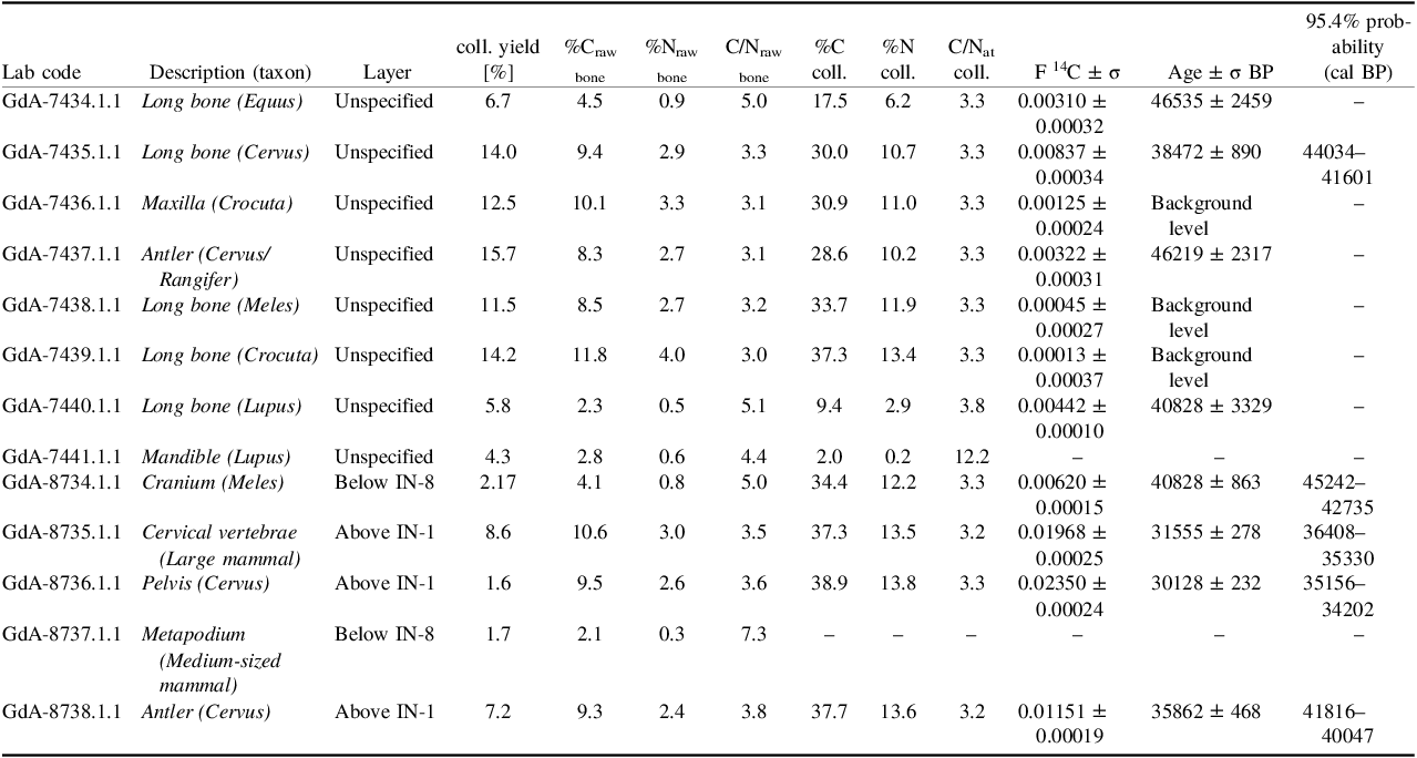
The first samples were collected in a preliminary, non-stratigraphic manner as part of an initial pilot phase. Prior to chemical preparation, each specimen was examined under the microscope and photographed for documentation. Only bones of selected species that assumed not to exceed the radiocarbon dating range were selected for dating. To complement the study, additional samples from the known stratigraphic context (given in the Table 1) were collected for the AMS analysis. All samples were prepared and measured by using the same, consistent protocol.
14C results and collagen quality indicators. This table presents analytical information for each sample, including the laboratory code, sample description, identified species and localisation of the sample. Indicators of collagen preservation are provided through percent collagen yield (coll. yield [%]), unpretreated bone carbon and nitrogen contents (%Craw, %Nraw), and the corresponding raw C/N ratio. The final purified collagen (coll.) measurements include carbon and nitrogen content and the atomic C/N ratio (C/Nat) used to assess collagen integrity. Reported radiocarbon data comprise the background-corrected fraction modern carbon (F14C) with its associated analytical uncertainty (σ), the conventional 1⁴C age (BP) with corresponding uncertainty (σ) and calibrated age ranges expressed as the 95.4% (2σ) confidence interval.

Table 1. Long description
The table presents analytical information for each bone sample, including laboratory code, description, species, and sample location. It details collagen preservation indicators such as percent collagen yield, carbon and nitrogen contents, and atomic C/N ratio. Radiocarbon data includes fraction modern carbon, conventional radiocarbon age, and calibrated age ranges. The table has 14 rows and 16 columns. Column headers include Lab code, Description (taxon), Layer, coll. yield [percent], %Craw bone, %Nraw bone, C/Nraw bone, %C coll., %N coll., C/Nat coll., F 14C ± σ, Age ± σ BP, and 95.4% probability (cal BP). Row 1: Lab code G dA-7434.1.1, Description Long bone (Equus), Layer Unspecified, coll. yield 6.7 percent, %Craw bone 4.5, %Nraw bone 0.9, C/Nraw bone 5.0, %C coll. 17.5, %N coll. 6.2, C/Nat coll. 3.3, F 14C 0.00310 ± 0.00032, Age 46535 ± 2459 BP. Row 2: Lab code G dA-7435.1.1, Description Long bone (Cervus), Layer Unspecified, coll. yield 14.0 percent, %Craw bone 9.4, %Nraw bone 2.9, C/Nraw bone 3.3, %C coll. 30.0, %N coll. 10.7, C/Nat coll. 3.3, F 14C 0.00837 ± 0.00034, Age 38472 ± 890 BP. Row 3: Lab code G dA-7436.1.1, Description Maxilla (Crocuta), Layer Unspecified, coll. yield 12.5 percent, %Craw bone 10.1, %Nraw bone 3.3, C/Nraw bone 3.1, %C coll. 30.9, %N coll. 11.0, C/Nat coll. 3.3, F 14C 0.00125 ± 0.00024, Age Background level. Row 4: Lab code G dA-7437.1.1, Description Antler (Cervus/Rangifer), Layer Unspecified, coll. yield 15.7 percent, %Craw bone 8.3, %Nraw bone 2.7, C/Nraw bone 3.1, %C coll. 28.6, %N coll. 10.2, C/Nat coll. 3.3, F 14C 0.00322 ± 0.00031, Age 46219 ± 2317 BP. Row 5: Lab code G dA-7438.1.1, Description Long bone (Meles), Layer Unspecified, coll. yield 11.5 percent, %Craw bone 8.5, %Nraw bone 2.7, C/Nraw bone 3.2, %C coll. 33.7, %N coll. 11.9, C/Nat coll. 3.3, F 14C 0.00045 ± 0.00027, Age Background level. Row 6: Lab code G dA-7439.1.1, Description Long bone (Crocuta), Layer Unspecified, coll. yield 14.2 percent, %Craw bone 11.8, %Nraw bone 4.0, C/Nraw bone 3.0, %C coll. 37.3, %N coll. 13.4, C/Nat coll. 3.3, F 14C 0.00013 ± 0.00037, Age Background level. Row 7: Lab code G dA-7440.1.1, Description Long bone (Lupus), Layer Unspecified, coll. yield 5.8 percent, %Craw bone 2.3, %Nraw bone 0.5, C/Nraw bone 5.1, %C coll. 9.4, %N coll. 2.9, C/Nat coll. 3.8, F 14C 0.00442 ± 0.00010, Age 40828 ± 3329 BP. Row 8: Lab code G dA-7441.1.1, Description Mandible (Lupus), Layer Unspecified, coll. yield 4.3 percent, %Craw bone 2.8, %Nraw bone 0.6, C/Nraw bone 4.4, %C coll. 2.0, %N coll. 0.2, C/Nat coll. 12.2, F 14C not provided, Age not provided. Row 9: Lab code G dA-8734.1.1, Description Cranium (Meles), Layer Below IN-8, coll. yield 2.17 percent, %Craw bone 4.1, %Nraw bone 0.8, C/Nraw bone 5.0, %C coll. 34.4, %N coll. 12.2, C/Nat coll. 3.3, F 14C 0.00620 ± 0.00015, Age 40828 ± 863 BP. Row 10: Lab code G dA-8735.1.1, Description Cervical vertebrae (Large mammal), Layer Above IN-1, coll. yield 8.6 percent, %Craw bone 10.6, %Nraw bone 3.0, C/Nraw bone 3.5, %C coll. 37.3, %N coll. 13.5, C/Nat coll. 3.2, F 14C 0.01968 ± 0.00025, Age 31555 ± 278 BP. Row 11: Lab code G dA-8736.1.1, Description Pelvis (Cervus), Layer Above IN-1, coll. yield 1.6 percent, %Craw bone 9.5, %Nraw bone 2.6, C/Nraw bone 3.6, %C coll. 38.9, %N coll. 13.8, C/Nat coll. 3.3, F 14C 0.02350 ± 0.00024, Age 30128 ± 232 BP. Row 12: Lab code G dA-8737.1.1, Description Metapodium (Medium-sized mammal), Layer Below IN-8, coll. yield 1.7 percent, %Craw bone 2.1, %Nraw bone 0.3, C/Nraw bone 7.3, %C coll. not provided, %N coll. not provided, C/Nat coll. not provided, F 14C not provided, Age not provided. Row 13: Lab code G dA-8738.1.1, Description Antler (Cervus), Layer Above IN-1, coll. yield 7.2 percent, %Craw bone 9.3, %Nraw bone 2.4, C/Nraw bone 3.8, %C coll. 37.7, %N coll. 13.6, C/Nat coll. 3.2, F 14C 0.01151 ± 0.00019, Age 35862 ± 468 BP.
To remove surface contamination, the bones were mechanically cleaned and rinsed in deionized water using an ultrasonic bath. The outer layer was then removed, and each bone sample was manually crushed and sieved to obtain a homogeneous material with a grain size of approximately 0.2 mm. From each bone, a subsample of raw material was taken for pre-combustion analysis to determine the C/N ratio of the bone without chemical preparation to estimate the collagen yield.
The collagen extraction was performed using Longin method (Longin Reference Longin1971), modified by Piotrowska and Goslar Reference Piotrowska and Goslar2002. Firstly, the bone material was demineralised in 0.5M hydrochloric acid in room temperature until reaction ceased (minimum 8 hr). Then samples were rinsed in deionized water to neutral pH and treated with 0.1M sodium hydroxide solution in room temperature for one hour to remove humic acids. After rinsing, a second HCl bath was applied to eliminate any carbon dioxide absorbed during the previous step. To gelatinize the collagen, the samples were heated in pH=3 hydrochloric acid solution at 85 oC for 20 hours. After that, the solution was centrifuged and filtered using EzeeTM filters (Elkay, UK), which were precleaned in deionized water. The filtered collagen was freeze-dried to remove remaining water and ensure a material homogeneity for combustion.
The additional filtration with ultrafilters can be a valuable step in dating very old and poorly preserved collagen (Pawełczyk et al. Reference Pawełczyk, Niedziałkowska, Pawełczyk, Piotrowska and Sykut2024). However, the preliminary combustion indicated that, despite the expected “old” age of bones, the collagen is of good quality and shows no obvious signs of significant diagenetic alteration. Therefore, further purification steps such as ultrafiltration, may not be strictly necessary in that case.
Dry collagen was weighted into tin capsules and combusted in a Vario Micro Cube elemental analyser (Němec et al. Reference Němec, Wacker and Gäggeler2010; Wacker et al. Reference Wacker, Němec and Bourquin2010) to determine total carbon and nitrogen content and to estimate the atomic C/N ratio. The system was calibrated with certified standards of aspartic and glutamic acids. The resulting CO₂ gas was reduced to graphite using the AGE-3 automatic graphitization system (Němec et al. Reference Němec, Wacker and Gäggeler2010; Wacker et al. Reference Wacker, Němec and Bourquin2010) and the graphite targets were pressed into cathodes using a PSP manual press (IonPlus). Along with the samples the reference materials were also graphitized. Oxalic Acid II (NIST SRM4990C) was used as modern reference material, and Phthalic Anhydride (Sigma-Aldrich) was used as background material. F14C measured in the same magazine as unknown samples was 0.00341 ± 0.00011. The concentration of radiocarbon was measured in the graphitized samples using a MICADAS accelerator mass spectrometer in the Gliwice Radiocarbon Laboratory (Ustrzycka et al. Reference Ustrzycka, Piotrowska, Kłusek, Pawełczyk, Michczyńska, Michczyński, Kozioł and Jędrzejowski2025). Results were calibrated using IntCal20 calibration curve (Reimer et al. Reference Reimer, Austin, Bard, Bayliss, Blackwell, Ramsey, Butzin, Cheng, Edwards, Friedrich, Grootes, Guilderson, Hajdas, Heaton, Hogg, Hughen, Kromer, Manning, Muscheler, Palmer, Pearson, Plicht, Reimer, Richards, Scott, Southon, Turney, Wacker, Adolphi, Büntgen, Capano, Fahrni, Fogtmann-Schulz, Friedrich, Köhler, Kudsk, Miyake, Olsen, Reinig, Sakamoto, Sookdeo and Talamo2020) in OxCal v4.4 (Bronk Ramsey Reference Bronk Ramsey2009).
In the analysis of bones for 1⁴C dating, the C/N ratio serves as a key indicator of collagen quality, allowing the assessment of both preservation state and potential contamination. An optimal C/N ratio falls in the range of 2.9–3.6 (DeNiro Reference DeNiro1985) with a nitrogen content (%N) of between 5.5 and 17.3%, and carbon content (%C) between 15.3 and 47%. Additionally, a minimum collagen yield of 1% by weight is generally required for reliable radiocarbon dating; samples below this threshold are typically considered too poorly preserved for accurate analysis (Ambrose Reference Ambrose1990; van Klinken Reference van Klinken1999).
The C/N atomic ratios (C/Nat) were calculated from the equation:
 $${C \over N}at = {{\% C} \over {\% N}} \times {{14} \over {12}}$$
Elemental carbon and nitrogen concentrations (%C and %N) were calibrated using certified amino-acid standards (glutamic acid and aspartic acid). The measured values for these standards were: 40.79% C, 9.74% N for glutamic acid, and 36.13% C, 10.54% N for aspartic acid.
Optically stimulated luminescence (OSL) dating
For the OSL analysis, a sediment sample was collected from the layer IN-7. To determine the luminescence age, both the equivalent dose (De) and the dose rate must be established. Determining the luminescence age requires measuring both the equivalent dose (De) and the dose rate. The equivalent dose was measured using the OSL method, conducted on automated Risø TL/OSL DA-20 readers equipped with a 6 mm Hoya U-340 filter and a calibrated ⁹⁰Sr/⁹⁰Y beta source, following the single aliquot regenerative (SAR) protocol (Murray and Wintle Reference Murray and Wintle2000). For OSL measurements, quartz grains of the 45–63 µm fraction were used. The sediment sample from the cave was prepared under subdued red light in a darkroom following standard chemical procedures, including treatment with hydrochloric acid (HCl) and hydrogen peroxide (H₂O₂), density separation, and subsequent etching with hydrofluoric acid (HF) (Moska et al. Reference Moska, Bluszcz, Poręba, Tudyka, Adamiec, Szymak and Przybyła2021). Dose rate was determined by gamma-ray spectrometry, which measured the activity of natural radionuclides from the 2 3⁸U and 2 3 2Th decay chains and the ⁴⁰K isotope. Sample was sealed in γBeaker containers to prevent radon loss (Poręba et al. Reference Poręba, Tudyka, Walencik-Łata and Kolarczyk2020) and stored for three weeks to ensure equilibrium in the uranium decay series. Dose rate calculations followed the conversion factors proposed by Adamiec and Aitken (Adamiec and Aitken Reference Adamiec and Aitken1998). The final equivalent dose (De) value for a sample was calculated using the Central Age Model (CAM) (Galbraith et al. Reference Galbraith, Roberts, Laslett, Yoshida and Olley1999) implemented in the R package “Luminescence” (Kreutzer et al. Reference Kreutzer, Burow, Dietze, Fuchs, Schmidt, Fischer, Friedrich, Riedesel, Autzen and Mittelstrass2020).
Results and discussion
The preservation state of the material varied significantly across the assemblage. All of the samples yielded collagen above the minimum preservation threshold (>1%), indicating generally good or satisfactory preservation. However, some of the samples produced carbon and nitrogen concentrations, as well as C/Nat ratios, that fall outside the accepted ranges for reliable radiocarbon analysis. This variability is consistent with differential diagenesis within the cave environment and highlights the need for individual collagen quality screening before interpretation. Most of the samples examined produced a proper C/Nat value of 3.3.
Four out of thirteen samples did not meet the established quality criteria for collagen preservation. In particular, samples GdA-7434.1.1, GdA-7440.1.1, GdA-7441.1.1, and GdA-8737.1.1 yielded too low carbon and nitrogen concentrations, as well as elevated C/N ratios, that fall outside the acceptable ranges for reliable radiocarbon dating. However, first two of those samples were still measured for 14C concentration as part of the pilot phase of the project.
Radiocarbon dating was performed on eleven out of thirteen samples representing various species from Graniczna Cave. Among them only five (GdA-7735.1.1, GdA-8734.1.1, GdA -8735.1.1, GdA -8736.1.1, GdA -8738.1.1) yielded a result within the range of method. Among these samples, two of them that show the youngest age, were located at the shallowest layer of the studied fragment of the shaft. The remaining samples produced results that were either beyond the effective dating limit of radiocarbon or were excluded due to poor preservation of bones. The measurement results for the tested bone samples are listed in Table 1.
Samples GdA-7434.1.1, 7437.1.1, and 7440.1.1, yielded dates beyond the calibration range and require cautious interpretation due to high uncertainty. Samples GdA-7436.1.1, 7438.1.1, and 7439.1.1 were characterized by extremely low 14C content, giving values indistinguishable from Phthalic Anhydride background samples which is shown in Figure 4B. The calibration results for the samples that fell into calibration range are shown in Figure 4A and Table 1.
Calibrated 1⁴C age probability distributions for the samples yielding finite radiocarbon ages; B: Comparison of radiocarbon results for Phthalic Anhydride (first three samples) and samples GdA-7736.1.1, GdA-7738.1.1, GdA-7739.1.1. Black lines are representing F14C values with uncertainties (few measurements for every sample), and gray marking is made for average value for each sample. Values are in the range 0.0018– 0.0069.

Figure 4. Long description
Panel A: A probability distribution graph shows calibrated radiocarbon ages for different samples. The x-axis represents the calibrated date in calBP, ranging from 34000 to 52000. The y-axis is not labeled but shows probability distributions. Each sample, labeled as R_Date GdA with specific identifiers, has a distinct probability distribution curve. Panel B: A comparison graph displays radiocarbon results for Phthalic Anhydride and specific samples. The x-axis represents F14C values ranging from 0.0018 to 0.0069. The y-axis lists sample identifiers. Black lines represent F14C values with uncertainties for each sample, and gray markings indicate average values for each sample.
The OSL dating provided an age of 85.4 ± 5.3 ka (GdTL-5496), based on an annual dose of 2.67 ± 0.14 Gy/ka and an equivalent dose of 228.2 ± 7.4Gy. The OSL data reinforces the significance of the site as an important reference point for Late Pleistocene environmental and depositional processes.
The OSL age of approximately 85.4 ka reflects the last exposure of the sediments to sunlight providing a maximum age for the layer containing the osseous material. The radiocarbon dates obtained from the well-preserved samples point to middle part of the Late Pleistocene (circa 45–34 ka), with several results approaching or exceeding the effective limit of the method. The two methods complement each other: while the OSL date establishes the lower limits of the chronological framework (MIS5/MIS4 boundary, the radiocarbon data reveal that part of the faunal material is too old for precise radiocarbon dating).
The radiocarbon results provide valuable insights into the age and preservation history of the bone assemblage from the site. All samples contained more than 1% collagen, indicating that the material retained sufficient organic content for assessment. Importantly, five samples produced finite radiocarbon ages with good precision, allowing us to establish an absolute chronological baseline for part of the assemblage. Three samples produced 1⁴C activities indistinguishable from the laboratory phthalic anhydride background. Such results are typical for very old Pleistocene material and indicate that these bones exceed the finite dating range of the 1⁴C method rather than reflecting analytical problems. These background-level outcomes therefore contribute useful information by constraining the minimum antiquity of the assemblage.
Importantly, the results obtained are consistent with the analytical capacity of the Gliwice Radiocarbon Laboratory, which has previously processed comparably old samples (e.g., mammoth bone SIRI-C; Piotrowska et al. Reference Piotrowska, Tomczyk, Pawełczyk and Stanaszek2019) reaching the method’s background levels. This demonstrates the laboratory’s ability to reliably identify and distinguish between finite ages and background signals for Pleistocene material.
The obtained results are valuable contribution to future research and development of the chronology of the site.
Conclusions
When undertaking radiocarbon dating of bones, it is crucial to carefully assess the quality and preservation of the material. Even samples that initially appear poorly preserved or at the edge of a method, can provide chronological information that contributes to the interpretation of the entire site.
A part of the samples fell within the effective range of the method and dates, with the oldest approximately 45 ka PB and the youngest around 34 ka BP. Three samples with dates beyond the effective range exhibit internal homogeneity.
Importantly, while several cave sites within the Kraków–Częstochowa Upland (e.g., Biśnik, Perspektywiczna, Stajnia caves) have also yielded rich faunal assemblages, this site preserves an especially abundant and taxonomically diverse collection of large mammals. Its combination of high specimen numbers and a wide range of species highlights the scientific value of the material and strengthens the importance of the radiocarbon results, which provide crucial chronological context for understanding the timing and formation of such faunal accumulations.
Acknowledgments
Research supported from EU funds FSD - 10.25, Development of higher education focused on the needs of the green economy European Funds for Silesia 2021-2027: The modern methods of the monitoring of the level and isotopic composition of atmospheric CO2 (project no. FESL.10.25-IZ.01-06C9/23-00).