Introduction
Researchers have presented numerous hypotheses about the brain's basis for delusions, arguably the most characteristic symptom of psychotic disorders. One prominent line of thinking relates to the processing of salience (Kapur, Reference Kapur2003; Palaniyappan & Liddle, Reference Palaniyappan and Liddle2012).
Preliminary evidence links delusions to the cortical salience network (CSN) (Palaniyappan & Liddle, Reference Palaniyappan and Liddle2012), comprising the left and the right anterior insula (LAI and RAI) and the dorsal anterior cingulate cortex (dACC) (Menon & Uddin, Reference Menon and Uddin2010). Previous research linked structural abnormalities of CSN to the severity of delusions (Crespo-Facorro et al. Reference Crespo-Facorro, Kim, Andreasen, O'Leary, Bockholt and Magnotta 2000 ; Koutsouleris et al. Reference Koutsouleris, Gaser, Jager, Bottlender, Frodl, Holzinger, Schmitt, Zetzsche, Burgermeister, Scheuerecker, Born, Reiser, Moller and Meisenzahl2008; Cascella et al. Reference Cascella, Gerner, Fieldstone, Sawa and Schretlen2011; Palaniyappan et al. Reference Palaniyappan, Mallikarjun, Joseph, White and Liddle2011), whereas one functional study suggested a reward prediction error-related signal in the insula associated with a combined delusion and hallucination score (Gradin et al. Reference Gradin, Kumar, Waiter, Ahearn, Stickle, Milders, Reid, Hall and Steele2011). Delusions of reference are associated with the activation of the right insula (Menon et al. Reference Menon, Schmitz, Anderson, Graff, Korostil, Mamo, Gerretsen, Addington, Remington and Kapur2011), and the resting-state connectivity of CSN to the striatum was linked to the severity of delusions (Orliac et al. Reference Orliac, Naveau, Joliot, Delcroix, Razafimandimby, Brazo, Dollfus and Delamillieure2013). However, these cross-sectional studies cannot determine, whether and how CSN functioning contributes to the development of delusions. Functional magnetic resonance imaging (fMRI) studies in high-risk patients initially characterized brain functioning preceding conversion to psychosis (Sabb et al. Reference Sabb, van Erp, Hardt, Dapretto, Caplan, Cannon and Bearden2010; Allen et al. Reference Allen, Luigjes, Howes, Egerton, Hirao, Valli, Kambeitz, Fusar-Poli, Broome and McGuire2012, Reference Allen, Chaddock, Egerton, Howes, Barker, Bonoldi, Fusar-Poli, Murray and McGuire2015), but these studies did not specifically focus on salience processing or delusions.
Several neuroscientific findings provide the foundation for understanding CSN functioning in delusions (Palaniyappan & Liddle, Reference Palaniyappan and Liddle2012). Anterior insula (AI) and dACC involve von Economo neurons, assumed to result in the fast, intuitive assessment of complex situations (Allman et al. Reference Allman, Watson, Tetreault and Hakeem2005). AI may integrate homeostatic, environmental, hedonic, motivational, social, and cognitive information into a unified representation of the sentient self, the framework within which the salience of any stimulus is evaluated at present (Craig, Reference Craig2009). Such salience then contributes to the recruitment of the brain systems related to, for example, attention and learning (Palaniyappan & Liddle, Reference Palaniyappan and Liddle2012).
We used the strength of CSN activation at the baseline assessment to predict the development of the severity of delusions at a 2-month follow-up assessment in patients with first-episode psychosis (FEP). We measured CSN activation using fMRI as the patients evaluated clinical insight and general metacognitive statements. While both types of statements are salient, the clinical insight statements, such as ‘Some of my unusual experiences are due to psychosis,’ require evaluation of the medical view in the context of the ‘psychotic sentient self’. We, therefore, hypothesized that stronger CSN activation at baseline assessment in a ‘clinical insight task v. metacognition task’ comparison would predict a worsening of or less improvement in delusions at the 2-month follow-up assessment.
Method and materials
Subjects
Data of 28 FEP patients and 25 controls were available for the present study. One patient and two controls were excluded due to more than one voxel diameter head movement during scanning, resulting in 27 patients and 23 controls.
We recruited FEP patients aged 18–40 years during their initial contact with psychiatric care for psychosis from the catchment area of the Helsinki University Hospital. We defined psychosis as individuals scoring at least 4 on items assessing delusions or hallucinations from the Brief Psychiatric Rating Scale – Extended (BPRS-E; Ventura et al. Reference Ventura, Lukoff, Nuechterlein, Liberman, Green and Shaner1993). Controls were identified from the Population Register Centre and were assessed using the same protocol as for patients. We excluded controls with a lifetime history of a psychotic disorder from the study. For both patients and controls, we excluded individuals with any chronic neurological, endocrinological, or cardiovascular disease, and individuals with any condition that prevented an MRI.
Clinical study protocol
The methods used in the clinical assessment are described in detail in our earlier paper (Mäntylä et al. Reference Mäntylä, Mantere, Raij, Kieseppä, Laitinen, Leiviskä, Torniainen, Tuominen, Vaarala and Suvisaari2015). We assessed patients at baseline as soon as the patient was able to provide informed consent, with an additional 2-month follow-up assessment. The severity of psychotic symptoms was evaluated using the BPRS-E. For clarity, we label here the BPRS item ‘unusual thought content’ as the ‘delusion score’. A change in the delusion score at follow-up was defined as: score at 2-month follow-up minus score at baseline.
Diagnostic assessment was based on the Research Version of the Structured Clinical Interview for DSM-IV Axis I Disorders (SCID-I). A senior psychiatrist (J.S.) with the interviewer defined the diagnosis. In cases of uncertainty, a consensus diagnosis between the senior psychiatrists (J.S., O.M., and T.K.) was made.
fMRI
Brain images were acquired for patients 6 ± 5 (mean ± s.d.) days after the first clinical evaluation, excluding one subject who experienced a 69-day delay. The second clinical evaluation took place 80 ± 18 days after the scan.
Anatomical T1-weighted images were acquired with 1 mm isotropic voxels (see Mäntylä et al. Reference Mäntylä, Mantere, Raij, Kieseppä, Laitinen, Leiviskä, Torniainen, Tuominen, Vaarala and Suvisaari2015 for details). We acquired the blood oxygenation level-dependent functional images first using a GE Signa VH/i 3-T scanner (GE Healthcare, UK) (14 patients, 12 controls) and then after a prescheduled scanner change, using the Siemens Magnetom Skyra 3-T system (Siemens AG, Germany) (13 patients, 11 controls) at Aalto AMI Centre, Aalto NeuroImaging, Aalto University School of Science. Identical gradient-echo echo-planar imaging sequences (TR: 2300 ms; TE: 30 ms; flip angle: 75°; field of view: 24 cm; base resolution 64 × 64) were used for both scanners to obtain complete brain functional images in 40 contiguous slices. The voxel size stood at 3.75 × 3.75 × 4 mm.
We presented metacognition statements during the first fMRI session. Clinical insight statements were presented during the second, separate fMRI session, immediately following the first session, in order to avoid confusion among patients between each task. During the first imaging session, we used a total of 12 metacognition statements modified from the Self-reflection and Insight Scale (Grant, Reference Grant2002) and Metacognitions Questionnaire (Wells & Cartwright-Hatton, Reference Wells and Cartwright-Hatton2004) in four blocks of three statements. During the second session, we used 16 clinical insight statements modified from the Insight and Treatment Attitudes Questionnaire (McEvoy et al. Reference McEvoy, Apperson, Appelbaum, Ortlip, Brecosky, Hammill, Geller and Roth1989) and Insight Scale for Psychosis (Birchwood et al. Reference Birchwood, Smith, Drury, Healy, Macmillan and Slade1994) presented in four blocks of four statements (see Supplementary Table S1 for the statements). Each block of statements was interleaved with a 30-s resting period. During each session, participants were tasked with rating statements on a visual analog scale ranging from ‘fully disagree’ (left end of the scale, encoded 0) to ‘fully agree’ (right end of the scale, encoded 100). By pressing the left- or right-hand buttons, the cursor moved to the left or right. The answer was recorded 2 s after the last cursor movement. Participants received an explanation of the clinical meaning of psychosis, and were given a chance to practice using the rating tool before entering the scanner with statements similar (although not the same) to those used during the scan. Statements and the visual analog scale were displayed using Presentation software (Neurobehavioral Systems Inc., USA ) and projected on to a semi-transparent screen within the participant's visual field.
Preprocessing and analysis
An experienced neuroradiologist excluded organic pathologies from the anatomical images. Functional data were preprocessed in Statistical Parametric Mapping (SPM8) (http://www.fil.ion.ucl.ac.uk/spm/software/spm8/) with the aid of the Data Processing Assistant for Resting-State fMRI Advanced Edition (DPARSFA) version 2.3 (Chao-Gan & Yu-Feng, Reference Chao-Gan and Yu-Feng2010). Functional images were corrected for slice-timing, realigned with respect to the first image for movement correction, normalized to the Montreal Neurological Institute template, and smoothed with an 8-mm full-width at half maximum Gaussian kernel.
Data were analysed in SPM8 using the general linear model. Due to increasing evidence of comparability of fMRI data acquired with different scanners (Forsyth et al. Reference Forsyth, McEwen, Gee, Bearden, Addington, Goodyear, Cadenhead, Mirzakhanian, Cornblatt, Olvet, Mathalon, McGlashan, Perkins, Belger, Seidman, Thermenos, Tsuang, van Erp, Walker, Hamann, Woods, Qiu and Cannon2014; Gee et al. Reference Gee, McEwen, Forsyth, Haut, Bearden, Addington, Goodyear, Cadenhead, Mirzakhanian, Cornblatt, Olvet, Mathalon, McGlashan, Perkins, Belger, Seidman, Thermenos, Tsuang, van Erp, Walker, Hamann, Woods, Constable and Cannon2015), we pooled data from the two scanners. To confirm independence of the results from the scanner, we also conducted the main correlational analysis separately for each scanner's data. Regressors convolved with the haemodynamic response function were built for each task and resting period, separately, based on the individual answer times in the Presentation log-files. Individual contrast images were computed for the ‘metacognition task v. resting’ comparisons for both groups, and for the ‘clinical insight task v. resting’ and ‘clinical insight task v. metacognition task’ for the patients. Images for each contrast were entered into one-sample t tests – one for each group and contrast – to validate the involvement of CSN in the task. The patients’ ‘clinical insight task v. metacognition task’ contrast images were then entered into the regression analysis with the change in delusion scores (main analysis). The ‘clinical insight task v. metacognition task’ comparison was used to control for the effects of no interest, such as the visuomotor functioning, attention, and self-referential processing.
For control purposes, we included the following nuisance covariates through a series of regression analyses: baseline delusion score, antipsychotic equivalent doses (Andreasen et al. Reference Andreasen, Pressler, Nopoulos, Miller and Ho2010), the delay between the baseline assessment and scan, and ratings of the clinical insight statements. Finally, we compared the ‘metacognition task v. resting’ contrast images between groups with independent-sample t tests, and entered ‘clinical insight task v. metacognition task’ contrast images into one-sample t tests in the patient group.
Statistical methods
All SPM analyses were family-wise error (FWE)-corrected for multiple comparisons at the cluster level using the primary voxel-wise threshold p < 0.001 as recommended by Woo et al. (Reference Woo, Krishnan and Wager2014). To our knowledge, the present stimuli have not been previously used in studies of FEP. However, involvement of the AI and the dACC has been observed in healthy subjects during self-referential processing (Modinos et al. Reference Modinos, Ormel and Aleman2009; Hu et al. Reference Hu, Di, Eickhoff, Zhang, Peng, Guo and Sui2016), and the present tasks were assumed to be salient, especially for FEP patients. Therefore, the correction for multiple comparisons was applied to three regions of CSN (regions of interest; ROIs), whenever correction for the whole-brain volume did not result in statistically significant findings. These regions were derived from the Functional Imaging in Neuropsychiatric Disorders Lab website (findlab.stanford.edu/functional_ROIs.html) and constructed through an independent components analysis (Shirer et al. Reference Shirer, Ryali, Rykhlevskaia, Menon and Greicius2012). ROIs included RAI, LAI, and a cluster extending from the bilateral dACC to the bilateral supplementary motor area (ROI3).
We did not calculate the correlation coefficients based on the activation strengths of the most significant voxels. Instead, we used the principal eigenvalues (most typical values) extracted from the a priori CSN template regions to avoid any circularity in our analysis (Kriegeskorte et al. Reference Kriegeskorte, Simmons and Bellgowan2009).
Ethics statement
The Ethics Committee of the Hospital District of Helsinki and Uusimaa (257/12/03/03/2009), the institutional review boards of the National Institute for Health and Welfare, Helsinki, Finland, and the University of Helsinki approved the study protocol. All participants provided their informed written consent before participating. The patient's capacity to give informed consent was assessed by the treating psychiatrist.
Results
Clinical findings
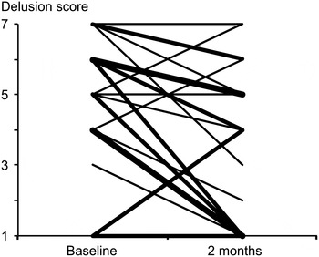
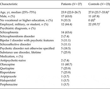
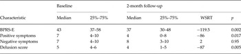
Table 1 presents the descriptive data for patients and controls, while Table 2 shows the clinical characteristics. Fig. 1 illustrates the changes in the delusion scores during the follow-up assessment. The median duration of medication before baseline assessment was 25 days (25% at 16 days, 75% at 43 days).
Individual changes in the delusion scores (Brief Psychiatric Rating Scale item ‘unusual thought content’) from the baseline assessment to the 2-month follow-up assessment. Thickness of the lines indicates number of cases with an identical trajectory for symptom severity (from one to three).

Descriptive data for first-episode psychosis patients and healthy controls

a Mann–Whitney U test, p = 0.046.
b Fisher's exact t test for difference, p = 0.002.
c Fisher's exact t test, p < 0.001.
Baseline and 2-month follow-up data for clinical characteristics of first-episode psychosis patients

BPRS-E, Brief Psychiatric Rating Scale – Extended; WSRT, Wilcoxon's signed rank test.
The sum of positive scores included BPRS-E items hallucinations, unusual thought content, bizarre behaviour, and conceptual disorganization. The sum of negative symptoms included BPRS-E item blunted affect and Scale for Assessment of Negative Symptoms items alogia, anhedonia and avolition. BPRS-E items were rescaled from 1–7 to 0–6 for the calculation of the sum scores.
Behavioural findings
The patients’ average ratings on the metacognition statements did not differ significantly from those of controls (p = 0.778). The patients’ ratings on clinical insight statements during scanning reached 63 ± 18, and were higher than those on the metacognition task (54 ± 10; p = 0.044).
The answer times of patients, measured as the time from the presentation of a statement to the end of answer for each item, reached a mean of 7.4 s for metacognition and 7.2 s for the clinical insight task. We found no difference between the two tasks (p = 0.684). Answer times did not correlate with a change in delusion score (p = 0.346, 0.973, and 0.112 for metacognition task, clinical insight task, and difference between the two tasks, respectively). The metacognition task answer times did not differ between patients and controls (p = 0.188).
Imaging findings
Compared to resting periods, CSN was activated during each task in patients and controls (metacognition v. rest for patients and controls, respectively: p = 0.001 and p = 0.001 for LAI, p = 0.002 and p = 0.004 for RAI, p < 0.001 and p < 0.001 for ROI3; insight v. rest in patients: p = 0.001 for LAI, p = 0.002 for RAI, p < 0.001 for ROI3), corrected for multiple comparisons within a priori ROI volumes.
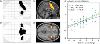
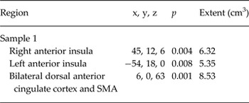
Stronger activation strength in the clinical insight task compared to the general metacognition task predicted less improvement or even worsening of delusions at 2-month follow-up (Table 3, Fig. 2). This finding was replicated with each scanner's data separately (Supplementary Fig. S1, Supplementary Table S2). The correlations remained significant after controlling for the confounding effects of the baseline delusion scores, the delay from the baseline clinical assessment to the scan, antipsychotic drugs dose equivalents, and patients’ ratings of clinical insight statements. On average, we observed no CSN activation difference in the ‘metacognition task v. resting’ contrasts between groups or in the ‘clinical insight task v. metacognition task’ contrasts within patients.
Activation strengths predicting a change in the delusion score. Brain images show the maximum intensity projections [(a) and (c), ‘glass brain’] and two MRI slices [(b) sagittal, (d) axial]. Black (a, c) and yellow (b, d) show the cortical salience network regions of interest. Grey and red show voxels for which the activity predicted the delusion score change (p < 0.001 at the voxel level, uncorrected for visualization). The y-axis of the scatterplot (e) displays the activation strength (in parameter estimates) for the comparison ‘clinical insight task v. general metacognition task’, extracted from the a priori regions (yellow). The x-axis shows the changes in delusion scores, where negative values reflect a decrease and positive values reflect an increase in the delusional severity. LAI, Left anterior insula; RAI, right anterior insula; ROI3; bilateral dorsal anterior cingulate cortex and supplementary motor area.

Brain regions for which activation predicted a change in the delusion scores

SMA, Supplementary motor area.
p values for correlations are family-wise-error-corrected for multiple comparisons in the whole-brain volume. See Fig. 2 for the unbiased correlation coefficients. Extent refers to the volume of contiguous voxels with p < 0.001 (uncorrected).
Discussion
Stronger AI activation in the ‘clinical insight task v. metacognition task’ comparison predicted less improvement to or a worsening of delusions using the 2-month assessment. The bilateral AI lies at the core of CSN, and an association between psychotic symptoms and CSN were found in previous cross-sectional studies (Crespo-Facorro et al. Reference Crespo-Facorro, Kim, Andreasen, O'Leary, Bockholt and Magnotta2000; Koutsouleris et al. Reference Koutsouleris, Gaser, Jager, Bottlender, Frodl, Holzinger, Schmitt, Zetzsche, Burgermeister, Scheuerecker, Born, Reiser, Moller and Meisenzahl2008; Cascella et al. Reference Cascella, Gerner, Fieldstone, Sawa and Schretlen2011; Gradin et al. Reference Gradin, Kumar, Waiter, Ahearn, Stickle, Milders, Reid, Hall and Steele2011; Jardri et al. Reference Jardri, Pouchet, Pins and Thomas2011; Menon et al. Reference Menon, Schmitz, Anderson, Graff, Korostil, Mamo, Gerretsen, Addington, Remington and Kapur2011; Palaniyappan et al. Reference Palaniyappan, Mallikarjun, Joseph, White and Liddle2011; Radaelli et al. Reference Radaelli, Poletti, Gorni, Locatelli, Smeraldi, Colombo and Benedetti2014). Importantly, our findings add to this literature by suggesting that CSN activation precedes the progression of symptoms. While this finding alone does not prove causality, it adds a new layer of evidence for a contribution of CSN functioning to the longitudinal course of delusions.
This is, to the best of our knowledge, the first time that the clinical insight task has been used in acutely psychotic patients to study CSN functioning. While the metacognition task focused on processing how an individual's mind works in general, the clinical insight task focused on processing whether an individual's experiences should be described as a mental illness. Patients’ ratings of the clinical insight statements suggest that they partially agreed with medical explanations. However, the relatively high symptom scores, especially for delusions, suggest that the competing psychotic reality was simultaneously present. This supports the notion that psychotic patients tend to maintain both the psychotic and medical realities at once (‘double bookkeeping’) (Henriksen & Parnas, Reference Henriksen and Parnas2014) and the assumption that patients processed the clinical insight statements in the context of a psychotic reality (i.e. ‘psychotic sentient self’). Such a psychotic reality may be associated with ‘aberrant salience’ (Kapur, Reference Kapur2003; Palaniyappan & Liddle, Reference Palaniyappan and Liddle2012), a ‘delusional atmosphere’, an ‘abnormal awareness of significance’, or an ‘anomalous self-experience’ (Henriksen & Parnas, Reference Henriksen and Parnas2014).
In addition to subjectively salient experiences (Craig, Reference Craig2009), AI is implicated in the processing of prediction error (Chase et al. Reference Chase, Kumar, Eickhoff and Dombrovski2015), i.e. discrepancies between the expected and actual outcomes that result in learning-related changes in internal representations. Distorted prediction-error signalling is linked to delusions (Corlett et al. Reference Corlett, Honey, Krystal and Fletcher2011). Supporting the role of CSN in a change to an individual's view of the world, insular activity during loss anticipation may predict loss-aversion learning (Samanez-Larkin et al. Reference Samanez-Larkin, Hollon, Carstensen and Knutson2008), and CSN activation during cognitive dissonance has been shown to predict an attitude change (van Veen et al. Reference van Veen, Krug, Schooler and Carter2009).
Abnormal salience-related learning alone may be insufficient to account for delusions; however, since an individual may remain capable of reasoning based on the available evidence, that questions the delusion. Accordingly, two-stage models were presented, including a defect in reasoning in addition to the primary basis of the delusion (Gilleen & David, Reference Gilleen and David2005; van der Gaag, Reference van der Gaag2006). Dysfunction in the CSN-related switching of appropriate cognitive systems (Menon & Uddin, Reference Menon and Uddin2010; Palaniyappan & Liddle, Reference Palaniyappan and Liddle2012; Palaniyappan et al. Reference Palaniyappan, Simmonite, White, Liddle and Liddle2013; Manoliu et al. Reference Manoliu, Riedl, Zherdin, Muhlau, Schwerthoffer, Scherr, Peters, Zimmer, Forstl, Bauml, Wohlschlager and Sorg2014) could account for such a compromised analytical reasoning regarding delusions.
In this framework, our findings may reflect CSN involvement in the evaluation of the salience of symptom-related experience and, correspondingly, the selection of action and updating of internal representations (Palaniyappan & Liddle, Reference Palaniyappan and Liddle2012). Within the framework of neuroscience, internal representations are the only access to reality and, therefore, the development of the severity of delusions may be explained by the psychotic updating of internal representations (Fletcher & Frith, Reference Fletcher and Frith2009).
CSN is linked with the mesostriatal dopaminergic system (Murray et al. Reference Murray, Corlett, Clark, Pessiglione, Blackwell, Honey, Jones, Bullmore, Robbins and Fletcher2008; Palaniyappan & Liddle, Reference Palaniyappan and Liddle2012; Raij et al. Reference Raij, Mäntylä, Kieseppä and Suvisaari2015) and, thus, the mesostriatal hyperdopaminergic state observed in psychosis (Howes et al. Reference Howes, Kambeitz, Kim, Stahl, Slifstein, Abi-Dargham and Kapur2012) may be linked to CSN functioning. Preliminary data suggests that CSN functioning may indeed contribute to the striatal dysfunction in psychosis (Raij et al. Reference Raij, Mäntylä, Kieseppä and Suvisaari2015).
The relatively small sample size stands as a limitation in our study and may have contributed to the lack of difference between groups and tasks. It should be noted, however, that the clinical insight task was the task of interest here and cannot be applied to healthy control subjects. Thereby, group comparison is of minor importance in the present study. The lack of a difference in activation between tasks within patients may reflect the average stabilizing state of patients being treated for psychosis. If all patients presented during the earlier phase of worsening delusions, CSN activation across all patients may have been stronger during the clinical insight task than during the metacognition task.
Patients experienced a short delay between symptom assessment and scanning. Considering relatively stable medication, it is unlikely that a prominent change in the delusional severity would have occurred between the first clinical evaluation and scanning. Furthermore, the correlation between CSN activation and the change in the severity of delusions was not affected by singling out the delay from clinical assessment to scanning. Correlates of symptoms other than delusions were not assessed in the present study due to inadequate variance in the symptom scores.
In conclusion, our findings link CSN functioning during clinical, symptom-related, insight processing to future development of delusion severity. Future studies should attempt to specify the association between psychotic symptoms other than delusions and CSN, and manipulate CSN functioning in order to affect symptoms. Follow-up studies of high-risk patients with appropriate salient stimuli should show how the emergence of delusions relates to CSN functioning. In the future, this type of protocol, optimized or combined with other predictors, may assist in identifying patients at risk for worsening or persistent psychosis.
Acknowledgements
Funding for this study was provided by Sigrid Juselius Foundation to J.S., the Finnish Cultural Foundation to J.S. and T.M., European Research Council Advanced grant (no. 232946 to Riitta Hari), the Jalmari and Rauha Ahokas Foundation and the Brain and Mind Doctoral Programme to T.M., and the University of Helsinki, the Academy of Finland (no. 251155), and the Finnish Medical Foundation to T.T.R
Supplementary material
For supplementary material accompanying this paper visit http://dx.doi.org/10.1017/S0033291716001057.