Most weeks, our multidisciplinary meeting pauses on the same kind of study: a patient with an enlarged right ventricle due to chronic pulmonary insufficiency, years after tetralogy of Fallot repair, but who has not yet reached the threshold we would act on. Disagreement is rarely about the measurements. It is about what they mean and when intervention is necessary. For years, we answered that with a single number: beyond a certain indexed volume, the chamber recovery was judged unlikely, and the valve replacement was necessary. Earlier guidelines were built largely around this number.Reference Baumgartner, De Backer and Babu-Narayan1,Reference Stout, Daniels and Aboulhosn2 The 2024 American Heart Association statement moves the assessment beyond volume, considering ejection fraction, exercise capacity, pressure load, and a validated risk score together.Reference Geva, Wald and Bucholz3 Even so, one group is still left unaccounted for: patients whose volumes remain questionable for decision-making.
Those borderline patients and decisions fill our meetings, and these are the issues on which we struggle to reach consensus. Setting a threshold does not end the argument because thresholds are never meant to be a key. The numbers everyone reaches for, an indexed end-diastolic volume above 160 mL/m2 and an end-systolic volume above 80 mL/m2, mark the point beyond which the ventricle is less likely to return to its normal size after valve replacement.Reference Oosterhof, van Straten and Vliegen4,Reference Ortiz-Garrido, Różewicz Juraszek, Gabbert, Jussli-Melchers and Voges5 These do not indicate a point where an intervention will definitely help or the ventricle is safe. Preoperative volume does not even predict reverse remodelling on its own; the shape of the right ventricle carries information that volume does not.Reference Thatte, Sleeper, Lu, Tang and Geva6 In our practice, the uncertain range runs from approximately 140 mL/m2 to 160 mL/m2 for the end-diastolic volume and 70 mL/m2 to 80 mL/m2 for the end-systolic volume. Within that range, no single number decides anything. This is the grey zone, and the patients in it are not the same.
What the statement does well is to widen the perspective beyond volume and to pay more attention to early changes: a three-percentage-point change in the peak right ventricular longitudinal strain is considered clinically meaningful to prompt further investigation by cardiac magnetic resonance.Reference Geva, Wald and Bucholz3 What it does not do is tell us how to manage when a patient is plainly worse than a year ago but has not crossed any formal threshold. A free-wall strain that is a little less negative each time,Reference Gao, Li and He7 a QRS that has widened across recordings,Reference Knauth, Gauvreau and Powell8 a fresh patch of late gadolinium enhancement,Reference Babu-Narayan, Kilner and Li9 a rising extracellular volumeReference Chen, Dusenbery, Valente, Powell and Geva10: any of these can carry meaning well before ejection fraction, peak oxygen uptake, or pressure cross the normal line. The statement is not blind to this deviation. An increase of 30 mL/m2 or more in indexed end-diastolic volume or a ten-point fall in either ventricle’s ejection fraction is considered clinically significant and sets monitoring surveillance intervals accordingly.Reference Geva, Wald and Bucholz3 In the grey zone, even a lesser fall may matter. A lower threshold could prove more clinically appropriate. Which cut-off should trigger earlier review is not yet established, though we believe a ≥5% decline in ejection fraction may be more suitable for the grey zone. Yet it does not say what that deviation should mean for the timing of replacement while the volumes still remain inside the grey zone. That is the patient volume serves worst: the one deteriorating quietly, so that by the time the numbers acknowledge a problem, the chance of ventricular recovery may already have passed.Reference Bokma, Geva and Sleeper11,Reference Bokma, Geva and Sleeper12 Left ventricular mechanics tell a similar story: rotational parameters are impaired in repaired tetralogy of Fallot and those show no recovery a year after valve replacement, even when the right ventricle is well remodelled.Reference Harrington, Ghelani and Thatte13 The tension between waiting for definitive criteria and acting early enough to be significant has been present in the timing literature for twenty years.Reference Geva14,Reference Leonardi, Perrone and Calcaterra15
There is a further problem that the statement does not address. Across our centres the advanced imaging methods on which this framework leans are neither evenly available nor equally taught. The Association for European Paediatric and Congenital Cardiology (AEPC) Imaging Working Group’s recent survey of European practice reveals this: cardiac magnetic resonance imaging and computed tomography are applied and taught very differently from one centre to another; less than two-thirds of centres train their fellows in either, and many trainees still travel abroad to learn them.Reference Ramcharan, Roest and Voges16 Echocardiographic practice differs too.Reference Voges, Cantinotti and Miller17 A scheme that assumes ready access to cardiac magnetic resonance, feature tracking, exercise testing, and tissue mapping is a different tool in a unit that works mostly from echocardiography, and even a well-equipped centre may wait months for a standard cardiac MRI, much less for tissue mapping or four-dimensional flow. A framework meant to be applied across diverse centres is most useful when it distinguishes the assessments available to every unit from those available only to some so that its guidance can still be followed where advanced imaging is limited.
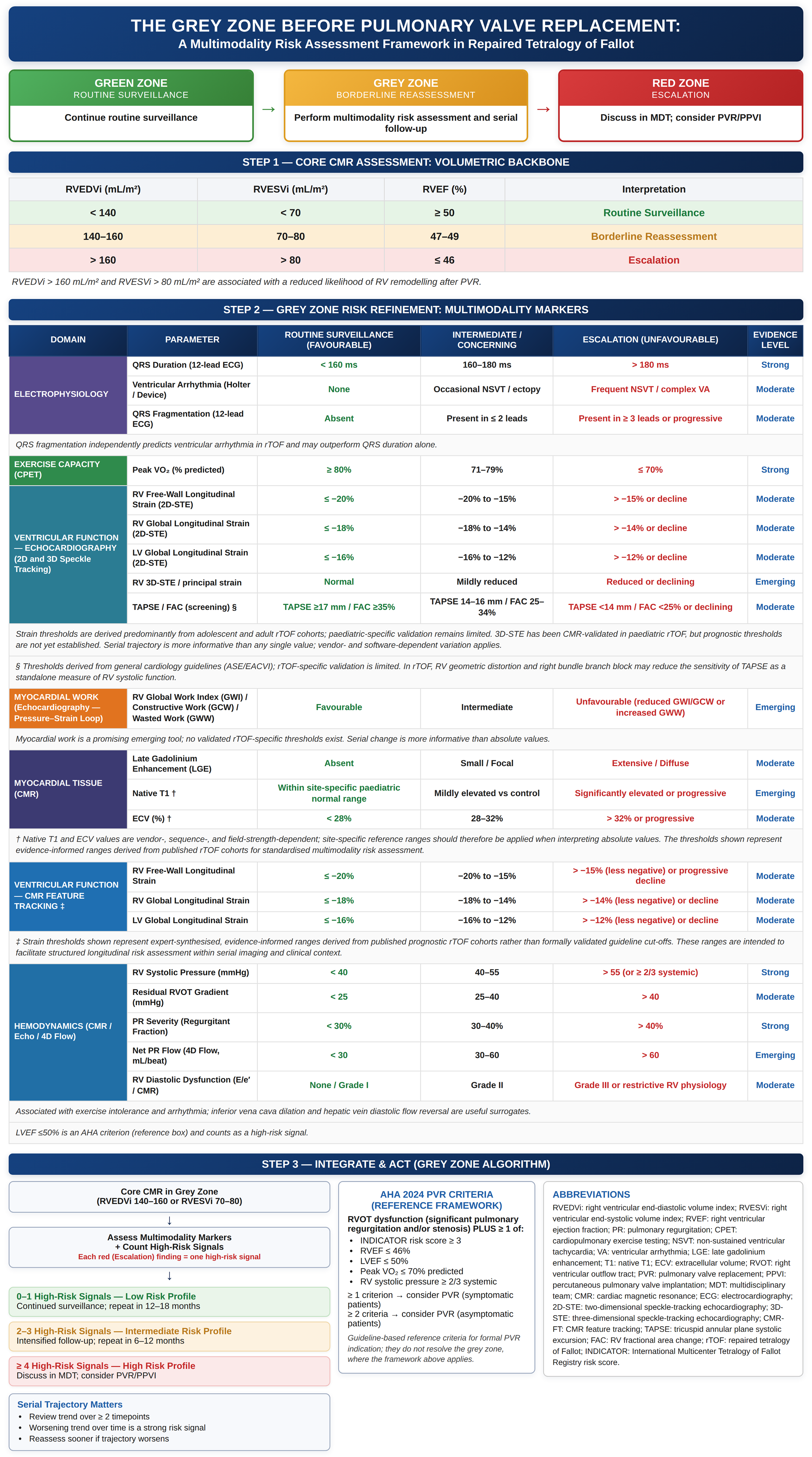
We are not proposing new cut-offs. In a borderline patient, we take the decision in steps (Figure 1). Volume is the starting point, but here it raises the question rather than answers it. We then weigh a range of markers to assess how risky it is to continue monitoring: strain by feature tracking or speckle tracking,Reference Gao, Li and He7,Reference Matsubara, Numata and Calderon-Anyosa18 exercise capacity, the rhythm substrate including QRS fragmentation,Reference Leonardi, Perrone and Calcaterra15 tissue characterisation,Reference Babu-Narayan, Kilner and Li9,Reference Chen, Dusenbery, Valente, Powell and Geva10,Reference Yim, Riesenkampff, Caro-Dominguez, Yoo, Seed and Grosse-Wortmann19 and loading conditions. The number of unfavourable markers gives an initial sense of risk, but the direction of travel is more informative: the same values worsening over serial studies mean far more than any single borderline result. Together, these markers determine the surveillance interval and when the case is reviewed again by the team. The result is a decision about surveillance and timing rather than a surgical instruction.
A multimodality risk-assessment framework for the “grey zone” before pulmonary valve replacement in repaired tetralogy of Fallot. Step 1 identifies the volumetric backbone by cardiac magnetic resonance. Step 2 refines risk within the grey zone using multimodality markers across ventricular function (feature tracking and two- and three-dimensional speckle-tracking echocardiography), exercise capacity, electrophysiology, myocardial work, tissue characterisation, and haemodynamics. Step 3 translates these signals into a management decision (routine surveillance, borderline reassessment, or escalation), shown beside the 2024 American Heart Association reference criteria. The framework is conceptual, supports longitudinal reassessment, proposes no new intervention thresholds, and requires prospective validation; the number of unfavourable markers in Step 3 is an illustrative guide to surveillance intensity rather than a validated score. The figure was prepared by the authors in PowerPoint with the assistance of a software tool; its content is the authors’ own.

Figure 1. Long description
The table is divided into three main steps for assessing risk before pulmonary valve replacement. Step 1 focuses on core cardiac magnetic resonance (CMR) assessment with volumetric backbone measurements. It includes RVEDVI, RVESVI, and RVEF with corresponding interpretation zones: Routine Surveillance, Borderline Reassessment, and Escalation. Step 2 refines risk within the grey zone using multimodality markers across various domains such as electrophysiology, exercise capacity, ventricular function, myocardial work, myocardial tissue, ventricular and atrial function, and hemodynamics. Each domain lists parameters, routing surveillance, intermediate concerning, escalation, and emergency levels. Step 3 integrates and acts based on the grey zone algorithm, assessing multimodality markers and counting high-risk signals to determine the surveillance profile. The table also includes abbreviations for better understanding.
The argument is most clearly illustrated in a standard study in two patients: the same indexed volumes, the same ejection fraction, yet neither meets any criteria for intervention. The first has steady free-wall strain, a narrow QRS, no fibrosis, and good exercise tolerance. The second has strain that has fallen study by study, a QRS that widens year to year, extensive late gadolinium enhancement, a high extracellular volume, and frequent ventricular ectopy. By volume you would watch both. By the whole picture you watch the first and discuss the second with the team to consider an earlier intervention decision, though nothing formal has been crossed. The framework does not make that decision; it records the reasoning behind it so that the next clinician can carry the trajectory forward at the following study.
There are three important caveats. First, most strain and tissue thresholds in daily practice were derived in adults and adolescents rather than validated prospectively in children; in tetralogy this matters less, since patients reaching valve replacement are nearly adolescent or older, and paediatric data are beginning to appear.Reference Matsubara, Numata and Calderon-Anyosa18,Reference Yim, Riesenkampff, Caro-Dominguez, Yoo, Seed and Grosse-Wortmann19 Native T1 and extracellular volume depend on scanner, sequence, and field strength and do not transfer across sites as fixed numbers.Reference Chen, Dusenbery, Valente, Powell and Geva10,Reference Yim, Riesenkampff, Caro-Dominguez, Yoo, Seed and Grosse-Wortmann19 Lastly, myocardial work and four-dimensional flow are still promising rather than proven and should be read that way. None of this is a reason to ignore the markers; it is a reason to let them inform clinical judgement rather than override it.
No single centre will be able to settle the grey zone with another threshold paper. The statement itself points in the same direction: its call for future research identifies myocardial strain and magnetic resonance tissue characterisation among the imaging biomarkers most in need of development and acknowledges that the risk models currently in use explain only about 25 to 30% of the variation in outcome.Reference Geva, Wald and Bucholz3 Closing this gap would require prospective, multicentre follow-up of borderline patients designed to show which combinations of markers predict the hard outcomes (death and sustained ventricular arrhythmia) and a ventricle that does not recover after intervention.Reference Bokma, Geva and Sleeper12,Reference Ghonim, Gatzoulis and Ernst20–Reference Ishikita, McIntosh and Hanneman23 That requires a single paediatric protocol, agreed across European centres and built to work in spite of the differences in imaging capacity that already exist; the need for such coordinated study has already been recognised, and high-volume centres are well placed to contribute.Reference Borrelli, Sasso and Sarubbi24 We present this framework as a way to initiate that work, not to finish it. The 2024 statement gave the field its firmest footing yet.Reference Geva, Wald and Bucholz3 The task now is to decide what to do with the part that was left grey.
Financial support
This work received no specific grant from any funding agency, commercial or not-for-profit.
Competing interests
The authors declare none.
Ethical standards
The two illustrative patients described in the text are fully conceptualised, anonymised teaching constructs. No patient data, imaging or identifiable clinical information were used; formal ethics approval and consent were therefore not required.