Implications
In this study, our data showed that acetate alters the process of lipid metabolism in rabbits via the regulation of adipocyte-specific transcription factors and steatolysis-related factors. Protein expression results showed that acetate GPR41/43, mitogen-activated protein kinase (MAPKs) signaling and AMPK signaling may be involved in this process. A better understanding of acetate-regulated lipid metabolism could be useful to study the interactions and their modulation of host physiology in response to nutrition in rabbit. They might also represent a novel link between gut microbiota and physiological processes.
Introduction
Short-chain fatty acids (SCFAs) are released following dietary fiber fermentation in the gastrointestinal tract, primarily in the cecum in rabbits. In addition to providing energy, SCFAs are known to have regulatory effects on energy metabolism. Previous studies found that acetate (major SCFAs, 70% to 80%) was a substrate for lipogenesis and adipocyte differentiation (den Besten et al., Reference den Besten, Bleeker, Gerding, van Eunen, Havinga, van Dijk, Oosterveer, Jonker, Groen, Reijngoud and Bakker2015). However, the regulatory mechanisms of acetate on lipid and adipocyte metabolism remain unclear.
Lipid deposition results from the dynamic balance among lipogenesis and lipolysis (Jurie et al., Reference Jurie, Cassar-Malek, Bonnet, Leroux, Bauchart, Boulesteix, Pethick and Hocquette2007), and they are regulated by metabolic enzymes and controlled by functional genes. Changes in their activities and gene expression can alter the rates of biosynthesis of lipid deposition (Zhang et al., Reference Zhang, Zhang, Wang, Dong, Tan, Zou, Peng, Xue, Wang and Dong2015). A constant positive energy balance leads to an excessive fat accumulation in adipose tissue. The previous study found that SCFAs treated cells showed increased levels of phosphorylated hormone-sensitive lipase (HSL) (Mora and Fullerton, Reference Mora and Fullerton2015). Adipocytes are specialized cells that store triglyceride for energy excess and release energy by lipolysis during energy shortage (Gregoire, Reference Gregoire2001). A constant positive energy balance leads to an excessive fat accumulation in adipose tissue. The adipogenesis sequentially regulated by several transcription factors. Li et al. (Reference Li, Yao and Jiang2014) found that SCFAs could stimulate the CCAAT/enhancer-binding proteins (C/EBPα) gene expression in the porcine adipocyte. In addition, they can also be controlled by many signaling pathways, such as adenosine monophosphate-activated protein kinase (AMPK) signaling (Hwang et al., Reference Hwang, Lee, Kim, Sung, Kim, Kim and Kwon2008), MAPKs signaling (Bost et al., Reference Bost, Aouadi, Caron and Binétruy2005; He et al., Reference He, Huang, Zhao, Feng, Shi, Wang and Wang2013), G protein-coupled receptor (GPR41)/GPR43 signaling (Choi et al., Reference Choi, Chung, Johnson, Go, Kim, Choi and Smith2013).
So we hypothesize that GPR41/43, AMPK or MAPKs singalings may be involved in acetate-regulated lipid metabolism. In this study, we investigated the effect of acetate injection on rabbit lipid metabolism, including lipid accumulation and expression of genes regulating adipogenesis and lipolysis, as well as several proteins involved in the adipogenesis, such as GPR41/43, AMPK or MAPK singalings.
Material and methods
Animals
New Zealand rabbits were individually housed in self-made cages (60×40×40 cm). Temperature and lighting were maintained according to commercial conditions. Diets were formulated according to the recommendations from the National Research Council (NRC, 1977) and de Blas and Mateos (Reference Dewulf, Cani, Neyrinck, Possemiers, Van Holle, Muccioli, Deldicque, Bindels, Pachikian, Sohet, Mignolet, Francaux, Larondelle and Delzenne1998) and pelleted using pressure. The diameter of the pellets was 4 mm. All of the rabbits received a starter diet containing 16% CP, 14% crude fiber, and 11 MJ/kg of digestible energy. All of the rabbits had free access to food and water during the rearing period. This study was approved by the Shandong Agricultural University and conducted in accordance with the ‘Guidelines for Experimental Animals’ of the Ministry of Science and Technology (Beijing, China).
Experimental protocol and sample collection
At 30 days of age, 80 New Zealand rabbits with similar BWs were randomly assigned to 1 of 4 groups (20 replicates per group, one rabbit per replicate). Each rabbit received a subcutaneous acetate injection (2 ml/injection, dissolved in saline) of 0 g/kg per day, 0.5 g/kg per day, 1.0 g/kg per day or 2.0 g/kg per day of body mass for 4 days. Acetate was purchased from Sigma (St. Louis, MO, USA). At the end of the last injection, eight rabbits from each group were fasted for 4 h and electrically stunned (70 V, pulsed direct current, 50 Hz for 5 s), and 10 ml of blood was immediately collected from the heart. Plasma was obtained following centrifugation at 400×g for 10 min at 4°C and stored at −20°C for subsequent analysis. The rabbits were sacrificed by cervical dislocation, and the shoulder adipose tissue was collected from neck and shoulder and cleaned of connective tissue. Half of the adipose tissue was collected, frozen in liquid nitrogen directly and then stored at −80°C for adipocyte histology analysis. The other part was washed several times with phosphate buffered saline, snap-frozen in liquid nitrogen and then stored at −80°C for gene and protein expression analysis. All the analysis work was finished within 1 month.
Measurements
The concentrations of leptin, adiponectin, triglyceride and glucose in plasma were measured spectrophotometrically using commercial diagnostic kits (Nanjing Jiancheng Bioengineering Institute, Jiangsu, China). Insulin was measured by a commercial radioimmunoassay (RIA) kit (RIA Insulin Kit, Union Biotechnology, Shenzhen, China). For adipocyte histology, the adipose tissue was visualized by Oil Red O staining. Briefly, tissues were immediately frozen in liquid nitrogen and cut in Leica CM-1850 cryostat microtome (Leica, Wetzlar, Germany). Then 3 μm thick sections were fixed in 4% formaldehyde for 60 min and stained with filtered 0.5% Oil Red O (Sigma-Aldrich, St. Louis, MO, USA) for 10 min, which was made by dissolving in isopropyl alcohol for 15 min at room temperature. Then the sections were stained with hematoxylin (Sigma-Aldrich) for 1 min, followed by photographed using an Olympus CX-41 phase contrast microscope (Olympus, Tokyo, Japan).
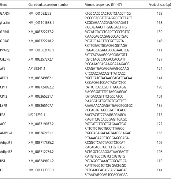
Gene expression in the adipose tissue was quantified using quantitative real-time PCR. Total RNA was isolated with Trizol (Invitrogen, San Diego, CA, USA). The quality and quantity of RNA were determined by agarose-gel electrophoresis and using a biophotometer (Eppendorf, Hamburg, Germany). RT reactions (10 µl) contained 500 ng of total RNA, 5 mmol/l of MgCl2, 1 µl of RT buffer, 1 mmol/l of deoxyribonucleoside triphosphate, 2.5 U of avian myeloblastosis virus, 0.7 nmol/l of oligo d(T) and 10 U of ribonuclease inhibitor (TaKaRa, Dalian, China). A real-time PCR analysis was conducted using the Applied Biosystems 7500 Real-time PCR System (Applied Biosystems, Foster, CA, USA). Each RT reaction served as a template in a 20 µl PCR reaction containing 0.2 μmol/l of each primer and SYBR green master mix (Takara). The primer sequences are listed in Table 1. The primers were designed for exon-intron junctions using Primer 5.0 software (Primer-E Ltd., Plymouth, UK). The real-time PCR reactions were performed at 95°C for 10 s of pre-denaturation, followed by 40 cycles consisting of denaturation at 95°C for 5 s and annealing and extension at 60°C for 40 s. A standard curve was plotted to calculate the efficiency of the real-time PCR primers. The mRNA levels of target genes were normalized to glyceraldehyde 3-phosphate dehydrogenase and β-actin (ΔCT). The ΔCT was calibrated against an average from the control checks. The number of target molecules relative to the control was calculated using
 $${\rm 2}^{{{\minus}\Delta \Delta C_{t} }} $$
. Therefore, all gene transcription results were reported as the n-fold difference relative to the control. The comparative CT method (
 $${\rm 2}^{{{\minus}\Delta \Delta C_{t} }} $$
) was used to quantitate the mRNA expression, in accordance with Livak and Schmittgen (Reference Livak and Schmittgen2001). The specificity of the amplification product was verified by a melting curve analysis and DNA sequencing.
Gene-specific primers used for the analysis of rabbit gene expression

GAPDH=glyceraldehyde 3-phosphate dehydrogenase; GPR41/43=G protein-coupled receptor 41/43; PPARγ=peroxisome proliferator-activated receptors; C/EBPα=CCAAT/enhancer-binding protein α; ADD1=differentiation-dependent factor 1; aP2=adipocyte protein 2; CPT 1/2=carnitine palmitoyltransferase 1/2; LEPR=leptin receptor; FAS=fatty acid synthase; ACC1=acetyl-CoA carboxylase 1; AMPKα=AMP-activated protein kinase α; AdipoR1/2=adiponectin receptor 1/2; HSL=hormone-sensitive lipase; LPL=lipoprotein lipase.
The samples obtained from the adipose tissue were homogenized on ice using a radioimmunoprecipitation assay buffer (50 mmol/l of Tris-HCl at pH 7.4, 1% of NP-40, 0.25% of sodium deoxycholate, 150 mmol/l of NaCl, 1 mmol/l of EDTA, 1 mmol/l of phenylmethylsulfonyl fluoride, 1 μg/ml of aprotinin, 1 μg/ml of leupeptin, 1 μg/ml of pepstatin, 1 mmol/l sodium of orthovanadate, and 1 mmol/l of sodium fluoride) and were centrifuged at 12 000×g for 5 min at 4°C. Protein concentrations were determined using a bicinchoninic acid assay kit (Beyotime, Jiangsu, China). After boiling at 100°C, the protein extracts (40 μg) were electrophoresed in 7.5% to 10% of SDS polyacrylamide gels, following the procedure described by Laemmli (Reference Laemmli1970). The separated proteins were transferred onto nitrocellulose membranes at 100 V at 4°C. Thereafter, the membrane was stripped to assay the phosphorylated proteins and Tubulin, separately. The membranes were blocked and immunoblotted with the following primary antibodies: phospho-AMPKα Thr172, AMPKα, phospho-p38 MAPKThr180/Tyr182, p38 MAPK, phospho-ERK1/2Thr202/Tyr204, ERK1/2, phospho-JNKThr183/Tyr185 and JNK. Protein detection was performed using HRP-labeled goat anti-mouse IgG (H+L) secondary antibody by enhanced chemiluminescence, using western blotting detection reagents (Beyotime). Monoclonal mouse anti-Tubulin antibody was used as a loading control. Western blots were developed and quantified using BioSpectrum 810 with VisionWorksLS 7.1 software (UVP LLC, Upland, California, USA).
Statistical analysis
The data were presented as the mean±SEM. The homogeneity of variances among the treatments were confirmed using Bartlett’s test. All of the data were subjected to a one-way ANOVA analysis to test the main effect of the treatment (n=8). When the main effect of the treatment was significant, the differences between the means were assessed by Duncan’s multiple range analysis. P<0.05 was considered statistically significant.
Results
Shoulder adipose accumulation and plasma variables
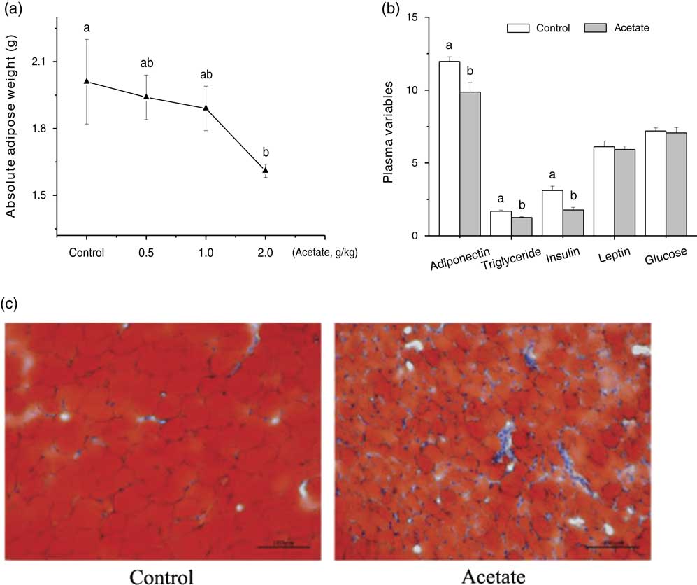
The shoulder adipose accumulation was measured to evaluate the effect of acetate injection on subcutaneous lipogenesis. It indicated that acetate induced a dose-dependent decrease in shoulder adipose (Figure 1a and c, P<0.05). In plasma, although acetate treatment significantly decreased the plasma adiponectin, triglyceride and insulin concentrations (Figure 1b, P<0.05), acetate injection did not alter the plasma leptin and glucose concentration.
Effect of acetate treatment (2.0 g/kg)on lipid deposition and plasma variables in rabbits. Rabbits received a subcutaneous acetate injection (2 ml/injection, dissolved in saline) of 0 g/kg per day, 2.0 g/kg per day of body mass for 4 days. (a) lipid deposition, (b) plasma variables (Leptin, ng/ml; adiponectin, mg/l; triglyceride, mM; glucose, mM; insulin, μIU/ml) and (c) oil red O staining of adipose histology (200×). The values are expressed as the mean±SEM (n=8). ab=Means differed significantly (P<0.05).

Genes expression related to lipometabolism
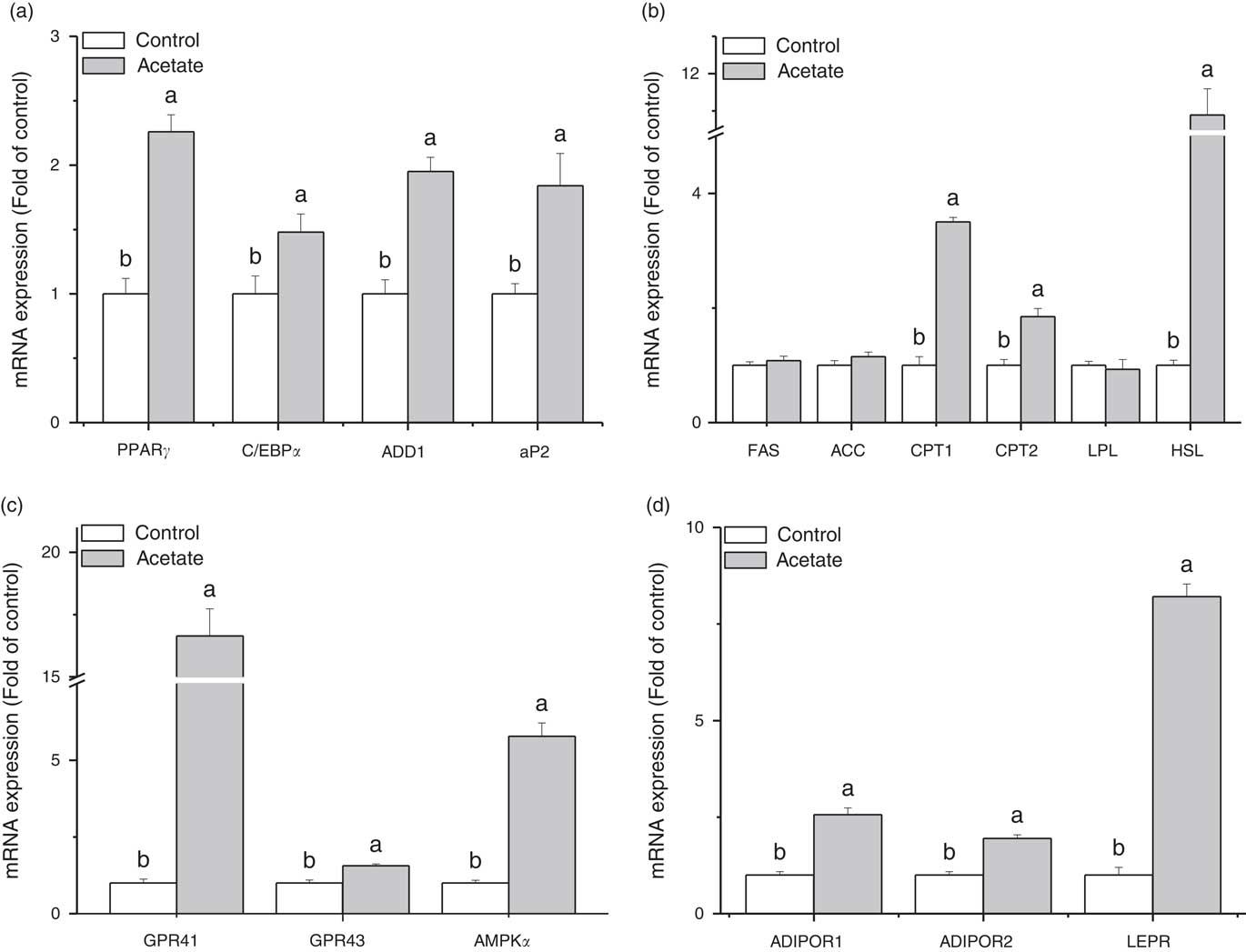
We determined the lipid metabolism-relative gene expressions in adipose tissue. The results showed that gene expressions of peroxisome-activated receptor gamma (PPARγ), C/EBPα, differentiation-dependent factor 1 (ADD1), aP2, AMPKα1, carnitine palmitoyltransferase 1 (CPT1), CPT2, GPR41, GPR43, AdipoR1, AdipoR2 and Leptin receptor were significantly upregulated by acetate injection (Figure 2, P<0.05). Compared with the control, the acetate treatment had no effect on the mRNA levels of fatty acid synthase (FAS) and acetyl-CoA carboxylase (ACC1) (Figure 2b, P>0.05).
The effect of acetate treatment (2.0 g/kg) on the mRNA expression level in shoulder adipose tissue of rabbits. Rabbits received a subcutaneous acetate injection (2 ml/injection, dissolved in saline) of 0 g/kg per day, 2.0 g/kg per day of body mass for 4 days. (a) PPARγ, C/EBPα, aP2 and ADD1; (b) FAS, ACC, CPT1, CPT2, LPL and HSL; (c) GPR41, GPR43 and AMPKα1; (d) AdipoR1, AdipoR2 and LEPR. The values are expressed as the mean±SEM (n=8). ab=Means differed significantly (P<0.05). PPARγ=peroxisome proliferator-activated receptors; C/EBPα=CCAAT/enhancer-binding protein α; ADD1=differentiation-dependent factor 1; aP2=adipocyte protein 2; FAS=fatty acid synthase; ACC=acetyl-CoA carboxylase; CPT=carnitine palmitoyltransferase 1; LPL=lipoprotein lipase; HSL=hormone-sensitive lipase; GPR41/43= G protein-coupled receptor 41/43; AMPKα=AMP-activated protein kinase α; LEPR=leptin receptor; AdipoR=adiponectin receptor.

Protein expression related to lipometabolism
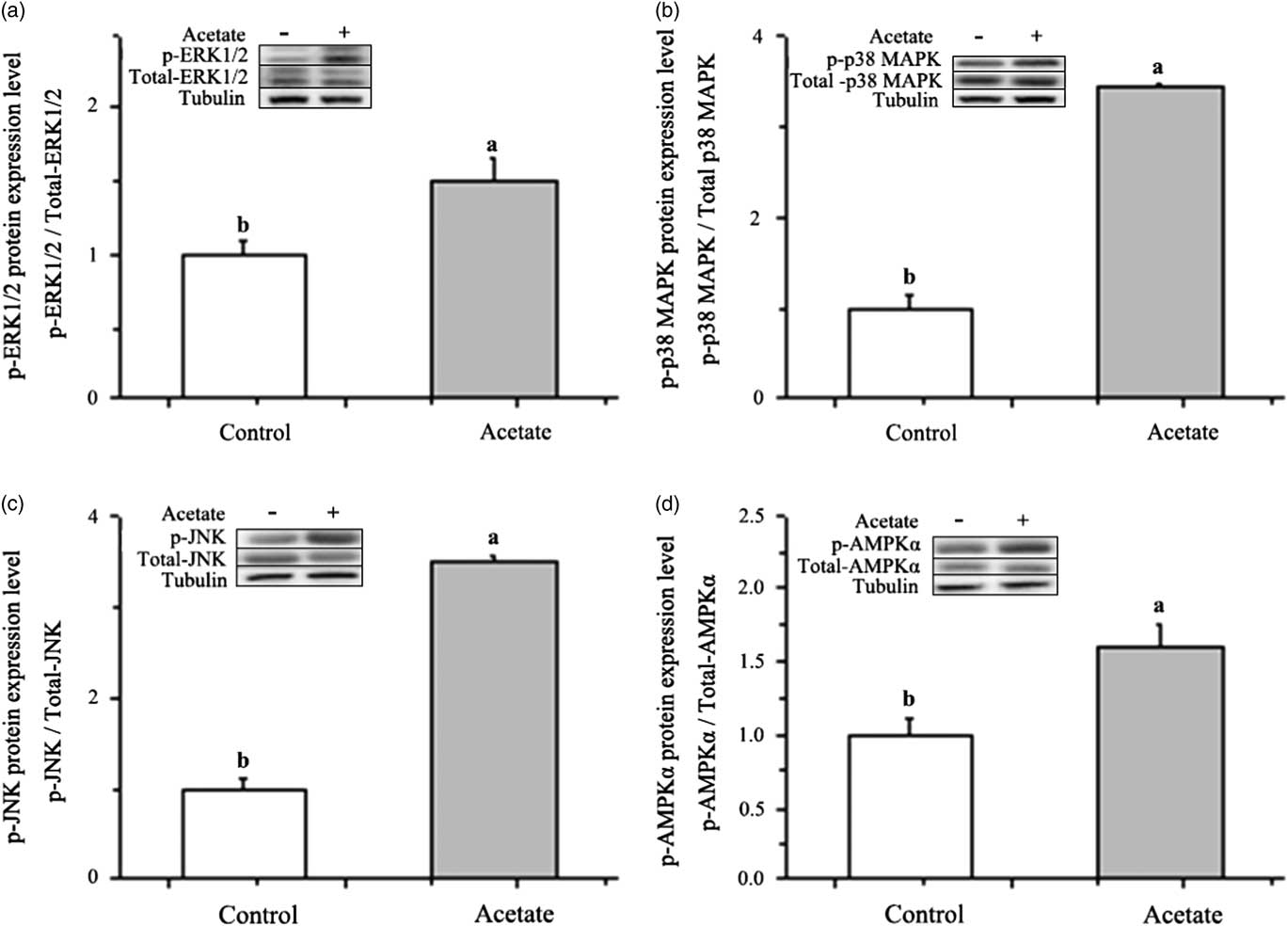
The results for Western blot analysis were shown in Figure 3 and Supplementary Material Figure S1. As AMPK plays an important role in the regulation of lipid metabolism and adipocyte differentiation and MAPKs also play an important role in many essential cellular processes including adipocytes differentiation, we detected the effect of acetate treatment on AMPK and MAPKs protein expression. Compared with the control, acetate injection significantly unregulated the phosphorated protein levels of AMPKα, ERK1/2, JNK and p38 MAPK (Figure 3, P<0.05; Supplementary Material Figure S1), and had no significant effect on the total protein expression of any one.
The effect of acetate treatment (2.0 g/kg) on protein expression level in shoulder adipose tissue of rabbits. Rabbits received a subcutaneous acetate injection (2 ml/injection, dissolved in saline) of 0 g/kg per day, 2.0 g/kg per day of body mass for 4 days. Protein expression were measured by Western blotting. (a) Phospho-AMPKα Thr172, (b) phospho-p38 MAPKThr180/Tyr182, (c) phospho-JNKThr183/Tyr185, and (d) phospho-ERK1/2Thr202/Tyr204. The values are expressed as the mean±SEM (n=8). ab=Means differed significantly (P<0.05). AMPKα=AMP-activated protein kinase α; ERK1/2=extracellular signaling-regulated kinases 1 and 2; p38 MAPK=p38 mitogen-activated kinase; JNK=c-jun NH2 terminal kinases.

Discussion
In the current study, we investigated which signaling pathways were involved in the regulation of acetate on lipid metabolism in rabbit adipose tissue. Our data demonstrated that (1) acetate increased the gene expressions of adipocyte-specific transcription factors (PPARγ, C/EBPα, aP2 and ADD1) and steatolysis-related factors (HSL, CPT1 and CPT2); (2) acetate treatment upregulated the gene expressions of GPR43, GPR41, AMPKα1, AdipoR1, AdipoR2 and LEPR; (3) acetate injection increased the phospholated protein levels of AMPKα, p38 MAPK, JNK and ERK1/2. These results indicated that GPR41/43, AMPKα and MAPKs signaling pathways may be associated with the regulation of acetate in lipid metabolism in rabbit adipose tissue.
Acetate promoted the lipid steatolysis and upregulated adipocyte differentiation factors genes expression
Recent evidence further revealed that SCFAs mediated a spectrum of physiological functions as signaling molecules (den Besten et al., Reference den Besten, Bleeker, Gerding, van Eunen, Havinga, van Dijk, Oosterveer, Jonker, Groen, Reijngoud and Bakker2015). Several studies established that SCFAs could regulate important signaling pathways involved in energy expenditure and homoeostasis (Kimura et al., Reference Kimura, Ozawa, Inoue, Imamura, Kimura, Maeda, Terasawa, Kashihara, Hirano, Tani, Takahashi, Miyauchi, Shioi, Inoue and Tsujimoto2013). Although acetate treatment did not alter the plasma glucose level in the present study, significantly decreased the insulin level. The result was in consistent with the previous study (Armstrong et al., Reference Armstrong, Wiggam, Ennis, Sheridan, Traub, Atkinson and Bell2001), which suggest acetate can affect the insulin sensitivity and secretion. In addition, the results may be relative to the decreased adipose tissue weight. In line with the findings in other species (Li et al., Reference Li, Yao and Jiang2014; Hu et al., Reference Hu, Kyrou, Tan, Dimitriadis, Ramanjaneya, Tripathi, Patel, James, Kawan, Chen and Randeva2016), we found that the decrease of absolute weight of adipose tissue was related to the increased HSL gene expression. Studies provided evidence that HSL, through possessing triacylglycerol lipase activity, appeared to be the rate-limiting enzyme for cholesteryl ester and diacylglycerol hydrolysis in adipose tissue, and it is essential for complete hormone stimulated lipolysis (Kraemer and Shen, Reference Kraemer and Shen2006). CPT1 and CPT2 mRNA levels were increased and adipose cell size was reduced after the acetate treatment, which suggested an enhanced mitochondrial up-take of long chain acyl-CoA for β-oxidation. But we also found that acetate up-regulated the adipocyte-specific transcription factors PPARγ, C/EBPα, aP2 and ADD1 expressions in rabbits adipose tissue, which implied that acetate maybe stimulate the process of adipocyte differentiation.
G protein-coupled receptor 41 and 43 is involved with the regulation of acetate in adipocyte differentiation factors genes expression
G protein-coupled receptor 41 and 43, also known as free fatty acid receptor (FFAR) 3 and 2, respectively, are expressed in adipose tissue (Dewulf et al., 2011; Li et al., Reference Li, Yao and Jiang2014). In 3T3-L1 cells, GPR43 siRNA decreased the expression of PPARγ (Hong et al., Reference Hong, Nishimura, Hishikawa, Tsuzuki, Miyahara, Gotoh, Choi, Feng, Chen, Lee, Katoh, Roh and Sasaki2005). Hong et al. (Reference Hong, Nishimura, Hishikawa, Tsuzuki, Miyahara, Gotoh, Choi, Feng, Chen, Lee, Katoh, Roh and Sasaki2005) found that the stimulatory effect of acetate and propionate on adipocyte differentiation in 3T3-L1 cells depended on GPR43, but not GPR41. The adipogenic effect of SCFAs on porcine stromal vascular fraction was not mediated through GPR41 and 43 (Li et al., Reference Li, Yao and Jiang2014). However, in our experiment, the GPR41 and GPR43 could be activated by acetate, which suggested that GPR41 and GPR43 may be involved in acetate caused the upregulation of adipocyte differentiation factors in rabbit adipose tissue. In addition, the regulation of acetate in GPR41 and GPR43 is species-specific.
Mitogen-activated protein kinases signaling pathways are involved in the acetate-regulated adipocyte differentiation factors genes expression
The mitogen-activated protein kinases (MAPKs) of ERK, p38 MAPK and JNK are intracellular signaling pathways that play a pivotal role in many essential cellular processes, such as proliferation and differentiation. Mitogen-activated protein kinases are activated by a large variety of stimuli, and one of their major functions is to connect cell surface receptors to transcription factors in the nucleus, which consequently triggers long-term cellular responses.
Recent results led to the conclusion that the function of ERKs in adipogenesis was multiple. Sale et al. (Reference Solinas, Vilcu, Neels, Bandyopadhyay, Luo, Naugler, Grivennikov, Wynshaw-Boris, Scadeng, Olefsky and Karin1995) demonstrated that the ERKs were required for the differentiation of 3T3-L1 fibroblasts to adipocytes. The adipocyte-specific transcription factor PPARγ is a substrate of ERKs. In addition, ERK activity is necessary for the expression of the crucial adipogenic regulators C/EBPα and PPARγ. In our experiment, acetate injection increased the phospho-ERK1/2 protein level in rabbit adipose tissue, suggesting that ERKs signaling was involved in the adipocyte differentiation caused by acetate. The upregulated adipocyte-specific transcription factors PPARγ, C/EBPα, aP2 and ADD1 may also be mediated through ERKs signaling.
Phosporylation of p38 MAPK regulates diverse biological processes (Uddin et al., Reference Uddin, Ah-Kang, Ulaszek, Mahmud and Wickrema2004). Engelman et al. (Reference Engelman, Berg, Lewis, Lin, Lisanti and Scherer1999) were the first to describe a positive role for p38 MAPK in adipogenesis. They showed that the addition of the p38 inhibitors early in 3T3-L1 differentiation decreased adipocyte formation. p38 MAPK chemical inhibitors treatment decreased the bone morphogenetic protein-2-induced adipocyte formation, this defect was associated with a decrease in PPARγ and C/EBPβ transactivation activities (Hata et al., Reference Hata, Nishimura, Ikeda, Yamashita, Matsubara, Nokubi and Yoneda2003). In the present study, the phospho-p38 MAPK protein was upregulated by acetate compared with the control, which suggested the p38 MAPK badger with adipocyte differentiation and the up-regulation of adipocyte-specific transcription factors (PPARγ, C/EBPα, aP2 and ADD1).
Several papers showed that JNK and the scaffold protein JNK interacting protein 1 (JIP1) were implicated in the development of obesity (Pal et al., Reference Pal, Febbraio and Lancaster2016; Rorato et al., Reference Rorato, Borges, Uchoa, Antunes-Rodrigues, Elias and Elias2017). The invalidation of JNK led to the resistance to high-fat diet induced obesity. Furthermore, JNK1 and JIP1 deficiencies provided partial resistance to obesity (Solinas et al., Reference Sakakibara, Yamauchi, Oshima, Tsukamoto and Kadowaki2007). Our results showed that acetate increased the phospho-JNK protein levels, which was in accordance with the findings in caski cells (Woo et al., Reference Woo, Lim, Kim, Suh, Min, Chang, Lee, Park and Kwon2004). Although there was no evidence for a role of this pathway in adipocyte differentiation, we could not exclude the possibility of JNK signaling participating in the process of acetate caused adipocyte differentiation.
Adenosine monophosphate-activated protein kinase signaling pathways is associated with acetate caused steatolysis
Adenosine monophosphate-activated protein kinase is the central component of a cellular signaling system that monitors cellular energy change, acting as ‘metabolic master switch’ to regulate ATP concentrations in the face of stress. In some cells, such as colonocytes, SCFAs are efficiently oxidized to generate ATP, and the generated ATP not only fuels but also regulates, various biological processes in these cells by inhibiting the activity of AMPK (Hardie et al., Reference Hardie, Ross and Hawley2012). However, acetate increased AMPKα1 mRNA and the phospho-AMPKα protein levels in the present study, which was consistent with the findings in rat hepatocytes (Sakakibara et al., Reference Sale, Atkinson and Sale2006). These results suggested the effect of acetate on AMPK was tissue specificity. In addition, the increased AMPK signaling may be related to the leptin and adiponectin signaling. Handy et al. (Reference Handy, Saxena, Fu, Lin, Mells, Gupta and Anania2010) found that leptin and adiponectin could increase phospho-AMPKα levels in hepatic stellate cells. Although acetate did not increase the plasma leptin and adiponectin concentrations, the LEPR, AdipoR1 and AdipoR2 mRNA levels were significantly upregulated after acetate treatment. Therefore, the leptin and adiponectin signaling were enlarged in rabbits’ adipose tissue. It remains to be determined whether or not this was because liver and adipose tissue could not efficiently use SCFAs for ATP production.
On activation, AMPK inhibits adipocyte differentiation in 3T3-L1 cells (Lee et al., Reference Lee, Kang, Bae and Yoon2011) and in adipocyte-derived human mesenchymal stem cells (Lee et al., Reference Lee, Cho, Bui and Kang2014). The AMPK activity levels remain constant throughout the process of adipogenesis in C3H10T1/2 cells (Chen et al., Reference Chen, Brooks, Houskeeper, Bremner, Dunlop, Viollet, Logan, Salt, Ahmed and Yarwood2017), while AMPK activator, AICAR, did not block SCFAs-induced expression of PPARγ or CEBPα mRNA in stromal vascular fraction of the porcine subcutaneous fat (Li et al., Reference Li, Yao and Jiang2014). With regards to stage-specific effects, Pantovic et al. (Reference Pantovic, Krstic, Janjetovic, Koci, Harhaji-Trajkovic, Bugarski and Trajkovic2013) demonstrated that there is a coordinated time-dependent activation of different signaling pathways during the osteogenic differentiation of hMSCs, which is AMPK-dependent in the early stages of differentiation followed by late stage activation of the Akt/mTOR signaling pathway. Given the results presented here, it could be argued that similar mechanisms might regulate adipogenesis in acetate-induced adipocyte differentiation of rabbit in the present study. In addition, AMPK may be involved in fat oxidation. It was reported that the activation of AMPK could increase CPT1 activity, resulting in increased fatty acid oxidation.
Conclusion
In conclusion, acetate up-regulated the adipocyte-specific transcription factors (PPARγ, C/EBPα, aP2 and ADD1), which were associated with the activated GPR41/43 and MAPKs signaling. Meanwhile, acetate decreased fat content via the up-regulation of the steatolysis-related factors (HSL, CPT1 and CPT2), and AMPK signaling may be involved in the process.
Acknowledgments
This work was supported by the Postdoctoral Science Foundation of China (2015M580601), Modern Agro-industry Technology Research system (CARS-43-B-1), Funds of Shandong ‘Double Tops’ Program (2017, 2017003) and Youth Science and Technology Innovation Fund of Shandong Agricultural University (2015–2016).
Supplementary material
To view supplementary material for this article, please visit https://doi.org/10.1017/S1751731117003275