Introduction
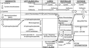
Formation of the chitin-free walls of the egg capsule (ootheca) involves intermingling of the secretions of both the left and right colleterial sexual accessory glands of this cockroach in the genital vestibulum. As depicted in fig. 1, the larger left gland (LCG) secretes a 4-o-β-D glucoside of a 3 hydroxybenzoate, crystals of calcium oxalate (Stay et al., Reference Stay, King and Roth1960), several proteins (oothecins – Pau et al., Reference Pau, Weaver and Edward-Jones1986) and a latent phenol oxidase, whereas the smaller right gland (RCG) exudes a β-D-glucosidase enzyme (Brunet & Kent, Reference Brunet and Kent1955; Wyatt & Davey, Reference Wyatt and Davey1996), as well as an ‘activator’ for the latent LCG phenoloxidase, (Whitehead et al., Reference Whitehead, Brunet, Kent and Snowball1965; Sugumaran & Nellaiappan, Reference Sugumaran and Nellaiapan1990). The copper-containing o-diphenoloxidase (not a tyrosinase: Whitehead et al., Reference Whitehead, Brunet and Kent1960) catalyses the synthesis of an o-benzoquinone from the 3,4-diphenol (catechol), which then cross-links the oothecins to form the walls of the egg case. Willis & Brunet (Reference Willis and Brunet1966) showed that the corpus allatum controls the function of the LCG but seemingly not the smaller RCG. Regulation of the synthesis of oothecins in the LCG of cockroaches is governed by juvenile hormone (JH). (Willis & Brunet, Reference Willis and Brunet1966; Tobe et al., Reference Tobe, Ruegg, Stay, Baker, Miler and Schooley1985; Pau et al., Reference Pau, Weaver and Edward-Jones1986).
Flow diagram depicting the rôle haemocytes play in synthesis from L-p-tyrosine of o-benzoquinone which cross-links oothecins to form sclerotin which hardens the oothecal walls.

This article outlines the rôle of certain haemocytes, found to be adhering to the LCG but not to RCG (Whitehead, Reference Whitehead1970a), which are responsible for the decarboxylation and deamination of tyrosine, the first two steps in the biosynthetic pathway (fig. 1) from tyrosine to o-benzoquinones, required for cross-linking oothecins to form sclerotin.
Materials and methods
The insects
Cockroaches were reared and females dissected as described (Brunet & Kent, Reference Brunet and Kent1955; Whitehead et al., Reference Whitehead, Brunet, Kent and Snowball1965). When adult females were injected in their abdomens on the day they emerged, the consequence of the treatment was decided by dissecting out the colleterial glands (LCG or RCG) seven days later.
Separation of tyrosine metabolites
Paper chromatography (using n-Butanol:glacial Acetic acid:water – 4:1:1 (BAW); 2% glacial acetic acid or n-Propanol/N-HCl – 3.1) and high voltage electrophoresis (HVE for 40 min at pH 6.5) was employed to separate the metabolites which formed from L-p-tyrosine as previously described (Whitehead, Reference Whitehead1969, Reference Whitehead1970a,Reference Whiteheadb). Detection of the metabolites relied on spraying the paper with (i) 0.4% (w/v) ninhydrin in n-butanol saturated water followed by heating for 10 min at 110°C or (ii) equal volumes of 1% (w/v) ferric chloride+1% (w/v) potassium ferricyanide as described (Whitehead et al., Reference Whitehead, Brunet, Kent and Snowball1965).
To identify the metabolites formed, 1 cm strips from the chromatograms or electropherograms were immersed in scintillant and counted in an LKB 1210 model spectrometer. In addition, the strips cut from the electropherograms were run through a Geiger-Muller strip counter to locate and identify the labeled metabolites arising.
β-D-glucosidase activity in the secretion from the RCG of cockroaches injected on the day of moulting with various doses of ecdysone was measured fluorimetrically in an Aminco-Bowman spectrometer using 4-methylumbelliferyl-β-D-glucopyranoside (from BDH) as the substrate by the method previously described (Whitehead, Reference Whitehead, Novak and Slama1965).
Incubation of tissues during in vitro experiments
(i) Haemocytes adhering to dissected LCGs were washed from the glands with isotonic insect saline (0.9% NaCl+0.01% CaCl) and incubated with 1 μCi (U-14C) L-tyrosine or (2-14C) tyramine for one hour.
(ii) At the same time, the gland, before and after washing, was also incubated for 1 h with these two substrates dissolved in 0.3 M sodium phosphate buffer (pH 6.8).
(iii) Haemolymph (100 μl) from females was incubated with 2 μCi [U-14C] L-p-tyrosine in pH 6.8 acetate buffer for 30 min at 37°C, after which the labeled metabolites found in the supernatant were separated by chromatography (BAW – 4.1.1). The labeled metabolites found on the dried paper in two regions at Rf%, 71–74 and 61–63, were subjected to HVE followed by assessment of activity on 1 cm strips using a scintillation counter.
Electron microscopy (EM)
Scanning EM of LCG was performed (C.P. dried, Au coating±450  by personnel in the Stereoscan Microscope Unit at Bedford College in London. LCG, with adhering haemocytes, were fixed in glutaraldehyde, stained with Osmium tetroxide (OsO4) then set in acrylamide before thin sections, for appraisal by EM in the Zoology Department, were cut using an ultra microtome.
Materials
Chemicals were obtained from L. Light & Co. Ltd, Colnbrook, Bucks or from BDH Ltd, London. Decarboxylase enzyme inhibitors were sourced from Hoffman la Roche, Basel (STHB hydrazine; RO 4-4602) or from Merck, Sharp and Dohme, Rahway NJ, USA (MK-485). DL-α-methyl DOPA and dimethyl tyrosine (DMT) were supplied by Kodak Ltd, Liverpool. Marsilid (iproniazid) came from Sigma Chemicals Inc. The inhibitors were formulated and injected with (U-14C) tyrosine into the abdomens of adult females as previously described (Whitehead, Reference Whitehead1974b). Scintillation fluid NE250 was sourced from Nuclear Enterprises Ltd, Edinburgh.
Radioactive chemicals
UDPG-14C, (U14C) L-p-tyrosine, (2-14C) tyramine and (3H) octopamine were obtained from the Radiochemical Centre, Amersham.
Hormones
Ecdysone was a gift from Dr Jan Koolman, Marburg. A juvenile hormone analogue (methoprene) was sourced from Calbiochem Inc.
Results
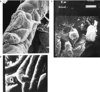
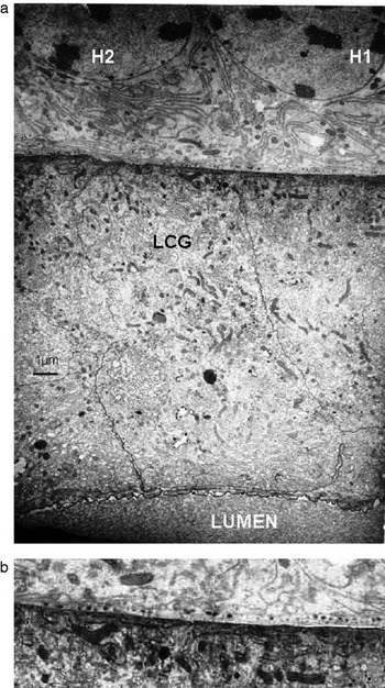
Microscopic examination of dissected colleterial glands -LCG- (fig. 2a) revealed numerous cells adhering to the surface of the glands. As these cells were removed (lower photo; fig. 2b) by rinsing the glands in insect saline, it was assumed that they were haemocytes (fig. 2c). Surprisingly, these cells were not present on RCGs. That the junction between adhering haemocytes and the LCG membrane was tight was confirmed by electron microscopy (fig. 3a). Granules, which stain heavily with OsO4, were seen underlying haemocytes at the junction between them and the gland (fig. 3b).
(a) Scanning EM of the colleterial gland (LCG) showing adhering haemocytes before the gland was washed and rinsed with isotonic insect saline. (Scale: diameter of a tubule±40 μm). (b) Scanning EM of LCG after washing of the gland with insect saline to remove adhering haemocytes. (Scale: diameter of a tubule±40 μm). (c) Scanning EM of several haemocytes adhering to the colleterial gland (LCG).

(a) Thin section EM showing two haemocytes (H1, H2) adhering to the surface of the colleterial gland (LCG). The ‘lumen’ of the LCG cell is shown (see also fig. 1). (b) Enlargement from (a) showing that OsO4 dense granules can be seen underlying the junction between adhering haemocytes and LCG.

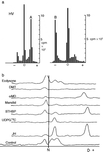
Two enzymes, Tyrosine decarboxylase (Whitehead, Reference Whitehead1969: EC.4.1.1.25) and a monoamine oxidase (MAO), were detected in haemocytes adhering to the LCG outer surface. These enzymes were also present in haemolymph from females (fig. 4a) taken seven days after emergence.
(a) Quantitative detection, using a scintillation counter, of (B) tyramine (right hand trace) and of (A) a phenolic acid (left hand trace) both derived from (U14C) tyrosine. A and B were formed in female haemolymph after incubation at 37°C for 30 min. HVE was employed at 4 KV and pH 6.5 for 40 min to quantify the metabolites present on 1 cm strips after they were first separated into two zones, Rf% 71–74 (A) and 61–63(B), by chromatography in n-Butanol acetic acid water, 4.1.1 and eluted from the paper prior to HVE. (b) Scanning of HV electropherograms shows qualitative evidence that injecting an analogue of juvenile hormone (JH) into recently emerged females enhances the synthesis in LCG of the glucoside of dihydroxybenzoate and a phenolic acid from 14C- tyrosine seven days later. Injection of enzyme inhibitors marsilid and dimethyl tyrosine (0.1 μmole; see table 2) or ecdysone (25 or 100 ng) with tyrosine depresses synthesis of a phenolic acid migrating to the anode. Separation on paper was achieved in 40 min by HVE electrophoresis (4 KV) at pH 6.5.

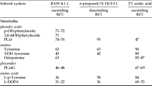
Proof that the electrophoretically basic substance formed from p-tyrosine was indeed tyramine was deduced from its behaviour (table 1) in three different chromatographic solvents systems and from its mobility ‘m’=−1.15 at pH 6.5 with ε-DNP-lysine being neutral. The right-hand electropherogram shown in fig. 4a depicts quantitatively the conversion by haemocytes of tyrosine to the basic amine (B) tyramine.
Separation by chromatography, using three solvent systems and Whatman no. 1 paper (21×50 cm), of L-p-tyrosine metabolites found in female cockroaches after carrying out in vivo or in vitro experiments with LCG with or without adhering haemocytes.

The decarboxylase and MAO enzymes were absent in cells of the gland itself when it was washed in insect saline to remove adhering haemocytes. In the absence of the haemocytes, the phenolic glucoside was not synthesized from tyrosine by the isolated gland, (Whitehead, Reference Whitehead1970a, Reference Whitehead1974a).
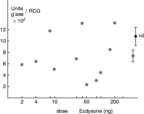
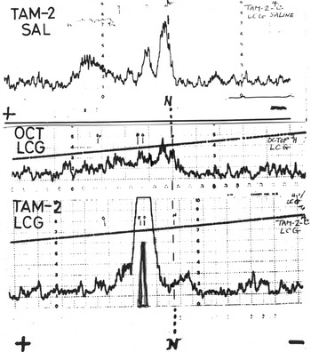
Dissected LCGs, which had not been rinsed with saline, when incubated with 1 μg JH (methoprene) and 1 μCi (U-14C) tyrosine, showed an enhanced ability to decarboxylate and deaminate tyrosine (fig. 4b, table 2), whereas added α-ecdysone had little effect. By contrast (Whitehead, Reference Whitehead1974a), injecting α-ecdysone (25–100 ng) into the abdomen on the day of emergence of the adult female significantly (P<0.001) inhibited synthesis later of the phenolic glucoside in the LCG (table 2) but surprisingly not of the β-glucosidase (fig. 5) present in the RCG. Injecting inhibitors of the decarboxylase, monoamineoxidase and monooxygenase enzymes together with (U-14C) tyrosine (table 2) into recently moulted females reduced later synthesis of the phenolic glucoside, thus reinforcing the claim that these enzymes were major players in the overall regulation of sclerotin formation required to harden the oothecal walls (fig. 1). Furthermore, p-hydroxyphenylacetic acid was shown earlier to be derived from (1-14C) tyramine and (G-3H) tyramine (Whitehead, Reference Whitehead1970a), or in the present case from (2-14C) tyramine, but not from tritiated octopamine, (middle trace of fig. 6) when the labeled amines were incubated for one hour with haemocytes washed from LCGs with saline (upper trace of fig. 6). The left-hand trace of fig. 4a indicates that in haemolymph from females, tyrosine itself can give rise to a phenolic acid.
Injection of ecdysone (2–200 ng) did not inhibit biosynthesis of the β-glucosidase enzyme found in homogenates of the right colleterial gland (RCG).

Qualitative electropherogram (lower trace) showing separation by HVE (4 KV) of labeled metabolites arising in LCG+adhering haemocytes (TAM-2 LCG) from 2-14C- tyramine migrating towards the anode at pH 6.5. The middle trace shows 3H-octopamine is not metabolized by LCG, while the upper trace (TAM-2 SAL) shows that haemocytes washed from LCG with saline can deaminate tyramine.

Effect of injecting enzyme inhibitors or ecdysone dissolved in saline on in vivo biosynthesis of a phenolic glucoside in seven days from (U-14C) L-p-tyrosine in LCGs of female P. americana cockroaches.

a The 14C labelled dihydroxyphenolic-4-β-D-glucoside was separated from other metabolites by paper chromatography in n-butanol:acetic acid:water (4:1:1 BAW) before counting in a LKB liquid scintillation spectrometer.
b Percent inhibition was calculated from the average of measurements taken after injection of 14C-tyrosine into the abdomens of three cockroaches.
Discussion and conclusions
It is proposed that, unlike the situation pertaining in newly-moulted cockroaches, (Whitehead, Reference Whitehead1969), where dopamine is formed in haemocytes from tyrosine and tyramine in the presence of the neuropeptide tanning hormone (secreted by the terminal abdominal ganglion), here – in the adult female cockroach – tyramine is oxidized by the MAO in haemocytes adhering to LCG to give p-hydroxyphenyl acetic acid (left-hand trace of fig. 4a, upper trace of fig 6), which is later hydroxylated by a monooxygenase in the gland to give homoprotocatechuate (3,4-dihydroxyphenylacetate: Koeppe & Mills, Reference Koeppe and Mills1974), the precursor of the dihydroxybenzoate (fig. 1). Haemocytes adhering to LCGs apparently lack the monooxygenase enzyme to hydroxylate the monophenol, whereas we know that the LCG does secrete a copper-containing phenoloxidase enzyme (Brunet & Kent, Reference Brunet and Kent1955; Whitehead et al., Reference Whitehead, Brunet and Kent1960, Reference Whitehead, Brunet, Kent and Snowball1965). That the enzyme exists in a latent, particulate form until it is activated by the secretion from the RCG (Whitehead et al., Reference Whitehead, Brunet, Kent and Snowball1965; Sugumaran & Nallaiappan, Reference Sugumaran and Nellaiapan1990) is illustrated in fig. 1. Activation by fatty acids or phospholipids (Sugumaran & Nallaiappan, Reference Sugumaran and Nellaiapan1990) enables the enzyme to give rise to o-benzoquinones once the secretions from both glands intermingle in the vestibulum. Exactly how the secretion from the RCG carries out this important function needs to be investigated further.
Regarding the final steps in the biosynthetic pathway from tyrosine, UDPG and the appropriate transferase gives rise to the 4-β-D-glucoside of dihydroxyphenylacetate (homoprotechuate) responsible for protecting the diphenol from oxidation by the o-diphenoloxidase present in the LCG secretion (fig. 1). It is assumed that the phenylacetate gives rise to the benzoate, but is not clear exactly when and how this occurs. Further study is needed.
Finally, it is proposed that synthesis of the oothecal sclerotizing agent, whether dihydroxybenzylalcohol (Pau & Acheson, Reference Pau and Acheson1968; Sugumaran et al., Reference Sugumaran, Semensi, Dali and Nellaiappan1991) or a derivative of dihydroxyphenylacetate (Koeppe & Mills, Reference Koeppe and Mills1974), is controlled by the action of JH acting on haemocytes adhering to the larger LCG. On the other hand, JH could possibly act on developing LCG cells to promote binding of the specialized haemocytes. Ecdysone appears to somehow negate this.
It has already been shown by Friedel et al. (Reference Friedel, Feyereisen, Mundall and Tobe1980) that 20-OH-ecdysone has an allostatic effect in the cockroach D. punctata. In the spruce budworm, 20-OH-ecdysone suppresses production and/or release of JH I (Kathedi et al., Reference Kathedi, Perera, Zheng, Feng, Krell, Retnakaran and Palli2004). It is of interest here to recall that transplanting LCG into male cockroaches (Shaaya & Bodenstein, Reference Shaaya and Bodenstein1995) shuts off the synthesis of the phenolic glucoside but not of oothecin production. Perhaps males lack the specific haemocytes shown in this paper to be required for LCG to synthesize the phenolic glucoside? This parabiosis experiment should definitely be repeated.
Injection of α-ecdysone and inhibitors of decarboxylase and amineoxidase enzymes into recently moulted cockroaches inhibits synthesis in LCG of hydroxybenzoates (table 2) from 14C-tyrosine. Similarly, it was found that 20-OH-ecdysone inhibits uterine milk gland function in the female tsetse fly Glossina m. morsitans (Whitehead, Reference Whitehead1981), but topical application of JH III on days 2, 4 and 6 of the pregnancy cycle did not reverse this inhibition completely (Whitehead, Reference Whitehead1984, Reference Whitehead and Hedin1985).
Scanning electron microscopy (SEM) has demonstrated the presence of haemocytes closely associated with the larger LCG (fig. 2a, c); but, significantly for this study, they are absent on the RCG.
Thin section EM (fig. 3a) shows that the junction (fig. 3b) between adhering haemocytes and the left gland is indeed proximal. The OsO4 dense granules, seen attached closely to the outer membrane of the haemocytes (fig. 3b), were initially seen to originate from the Golgi apparatus within the LCG; they might, therefore, somehow be involved in initiating association of the haemocytes to the gland. Ultimately, JH may be responsible for the generation of binding sites on the LCG to which haemocytes can adhere; it appears that ecdysone reverses or antagonizes this activity or function. That ecdysteroids can act as anti-gonadotrophins in insects (or have an allatostatic effect: Friedel et al., Reference Friedel, Feyereisen, Mundall and Tobe1980) has been well documented (Whitehead, Reference Whitehead1974a, Reference Whitehead1981; Davey, Reference Davey1982, Reference Davey1993).
This incomplete study is published to encourage cell biologists to investigate the rôle certain haemocytes can play in the function of sexual accessory glands in those Orthopteroid insects, for example the Blattodea, which produce capsules to protect their eggs.
Acknowledgements
Support by the Agricultural Research Council and funds from the Overseas Development Administration (Grant R 2956) is gratefully acknowledged. Dr Peter Brunet encouraged this investigation. Dr Robin Offord of the Biochemistry Department kindly provided the facility for high voltage electrophoresis (HVE). Barbara Luke ably undertook the EM study and Monica Williams ably assisted the investigation.
Supplementary material
The online figure can be viewed at http://journals.cambridge.org/ber