Introduction
Clinostomum is a digenetic trematode that naturally parasitizes the throat and oesophagus of piscivorous birds, specially buff-backed herons (Ardeola ibis ibis), producing serious illness (Yamaguti, Reference Yamaguti1971; Aohagi et al., Reference Aohagi, Shibahara and Machida1992). Herons have an important role in the biological control of agricultural pests such as insects, molluscs and earthworms (Aboel Hadid & Lotfy, Reference Aboel Hadid and Lotfy2007). Moreover, metacercariae of Clinostomum parasitize fish, producing yellow grub disease (Silva-Souza & Ludwig, Reference Silva-Souza and Ludwig2005; Abd El-Galil et al., Reference Abd El-Galil, Kamel, Abd El-Rahman and Fouda2007), where growth rate, survival and reproductive fitness have been reported to be decreased in infected fish (Barber et al., Reference Barber, Hoare and Krause2000). Additionally, the presence of cysts renders fish unacceptable to consumers (Eiras, Reference Eiras1994). Most importantly, the disease can infect humans, causing pharyngitis and/or laryngitis if fish are consumed raw (Chung et al., Reference Chung, Kong and Moon1995; Shirai et al., Reference Shirai, Matsubara, Ohnishi, Nishiyama, Miyamoto, Watanabe, Harada, Kadota and Kohno1998; Kitagawa et al., Reference Kitagawa, Oda, Totoki, Washizaki, Oda and Kifune2003). These facts render the control of this parasite of great importance.
Irradiation has been used successfully to attenuate the infective stage of many parasites, and these have been used as vaccines against a number of parasites including Schistosoma (Dean, Reference Dean1983; Richter et al., Reference Richter, Harn and Matuschka1995; Coulson, Reference Coulson1997), Fasciola (Younis et al., Reference Younis, Yagi, Haroun, Gameel and Taylor1986; A/Gadir et al., Reference A/Gadir, Haroun and Gameel1987; Creaney et al., Reference Creaney, Spithill, Thompson, Wilson, Sandeman and Parsons1995), Taenia (Konno et al., Reference Konno, Oku, Sakai and Kamiya1997; Lagapa et al., Reference Lagapa, Konno, Oku, Nonaka and Kamiya2001), Ancylostoma (Menon & Bhopale, Reference Menon and Bhopale1985), Necator americanus (Jian et al., Reference Jian, Jun-Min, Hai-Chou, Hui-Qing, Hai-Nan, Hotez, Bin and Shu-Hua2006) and Trichenella (Nakayama et al., Reference Nakayama, Inaba, Nargis, Chisty, Ito and Kamiya1998). Accordingly, this study was undertaken to evaluate the effectiveness of UV-irradiation in attenuating C. complanatum metacercariae and evaluate their potential use as a vaccine.
Materials and methods
Birds
Adult buff-backed herons were used as final host for C. complanatum according to Aboel Hadid & Lotfy (Reference Aboel Hadid and Lotfy2007). Birds were caught from different villages in Beni-Suef province and reared in metal wire cages. Faeces were examined daily for at least 1 week to detect any parasitic infection. Birds that were found to harbour any parasites were excluded from the experiment. Parasite-free birds were maintained under suitable living conditions of fixed temperature range from 25°C to 28°C and 12 h of light per day. The birds' meals consisted of fresh beef meat and acidified tap water.
Parasitological examination
To detect trematode infections, 2 g of collected bird faeces were mixed with 10 ml tap water. The mixture was then strained using a stainless-steel strainer to remove large debris and centrifuged for 5 min at 1500 rpm. The supernatant was decanted and two drops from the sediment were transferred to a glass slide and examined using a 40 × light microscope. Three slides were examined for each sample (Hendrix, Reference Hendrix1998).
Eggs of cestodes and nematodes, as well as oocysts of protozoa, were screened using a centrifugal flotation technique. The collected faeces were mixed well with 10 ml tap water and sieved into a 20 ml centrifuge tube. The tube was then filled with saturated salt solution and centrifuged at 1500 rpm for 5 min. The surface film, which may contain parasite eggs or protozoan cysts, was collected by touching the surface gently with a wire loop, transferring it to a glass slide, covering and examining under a light microscope at 40 × magnification (Hendrix, Reference Hendrix1998).
Collection of encysted Clinostomum metacercariae
Oreochromis niloticus fish were caught from a semi-closed tributary of the Nile River at Beni-Suef province, Egypt. First, the buccal cavity, branchial cavity, branchiostegal muscle and gills of each fish were examined macroscopically for the presence of encysted Clinostomum metacercariae. Cysts were removed carefully, counted and placed in sterile Petri dishes containing 0.85% saline. Before experimental infection of A. ibis ibis, specimens of metacercariae were preserved, stained with acetocarmine and examined microscopically to ensure their identity (Amato et al., Reference Amato, Boeger and Amato1991).
UV-irradiation of encysted metacercariae
Twenty millilitres of 0.85% saline containing 100 cysts (5 cysts/ml) were poured into a clean glass dish and placed at the centre of a closed carton box (30 × 28 cm). A cyst suspension was exposed – through an opening in the centre of the box's upper side – to 254 nm UV light from a VL6-LC UV lamp (ETS Vilber-Louramat, Marne La Vallee, Cedex, France) held above the box. The distance between the UV lamp and the surface of the cyst suspension was 24 cm and exposure time was either 30 or 60 min. Light intensity at the bottom of the dish was estimated to be greater than 90% of that at the surface (Dean et al., Reference Dean, Murrell and Xu1983).
Experimental design
Thirty-five adult A. ibis ibis were divided equally into seven groups: group A, infection-free birds as negative control; group B, birds infected with 20 non-irradiated Clinostomum cysts; group C, birds infected with 20 cysts pre-exposed to UV light for 30 min; group D, birds infected with 20 cysts pre-exposed to UV light for 60 min; group E, birds infected with 20 cysts pre-exposed to UV light for 60 min then challenged by 20 normal cysts 8 days post-vaccination; group F, birds kept infection-free for 8 days, then infected with 20 normal cysts and kept as a positive control; and group G, birds infected with 20 normal cysts then after 8 days re-infected with another 20 normal cysts.
Experimental infection and worm recovery
Viable encysted metacercariae were washed three times with 0.85% saline and inoculated into the buccal cavity of birds. Any bird that spitted out any cyst was excluded from the experiment. On the sixth day post-infection, faecal examination was carried out daily, according to Hendrix (Reference Hendrix1998), to record parasite eggs. After 8 days post-infection, birds of groups B, C and D were sacrificed, while birds of groups E, F and G were sacrificed after 16 days from the first infection (8 days of post-challenge infection). The oesophagus, throat and buccal cavity of each bird were examined for the presence of adult worms. When found, worms were removed aseptically from each bird and counted. The worm burden of each group was calculated as mean ± SD. Reduction percentage in worm burden of vaccinated or vaccinated-challenged groups was calculated according to the following equation:

In vitro worm fecundity
Adult worms were cultured in vitro according to the method of Sanderson et al. (Reference Sanderson, Bartlett and Whitfield2002). Briefly, adult worms were washed three times with 0.85% saline in a sterile Petri dish. Worms were then placed in a sterile Petri dish with Eagle's minimum essential medium (MEM) (one worm/ml) containing 10% fetal calf serum (FCS), 0.15% sodium bicarbonate, 2 mml-glutamine, 1 mm sodium pyruvate and 50 Uml− 1 penicillin–streptomycin (Sigma-Aldrich, Poole, Dorset, UK). For recovering the eggs, dishes were then incubated at 37°C for 24, 48 and 72 h. Eggs, laid by worms that were recovered from each bird at each time interval, were collected and counted according to Soulsby (Reference Soulsby1982). Briefly, eggs were resuspended in a known volume and five replicates, each of 200 μl, were counted using McMaster slides. Worm fecundity and percentage of egg abnormalities for each bird at each time interval were then calculated.
Egg hatching
Eggs from each group were incubated in sterile Petri dishes containing 0.85% saline with porous slofane sheet covering for aeration in an incubator at 29°C for 2 weeks with daily shaking. After 8 days post-incubation, eggs were examined daily to record the embryonation and released free miracidia.
Blood sampling
Before sacrificing the birds, blood samples were collected from the heart or wing veins. Blood samples were kept at room temperature for 2 h, then overnight at 4°C. Samples were then centrifuged for 10 min at 3000 rpm. Sera were collected and aliquots were taken and stored at − 70°C until used.
Soluble worm antigen preparation
Soluble worm antigen was prepared according to Salih et al. (Reference Salih, Bartlett and Voller1978). Briefly, viable adult worms were suspended at a concentration of 5 worms/ml in 0.01 m phosphate buffered saline (PBS) containing 5 mm phenyl methyl sulphonyl fluoride (PMSF), 1 mm Na2EDTA, 1 mm iodoacetamide and 1 mm aprotinin (Sigma-Aldrich) as protease inhibitors, and the pH was adjusted to 7.2. Worms were then exposed to three thaw–freeze cycles, then homogenized in a melting ice bath for 45 min. The resulting homogenate was centrifuged at 3500 rpm for 30 min at 4°C. The protein content of the supernatant was determined (Bradford, Reference Bradford1976). The supernatants were aliquoted and stored at − 70°C until used.
Passive haemagglutination test
The passive haemagglutination method was performed as described by Boyden (Reference Boyden1951). Briefly, tannic acid-treated sheep erythrocyte suspension was sensitized by 180 μg/ml soluble adult C. complanatum antigen. Serial dilutions of serum samples were prepared in a V-shaped microtitre plate according to Takatsy (Reference Takatsy1956) and 50 μl of sensitized erythrocytes were added to each well. The plate was then incubated at room temperature for 3 h. The well of the highest dilution that showed visible agglutination clearly was considered to be the end point of the test. Titres were calculated as the reciprocal of the antiserum dilution at the end point.
Statistical analysis
The data were analysed using one-way analysis of variance (ANOVA) using PC-STAT (University of Georgia, 1985). Results were expressed as mean ± SD. Values of P < 0.05 were considered statistically significant.
Results
Infection prevalence and intensity
Out of 400 examined fish (O. niloticus), 164 (41%) were found to be infected with C. complanatum metacercariae. Number of cysts counted per infected fish ranged from 3 to 10.
Effect of UV irradiation on the maturation of C. complanatum metacercariae
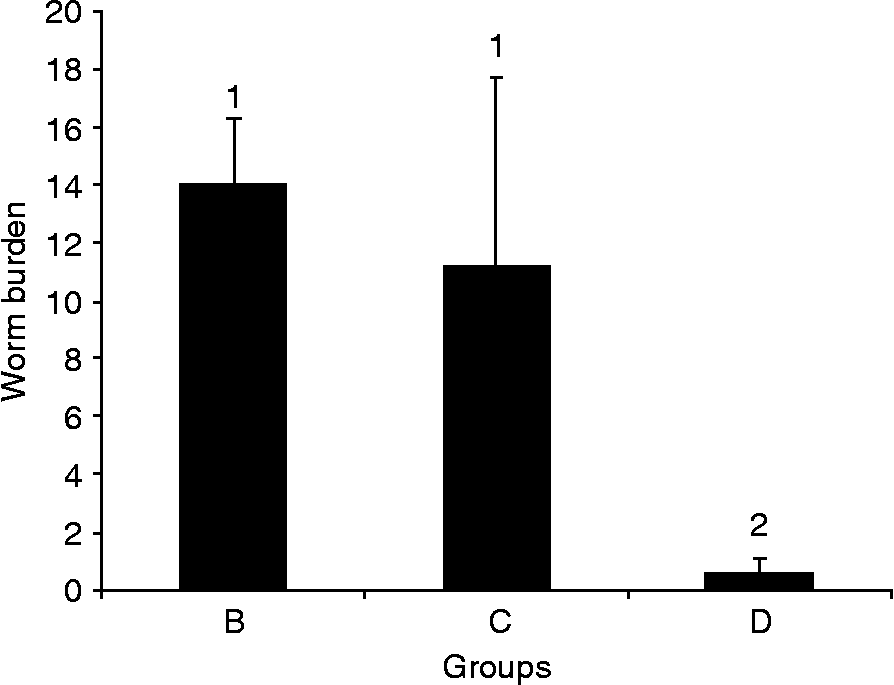
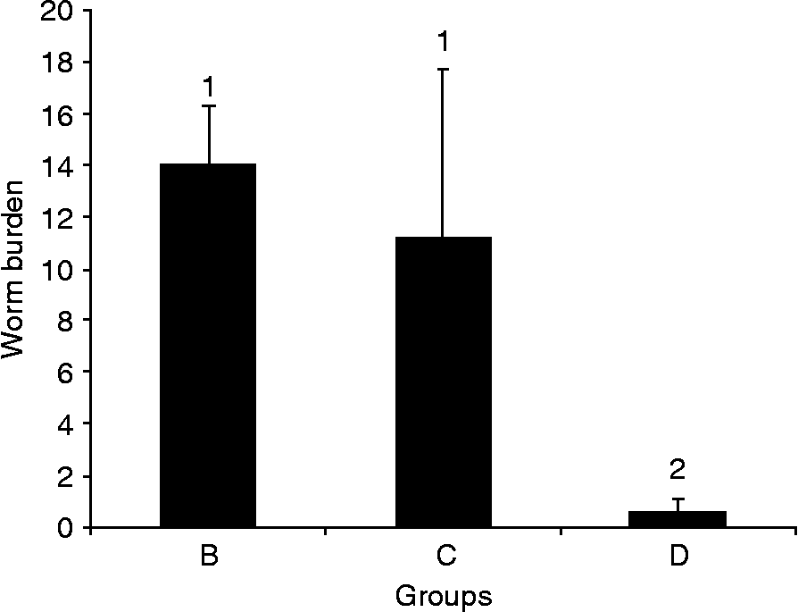
Infected birds with metacercariae pre-exposed to UV light for 60 min showed 95.7% and 94.6% significant (P < 0.01) reduction in worm burden when compared to infected groups with either normal or 30 min irradiated metacercariae, respectively. However, there was no significant difference (P>0.05) between worm burden in birds infected with either normal metacercariae or metacercariae pre-exposed to UV light for 30 min (fig. 1).
Effect of UV irradiation on the maturation of C. complanatum metacercariae. Worm burden of birds infected with 20 non-irradiated Clinostomum metacercariae (B); birds infected with 20 metacercariae pre-exposed to UV light for 30 min (C); and birds infected with 20 metacercariae pre-exposed to UV light for 60 min (D). Values represent the mean ± SD of five birds in each group. Histograms that share the same superscript number are not significantly different.
Protective capacity of 60 min UV-exposed metacercariae against challenge infection
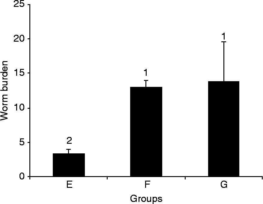
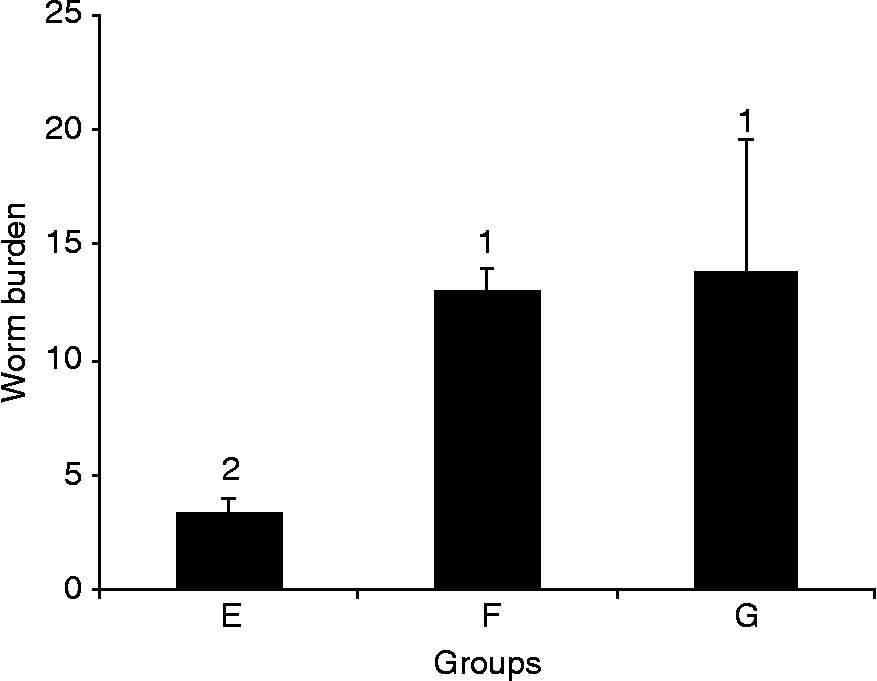
Vaccination of herons with metacercariae pre-exposed to UV light for 60 min resulted in 73.8% and 75.4% significant (P < 0.01) reduction in worm burden upon challenge infection compared to unvaccinated birds or those pre-infected with normal metacercariae, respectively. On the other hand, there was no significant difference (P>0.05) in worm burden between birds infected with non-irradiated cysts and re-infected after 8 days with normal ones, and those infected with normal cysts (fig. 2).
Protective capacity of 60 min UV-exposed metacercariae against challenge infection. Worm burden of birds infected with 20 metacercariae pre-exposed to UV light for 60 min then challenged by 20 normal metacercariae 8 days post-vaccination (E); birds kept infection-free for 8 days, then infected with 20 normal metacercariae (F); birds infected with 20 normal metacercariae and after 8 days re-infected with another 20 normal metacercariae (G). Values represent the mean ± SD of five birds in each group. Histograms that share the same superscript number are not significantly different.
Effect of metacercariae exposure to UV light on in vitro adult worm fecundity
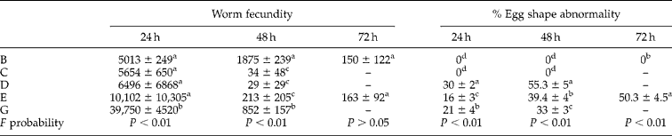
At 24 h after in vitro incubation, there was no significant difference (P>0.05) in fecundity among worms recovered from birds infected with normal, 30 min UV-exposed cysts, 60 min UV-exposed cysts and those vaccinated with 60 min UV-exposed cysts and challenged with normal ones. Meanwhile, worms recovered from birds infected and re-infected with normal cysts laid a significantly (P < 0.01) larger number of eggs than other groups (table 1).
Effect of metacercariae exposure to UV light on in vitro adult worm fecundity and percentage of egg shape abnormality.
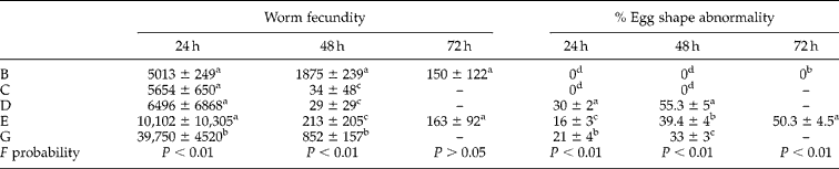
Worms recovered from birds infected with normal metacercariae (B); infected with 30 min UV-exposed metacercariae (C); infected with 60 min UV-exposed metacercariae (D); infected with 60 min UV-exposed metacercariae and challenged with normal metacercariae (E); and infected and re-infected with normal metacercariae (G) were cultured in vitro. The number of eggs laid by worms was counted at each time interval and the percentage of abnormality in egg shape was calculated. Data are expressed as mean ± SD of five birds in each group. Means that share the same superscript symbol are not significantly different.
After 48 h post-incubation, worms recovered from birds infected with 30 min UV-exposed cysts, 60 min UV-exposed cysts and birds vaccinated with 60 min UV-exposed cysts and challenged with normal cysts laid significantly (P < 0.01) fewer eggs compared to worms recovered from birds infected with either normal or infected and re-infected with normal cysts. Worm fecundity from birds vaccinated with 60 min UV-exposed cysts was lowest, while birds infected with normal cysts showed the highest value (table 1).
Fecundity of worms recovered from birds infected with 30 min UV-exposed cysts, 60 min UV-exposed cysts and birds infected and re-infected with normal cysts could not be estimated for a longer incubation period since all worms died before 72 h of incubation. In the meantime, worms recovered from birds infected with normal cysts and birds vaccinated with 60 min UV-exposed cysts and challenged with normal cysts remained alive after 72 h of incubation. There was no significant difference (P>0.05) in worm fecundity between both groups (table 1).
Effect of UV exposure on percentage of egg shape abnormality
Normal eggs were oval in shape, operculated, thin shelled, brown in colour and measured 105–120 × 70–85 μm in diameter. In addition, yolk material occupied the entire space, masking the embryonal cells. However, abnormal eggs were spherical with reduced size (75–85 μm in diameter), or complete absence of operculum, or normal shape and size but with abnormal yolk material distribution. There were no abnormalities in eggs laid by worms developed from normal metacercariae at 24, 48 and 72 h post-incubation. Similarly, eggs laid by worms developed from 30 min UV-exposed metacercariae at 24 and 48 h post-incubation showed no abnormalities. On the other hand, a significant (P < 0.01) number of abnormal eggs laid by worms developed from 60 min UV-exposed metacercariae at 24 and 48 h compared to other groups. Abnormal eggs laid by worms recovered from birds vaccinated with 60 min UV-exposed cysts and challenged with normal cysts increased significantly, from 16% at 24 h to 50.3% at 72 h post worm incubation (table 1).
Effect of metacercariae exposure to UV light on hatching capability
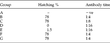
Seventy-eight per cent of eggs laid by worms developed from both normal and 30 min UV-exposed metacercariae hatched into miracidia. Similarly, 78% of eggs laid by worms recovered from birds infected and re-infected with normal metacercariae hatched into miracidia. However, only 1.5% of eggs produced by worms recovered from birds vaccinated with 60 min UV-exposed cysts and challenged by normal cysts hatched into miracidia. Interestingly, all eggs laid by worms developed from 60 min UV-exposed metacercariae did not hatch at all (table 2).
Effect of metacercariae exposure to UV light on hatching capability of eggs in vitro and humoral immunity of vaccinated herons.
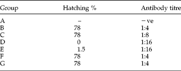
Hatching percentage of eggs laid in vitro by worms recovered from birds infected with normal metacercariae (B); infected with 30 min UV-exposed metacercariae (C); infected with 60 min UV-exposed metacercariae (D); infected with 60 min UV-exposed metacercariae and challenged with normal metacercariae (E); infection-free for 8 days, then infected with normal metacercariae (F); infected and re-infected with normal metacercariae (G) were calculated 2 weeks post-incubation. Antibody response of non-infected birds (A) and the other bird groups against soluble adult worm antigen was measured by passive haemagglutination.
Effect of UV-attenuated metacercariae vaccination on humoral immunity of A. ibis ibis
Both birds vaccinated with 60 min UV-exposed cyst and vaccinated-challenged birds showed higher antibody titre (1:16), while lower antibody titre (1:8) was detected in birds infected with 30 min UV-exposed cysts. The lowest antibody titre (1:4) was observed in birds infected with normal cysts and birds infected and re-infected with normal cysts. No visible agglutination was detected in non-infected birds (table 2).
Discussion
Exposure to UV light impaired the development of C. complanatum metacercariae into adult worms. This was effective upon increasing the exposure period from 30 to 60 min. Reduction in maturation in response to UV light exposure could be attributed to the harmful effects from the initiation of photochemical absorption by molecules including nucleic acids and various proteins, in addition to other molecules such as porphyrins, carotenoids and steroids (Harm, Reference Harm1980; Diffey, Reference Diffey1991). Nucleotide bases represent the chromophores (absorbing centres), with pyrimidine being comparatively sensitive to UV light (Smith, Reference Smith1966). Consequently, pyrimidine derivatives have been reported to be the major photoproducts following the absorption of UV photons by DNA (Errera, Reference Errera1958; Nabih & Abd El-Hamid, Reference Nabih and Abd El-Hamid1984). These photoproducts include cyclobutyl-type dimers (pyrimidine dimers), pyrimidine adducts (spore photoproducts), pyrimidine hydrates and DNA–protein cross-links (Patrick & Rahn, Reference Patrick, Rahn and Wang1976; Diffey, Reference Diffey1991). Damage to DNA and RNA molecules by absorbing energy in the UV range (maximum between 254–265 nm) includes building bridges that destroy molecular functions. While some damage can be repaired through photo-reactivation, post-replication and excision repair (Freifelder, Reference Freifelder1987; Lehmann & Norris, Reference Lehmann and Norris1990), some damage is permanent and cannot be repaired. This risks both survival and development of an organism (Auerbach, Reference Auerbach1976; Nilsson, Reference Nilsson1996).
While some 60 min UV-exposed metacercariae, however few, developed into adult worms, eggs laid by those worms were greatly reduced in number, and most of those eggs were abnormal in shape. Interestingly, all eggs (normal and abnormal) were unable to hatch into miracidia. The most likely explanation of these results may be related to the serious effect of UV light on DNA and RNA molecules of metacercariae. Accordingly, this resulted in retardation of development of metacercariae into adult worms, reduction of fecundity of developed worms and lowered hatchability of any eggs produced. On the other hand, the insignificant difference in egg abnormality and egg hatchability of worms developed from 30 min UV-exposed metacercariae compared to worms developed from normal metacercariae indicated the insufficiency of exposure to UV light for 30 min to attenuate metacercariae.
The use of irradiated infective stages to elicit immune responses to homologous infections has been acknowledged against many parasitic helminths. For example, vaccination of golden hamsters against Ancylostoma ceylanicum reduced worm burden to about 1% following homologous challenge infection (Menon & Bhopale, Reference Menon and Bhopale1985). Similar results have also been reported indicating the effective use of this method to vaccinate mice (Dean, Reference Dean1983; Kamiya et al., Reference Kamiya, Ozaki, Nakayama and Inaba1993; Dean et al., Reference Dean, Mangold, Harrison and Ricciardone1996; el Ridi et al., Reference el Ridi, Ozaki, Sato, Inaba and Kamiya1997), rats (Konno et al., Reference Konno, Oku, Sakai and Kamiya1997) and hamsters (Jian et al., Reference Jian, Jun-Min, Hai-Chou, Hui-Qing, Hai-Nan, Hotez, Bin and Shu-Hua2006) against Schistosoma, Taenia and Necator, respectively. In line with the previous reports, data presented in the current study indicated that exposure of C. complanatum metacercariae to UV light for 60 min is a powerful tool in attenuating metacercariae. The possible role of attenuated metacercariae as a candidate vaccine for herons was obvious from their protective effect against challenge infection by normal metacercariae 8 days post-vaccination (73.8% reduction in worm burden). This was further confirmed by the results of humoral immune response in challenged birds, where antibody response against soluble adult worm antigens was higher in vaccinated and vaccinated-challenged birds compared to unvaccinated-infected and infected/re-infected birds. The role of antibody in a radiation-attenuated vaccine model has been reported in number of studies, including murine schistosomiasis (Jankovic et al., Reference Jankovic, Wynn, Kullberg, Hieny, Caspar, James, Cheever and Sher1999) and treatment with anti-μ (Sher et al., Reference Sher, Hieny, Jame and Asofsky1982). In both studies, protection against challenge infections was reduced. The capability of mediating passive transfer of resistance to naive recipients against Schistosoma (McLaren & Smithers, Reference McLaren and Smithers1988; Richter et al., Reference Richter, Incani and Harn1993) further confirmed the role of antibody-mediated immune response in a radiation-attenuated vaccine model. Yet, questions of how antibody- or cell-mediated response contributes to resistance against challenge infection were not addressed in this article and further investigation is needed.
In conclusion, these data showed that exposure of C. complanatum metacercariae to UV light at 254 nm for 60 min impaired maturation of metacercariae into adult worms. Treating A. ibis ibis with irradiated metacercariae elicited high resistance against challenge infection. These data signify the effectiveness of UV-irradiated metacercariae as a potent vaccine against C. complanatum. We believe that application of this method might help in controlling yellow grub disease where UV light is cheap, convenient to perform and particularly relevant to use under field conditions (Shi et al., Reference Shi, Jiang, Han, Li and Ruppel1990; Kumagai et al., Reference Kumagai, Sato and Kamiya1992).