Introduction
Triatomines are important insects since all of them are potential vectors for Trypanosoma cruzi, the causal agent of Chagas disease, which affects 28 million people in Latin America of whom 15 million are already infected (WHO, 2007). Given that triatomines have a restricted diet, they host extracellular symbiont bacteria in their intestine, which provide them with vitamins and other nutrients, essential for the insect's development and normal growth (Beard et al., Reference Beard, Dotson, Pennington, Eichler, Cordon-Rosales and Durvasula2001). Most of the bacterial symbionts isolated from triatomines belong to the Nocardiaceae family, which includes the Rhodococcus and Nocardia genera. Rhodococcus rhodnii, Nocardia sp., Gordonia sp. and Rhodococcus equi-like bacteria have been reported as being symbionts, respectively, of Rhodnius prolixus Stål, Triatoma infestans Klug, Triatoma sordida Stål and Panstrongylus megistus Burmeister (all Hemiptera: Reduviidae) insect vectors (Eichler and Schaub, Reference Eichler and Schaub2002). Recently, Corynebacterium sp. has also been identified as a T. infestans symbiont (Durvasula et al., Reference Durvasula, Sundaram, Kirsch, Hurwitz, Crawford, Dotson and Beard2008).
Rhodnius ecuadoriensis Lent & León, distributed throughout Ecuador and northern Peru, is the second most relevant triatomine vector of Chagas disease in Ecuador (Villacís et al., Reference Villacís, Arcos-Terán and Grijalva2008). Depending on the region, it occupies intradomiciliary, peridomiciliary or sylvatic habitats (Grijalva et al., Reference Grijalva, Palomeque, Villacís, Black and Arcos-Terán2010; Villacís et al., Reference Villacís, Grijalva and Catalá2010). Nonetheless, little is known about the intestinal bacteria that this triatomine carries as well as the relationships between these bacteria and the parasites that the insect shelters and transmits. Such interactions influence the disease transmission and epidemiology as reported for Serratia marcescens, a bacterium found in the intestine of R. prolixus, which has anti-trypanolytic activity, thereby affecting initial T. cruzi development (Azambuja et al., Reference Azambuja, Feder and Garcia2004).
With the aim to provide some insights into the knowledge of the intestinal bacterial flora of R. ecuadoriensis, the insects' intestinal flora were cultured in conditions favouring the growth of bacteria from the Nocardiaceae family as a first step in identifying possible mutualistic symbiont bacteria in this vector of Chagas disease.
Materials and methods
Twenty parasite infection-free R. ecuadoriensis laboratory-reared adult insects (8 males and 12 females) were studied. Four adult specimens of R. prolixus were analysed as control (two males and two females). The insects, kept at 26 °C with 70–80% relative humidity, were fed on the blood of healthy Swiss mice and analysed 5 days later. Two dead R. ecuadoriensis nymphs from a different laboratory culture (kindly provided by Dr Nicolas Jaramillo, Universidad de Antioquia, Colombia) were additionally analysed.
The insects were decontaminated by washing them with 70% alcohol. After washing, they were dissected and their digestive tracts were extracted under sterile conditions. When possible, the faeces were also collected and analysed. Around 10–15 μl of digestive and rectal tract content or faeces were mixed with Brain Heart Infusion (BHI) broth, cultured on solid BHI, blood and Corynebacterium media (Oxoid, Cambridge, UK). Each media culture was incubated at both 26 and 37 °C for 8–10 days. The isolated strains were kept in refrigeration and in glycerolized stocks at − 70 °C. The R. equi CECT555 strain was used as control species (kindly provided by the Spanish Culture Collection at the Universidad de Valencia, Spain).
The morphology of the isolated bacteria was determined by macroscopic and microscopic observation, following Gram staining. Urease, catalase and carbohydrate assimilation tests were used for biochemical identification; the method described by Yassin (Reference Yassin2005) was used for the latter, where trehalose, xylose, sucrose, mannitol, sorbitol and citrate were used as carbon sources.
For molecular identification, the 16S rDNA sequence from two samples of each Rhodnius species was determined after being amplified and cloned. The obtained sequences were compared with those reported in the GenBank database using the BLAST-N tool. To confirm the identification of the other samples, a polymerase chain reaction restriction fragment length polymorphism (PCR-RFLP) assay based on the 16S rRNA-encoding gene was performed (Pavía et al., Reference Rodríguez, Pavía, Calderon and Montilla2005; Rodríguez et al., Reference Pavía, Calderon and Puerta2007). Finally, the obtained sequences together with those previously reported in GenBank, were aligned using the ClustalW software (version 1.81), and phylogenetic trees were constructed by the neighbour-joining method using the p-distance model and MEGA 3.1 software with bootstrap values obtained from 1000 pseudoreplicates (Saitou and Nei, Reference Saitou and Nei1987).
Results and Discussion
The following bacterial colonies were identified in the intestinal content samples and faeces of all nymphs and adults from both species of insects, without differences between males and females: rounded, smooth, pinkish-white or coral pink colonies corresponding to Gram-positive coccobacilli in fresh cultures and filamentous Gram-variable and Gram-positive coccobacilli in older cultures. Classical biochemical tests (i.e. urease and catalase production) identified the genus from the stained colonies as being Rhodococcus. The assimilation test for different carbon sources gave positive results for mannitol and sorbitol and negative ones for trehalose, xylose, citrate and sucrose. Such results correspond with those found for R. rhodnii bacterium isolated from R. prolixus and clearly differ from those of R. equi (Yassin, Reference Yassin2005). No other bacteria were isolated under the culture conditions followed in this study.
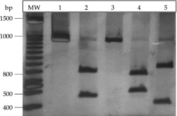
Analysis of the rDNA sequence obtained from R. ecuadoriensis (EF108418 GenBank access number) confirmed its identity as it was 100% identical to the 16S rRNA encoding gene from R. rhodnii, the bacterium isolated from R. prolixus (EU650780 GenBank access number). Moreover, PCR-RFLP results from all dissected insects and faecal samples showed an identical pattern to that of Rhodococcus bacteria isolated from R. prolixus and R. ecuadoriensis: 780 and 580 bp BamH1 fragments, 745 and 594 bp PstI fragments and 829 and 510 bp SacI fragments. No digestion was observed with HindIII endonuclease (Fig. 1).
Restriction fragment length polymorphism analysis of the amplification products obtained with the 16F/R primers. Silver-stained 6% PAGE showing the digestion profile for Rhodococcus rhodnii 16S rRNA-encoding genes isolated from Rhodnius ecuadoriensis. MW: 100 bp molecular weight marker (Invitrogen, California, USA). (1) Undigested PCR product, (2) PCR product digested with BamH1, (3) digested with HindIII, (4) digested with PstI and (5) digested with SacI.

BLAST-N analysis revealed 99% identity between the R. rhodnii sequence obtained in this study and the sequences reported previously as R. rhodnii in GenBank (DSM43959, DSM43336T, DSM43337T and ATCC 35071T strains). On the contrary, identity was just 97.8 and 96.9% with the respective Rhodococcus triatomae and Rhodococcus sp. sequences.
The R. rhodnii bacterium was therefore systematically isolated from the intestinal content and faeces from all R. ecuadoriensis nymphs and adults analysed. These findings did not result from a cross-contamination in our laboratory since the same bacterium was also isolated from specimens that had been reared in another laboratory. Furthermore, the insect colonies (kept separately according to species) were always manipulated at different times, and insects were never extracted from their hosts during feeding. Culture environmental controls were carried out frequently.
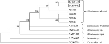
In the ClustalW alignment-based phylogenetic analysis, R. rhodnii sequences isolated from R. prolixus and R. ecuadoriensis were grouped together with other sequences from the same (previously reported) species (Fig. 2). The R. triatomae and Rhodococcus sp. sequences were located near to the R. rhodnii group but not within the same clade, which corresponds with the computed percentages of identity. As expected, R. equi and Nocardia sp. sequences were located further from the R. rhodnii group (Fig. 2). The fact that R. rhodnii bacteria isolated from triatomines were grouped apart from those from other sources can possibly be associated with their lifestyles. However, a larger amount of samples is needed for making definite phylogenetic deductions.
Phylogenetic analysis of Rhodococcus rhodnii ribosomal 16S RNA gene sequences isolated from Rhodnius prolixus and Rhodnius ecuadoriensis. ClustalW was used for aligning the obtained sequences and those previously reported in GenBank. The phylogenetic tree was constructed using MEGA software (version 4.0), using the neighbour-joining method. Bootstrap values were expressed in percentages; 1000 replicas were obtained. The R. rhodnii sequences isolated in this work are shown in boxes, and the species to which each sequence used for the analysis belongs are shown within brackets.

Although only specimens from laboratory colonies were analysed in our study, a similar result can be expected for wild insects, as the same bacterium was present in our laboratory-reared and field-caught R. prolixus individuals (Vallejo et al., Reference Vallejo, Guhl and Schaub2009). Finally, it is likely that R. rhodnii could have a beneficial symbiont effect in R. ecuadoriensis as it has in R. prolixus.
Acknowledgements
We would like to thank Nicolas Jaramillo from the Universidad de Antioquia for providing R. ecuadoriensis laboratory specimens. This study received financial support from Vicerrectoria Académica, Pontificia Universidad Javeriana, Bogota, project no. 0000407.