INTRODUCTION
When the cercaria (Schistosoma mansoni) penetrates the skin of the host, transformations occur in the membrane (Hockley and McLaren, Reference Hockley and McLaren1973) and the acetabular glands and their secretions (Stirewalt, Reference Stirewalt1974; Curwen et al. Reference Curwen, Ashton, Sundaralingam and Wilson2006). The schistosomulum which is produced during this process is the object of much research in vaccine preparation and drug development. For such research, large numbers of this stage of the parasite can be produced by mechanical methods rather than penetration through the skin, but it is not certain that the mechanical forms are equivalent in all properties to those from the skin. Brink et al. (Reference Brink, McLaren and Smithers1977) concluded that they were equivalent in most properties. Protasio et al. (Reference Protasio, Dunne and Berriman2013) has carried out a precise documentation of the difference in the transcriptome between those parasites (schistosomula) that have been prepared artificially (mechanically transformed schistosomula, MECH) and those that have been formed by penetration through the skin held in an in vitro system (skin penetrated schistosomula, PEN).The major differences between the two forms are the presence in the skin penetrated forms of more active mitochondria. In the work described here, we have compared these two forms of schistosomula, termed PEN and MECH for skin penetrated and mechanically transformed schistosomula respectively, with another stage, the schistosomula that remain in the skin, called the migratory stage (MIG). In a natural infection, such forms often reside in the skin for up to 3 days (Barbosa et al. Reference Barbosa, Pellegrino, Coelho and Sampaio1978; Crabtree and Wilson, Reference Crabtree and Wilson1985). Such forms have been studied for parasite permeability and in membrane internalization processes. The considerable differences that have been found in the surface membrane may have important implications for host–parasite communication.
MATERIALS AND METHODS
Mice
Swiss mice weighing between 30 and 40 g were used. All procedures involving animals were approved by the local Ethics Commission on Animal Use (CEUA) from Fiocruz (LW42/10).
Cercariae
Cercariae were obtained from S. mansoni infected Biomphalaria glabrata reared and maintained in the Mollusc room, Lobato Paraense, in Research Centre Rene Rachou /FIOCRUZ. The strain of S. mansoni used in these studies was the LE strain.
Production of schistosomula: In vitro
Apparatus designed by Clegg (Reference Clegg1965) as recently described by Protasio et al. (Reference Protasio, Dunne and Berriman2013) was used with skin samples derived from freshly sacrificed mice. Skin withdrawn from one mouse was used for four penetration chambers. Schistosomula were collected over a period of 2 h and designated penetrated forms (PEN).
Cercariae were placed on the skin and after 2 h the skin from the apparatus was removed and chopped into small pieces with scissors and incubated in RPMI medium for 2 h, during which time schistosomula trapped in the skin migrated out of the skin snips held in a cell strainer (BD Falcon). These were termed migratory forms (MIG). In some experiments the skin was retained after the exposure to cercariae and was given an intradermal injection of fluorescent dye before being chopped into small pieces as described above. Schistosomula were collected from the cell strainer as described above and examined, after washing, in the fluorescence microscope.
Schistosomula were produced mechanically by the method of Colley and Wikel (Reference Colley and Wikel1974), and by the vortex procedure (Ramalho-Pinto et al. Reference Ramalho-Pinto, Gazzinelli, Howells, Mota-Santos, Figueiredo and Pellegrino1974) after concentrating the cercariae on ice. These have been termed mechanical forms (MECH).
Fluorescent dyes
Probes to investigate membrane permeability
We were investigating whether membrane-impermeant molecules could enter the parasite after skin penetration, skin residence and skin migration and we chose the following compounds to do this: Lucifer Yellow CH lithium and potassium salt, molecular weight 457 Da (cat number: L-453), purchased from Invitrogen (Life Technologies do Brasil Ltda, Sao Paulo); Fluorescein isothiocyanate (FITC)-labelled dextrans of molecular weights, 10 kDa (Sigma-Aldrich cat number: FD10S) 20 kDa (Sigma-Aldrich cat number: FD20S) and 40 kDa (Molecular Probe/Life Technologies cat number: D-1844). All fluorescent chemicals are water soluble and were dissolved (1 mg mL−1) in RPMI 1640 medium (Gibco/Life Technologies cat number: 31800-022). Lucifer Yellow CH is a membrane-impermeant compound (Peracchia, Reference Peracchia1981), with a structure that enables it to be aldehyde fixed into tissues with which it comes into contact (Heinrichs, Reference Heinrichs1985).
Fluorescent dextrans (Fl dextrans) are often used to assess membrane integrity and to examine membranes for the sizes of membrane pores (Shimizu and Kawazoe, Reference Shimizu and Kawazoe1996).
Probes for intracellular organelles and membranes
The dyes used to stain specific organelles in the schistosomula are described in Table 1. These probes were incubated with schistosomula in 1 mL RPMI for 1 h at the concentrations shown. After incubation the schistosomula were washed three times in RPMI and examined under the fluorescence microscope.
List of probes to investigate intracellular organelles and membranes

References: Lysotracker red (Al-Adhami et al. Reference Al-Adhami, Noble, Sharaf, Thornhill, Doenhoff and Kusel2005); Mitotracker red (Poot et al. Reference Poot, Gibson and Singer1997); Monochlorobimane (Ribeiro et al. Reference Ribeiro, Coelho, Vieira, Powell and Kusel1998a , Reference Ribeiro, Coelho, Vieira, Watson and Kusel b ); Monodansylcadaverine (Al Adhami et al. Reference Al-Adhami, Noble, Sharaf, Thornhill, Doenhoff and Kusel2005); FM 143 (Ribeiro et al. Reference Ribeiro, Coelho, Vieira, Powell and Kusel1998a , Reference Ribeiro, Coelho, Vieira, Watson and Kusel b ).
Migrating schistosomula in living mice
Mice were anaesthetized with sodium pentobarbital-(Hypnol) (40 mg kg−1) and infected with 1000 cercariae over a period of 30 min.
After recovery the mice were housed for 24 and 48 h. Euthanasia was achieved by cervical dislocation, the region of skin penetrated by cercariae 24 or 48 h previously was injected (intradermal route) with Lucifer yellow or FITC-dextrans and the skin removed after 15 min and cut with scissors into small pieces about 4 mm square(snips). Schistosomula were recovered from the excised skin by incubating skin snips in RPMI in a cell strainer (BD Falcon) placed in a six-well plate. The schistosomula were washed with RPMI three times and observed by fluorescent microscopy.
Control incubations with skin
To investigate whether the presence of cut skin alone rather than the requirement to migrate in skin, was responsible for the alterations seen in the permeability and endocytosis of the schistosomula from skin, the uptake of fluorescent dextrans, Lucifer yellow or FM 143 was observed in MECH in the presence of mouse skin. The skin was cut into small pieces in the same manner as for the work with migrating schistosomula as the result of infection, and MECH added to the pieces in a 15 mL Falcon tube, in the presence of RPMI and the relevant dye, at 100 μg mL−1 (Dextrans and Lucifer yellow) and 5 μg mL−1 (FM143). After incubation for 1 h at 37 °C the mixture was placed in a cell strainer in 5 mL RPMI in a Falcon six-well plate. The results from this experiment are described in the results section below (Fig. 2e and f).
Fixation of schistosomula
Labelled schistosomula were fixed at 4 °C for 4 h in 4% paraformaldehyde. The 4% paraformaldehyde was prepared by dissolving the powder (Sigma) in RPMI 1640 at 60 °C.
Examination of parasites by fluorescence microscopy
All labelled parasites were viewed and photographed using a Zeiss microscope. This allowed the variability of the labelling of the parasite population to be observed, counted and recorded. Photographs were taken after observation of the parasites under the 10× and 40× objectives with a Cannon digital camera. For photography movement of the parasite was slowed by the application to the slide of sodium pentobarbitol effected (10 mL of 1 mg mL−1)
RESULTS
Labelling of parasites in vitro
Detection of intracellular organelles in schistosomula
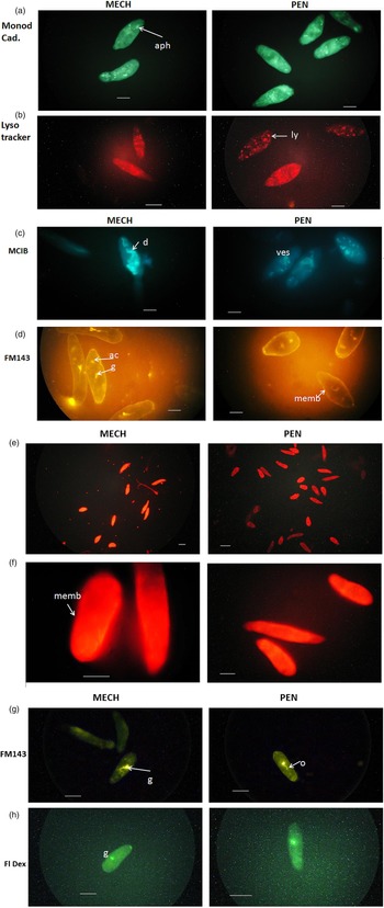
Examples of the staining of mechanically transformed (MECH) and skin transformed (PEN) schistosomula are shown in Fig. 1.
Interaction of mechanical (MECH) and skin penetrated (PEN) schistosomula with fluorescent dyes of various specificities. Size bar: 50 μm. (a) Monodansylcadaverine (monodcad.). Schistosomula were incubated in vitro with monodansylcadaverine (20 ng mL−1). Specific intense blue/green staining of autophagosomes: aph; (b) lysotracker red. Schistosomula incubated in lysotracker red (40 ng mL−1). Lysosome vesicles containing red dye (ly); (c) monochlorobimane (MClB) MECH. gl glutathione rich regions are remains of penetration glands (pre-acetabular gland duct(d) visible) PEN remains of penetration glands ves. glutathione rich vesicles; (d) FM 143 schistosomula incubated in FM 143(5 mg mL−1). There is some background fluorescence. Dye localized in surface membrane (memb) gut (g) and acetabulum (ac); (e, f) schistosomula incubated in mitotracker red (50 ng mL−1) (e) low magnification to show whole bodies of all schistosomula labelled with dye. (f) General staining of schistosomula except some dark regions in MECH; (g, h) schistosomula incubated in FM143 (g) to show gut (g) and oesophagus (o) labelling and Fl dextrans (h) to show gut labelling. No differences between MECH and PEN are observed. Size bar 50 μm in (g) and 80 μm in (h).

Auto-phagosomes and lysosomes
When skin PEN and MECH were compared, both forms showed strong labelling of autophagosomal vesicles (aph) by monodansylcadaverine (Fig. 1a) and of lysosomes (ly) by lysotracker red (Fig. 1b). The distribution of both kinds of organelle is very similar in MECH and PEN.
Glutathione rich regions
These were stained by monochlorobimane (Fig. 1c) and represent remnants of the penetration glands. These have been previously shown to be rich in glutathione (Ribeiro et al. Reference Ribeiro, Coelho, Vieira, Powell and Kusel1998a , Reference Ribeiro, Coelho, Vieira, Watson and Kusel b ). The regions appeared as vesicular structures and were strongly evident in MECH (Fig. 1c) and were still strongly evident in PEN (Fig. 1c).It is surprising to find these regions in PEN forms, since the penetration glands are evacuated during penetration (Stirewalt, Reference Stirewalt1974). Fragments forming vesicles persist for some time after penetration.
Surface membrane
When schistosomula were incubated with FM 143, the surface membrane (memb) was uniformly labelled and the gut (g) and acetabulum (a) also became labelled (Fig. 1d). Very few membrane-derived vesicles could be seen in either preparation, suggesting very little internalization of surface membrane was occurring under these conditions. No great differences in the intensity of labelling with FM 143 occurred in the surface in PEN forms when compared to MECH forms (Fig. 1d).
Mitochondria
Figure 1e show the staining of MECH and PEN with the mitochondrial specific dye, mitotracker. These low magnification images show that each schistosomulum is uniformly labelled. The distributions of the dye in the mitochondria are very similar, except in the MECH forms there is a distortion of the pattern by the presence of pre-acetabular glands (seen in bright field microscopy, not shown here) which form a dark region (Fig. 1f).
Probes for membrane and parasite permeability
Treatment of PEN, and MECH forms with Lucifer yellow, FITC (10, 20, 40 kDa) Dextrans (Fl Dextrans) and FM143 showed that gut labelling occurred with both MECH and PEN forms but few parasites internalized the label into other parts of the body. There were no differences between PEN and MECH. The results for FM 143 are shown in Fig. 1g with gut and oesophagus very clearly labelled, and Fl dextrans in Fig. 1h. Lucifer yellow in MECH and PEN appear similar to Fig. 2a (PEN) with gut and oesophagus labelled.
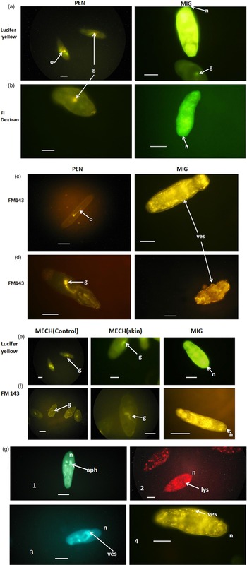
Interaction of PEN and schistosomula which have resided in the skin of mice (MIG) for 24 h with membrane impermeant and membrane internalization dyes. Size bar 50 μm. (a) PEN Schistosomula were incubated with Lucifer yellow (100 mg mL−1). The dye is found in the gut (g) and oesophagus (o) but not in the tissues of the parasite. MIG schistosomula migrated from skin exposed to Lucifer yellow. The dye has entered most of the tissues of the parasite; (b) PEN Schistosomula were incubated in fluorescent dextrans (Fl dextrans) of mol. wt. 10–40 kDa. Only the results for 10 kDa are shown as representative of all these dextrans. The dye has entered the gut but not the tissues of the schistosomulum. MIG schistosomula migrated from skin exposed to Fl dextrans. The schistosomulum is representative of all dextrans, and the dye has entered most of the tissues of the parasite. n, position of nephridiopore; (c, d) PEN schistosomula were incubated with FM 143 (5 μg mL−1). The dye has stained the surface membrane and entered the gut (g) and oesophagus (o), but little is found in the tissues of the parasite. (c, d) MIG schistosomula migrated from the skin exposed to FM 143. The schistosomulum shows numerous internal labelled vesicles (ves). In (d) MIG, the vesicles could withstand gentle crushing under the coverslip (ves); (e) schistosomula prepared mechanically (MECH), and MECH in the presence of skin (MECH skin). These were incubated in the presence of Lucifer yellow. No effect of skin on MECH forms was seen (g gut) The MIG forms show intense internal labelling Size bar is 50 μm; (f) schistosomula prepared mechanically (MECH), and MECH in the presence of skin (MECH skin). These were incubated in the presence of FM 143. No effect of skin on MECH forms was seen. The MIG schistosomula show great surface membrane internalization and intense internal labelling (n, position of nephridiopore). Size bar is 50 μm; (g) schistosomula to show internal organelles. Top left,1 MECH monodansylcadaverine, aph autophagosome; top right, 2 MECH lysotracker, lys lysosomes; bottom right, 3 MECH Monchlorobimane, ves. glutathione rich vesicles; bottom right,4 MIG FM 143, ves. endosomal vesicles.

From the results with fluorescent dyes with PEN and MECH schistosomula, no clear differences were observed between the two forms (supporting the conclusions of Brink et al. (Reference Brink, McLaren and Smithers1977) and Protasio et al. (Reference Protasio, Dunne and Berriman2013).
We then wished to compare the MECH and PEN forms with those schistosomula retained or migrating in the skin in their interaction with some fluorescent dyes.
Labelling of parasites recovered from infected mice
Lucifer yellow
Lucifer yellow was injected into the skin site of mice which had been exposed to cercariae 24 h previously. The excised skin was placed in the cell chamber (Cell strainer BD Falcon) and schistosomula migrated from the skin snips over a period of 3 h. The Lucifer yellow, in the majority of parasites showed a striking uptake into the internal body tissues (Fig. 2a, MIG). Some parasites showed uptake into the gut and minimal uptake into other body regions (labelled (g) in Fig. 2 MIG).
Fluorescent dextrans
FITC dextrans of molecular weight 4, 10, 20 and 40 kDa (together called Fl Dextrans) were injected into the skin sites of mice infected with cercariae 24 h previously. The majority of parasites showed massive uptake of the Fl dextrans of all molecular weights into the internal body tissues (Fig. 2b MIG).
FM 143
When FM 143 was injected into the skin of mice infected with cercariae 24 h previously, massive uptake occurred into the body structures (Fig. 2c MIG). This great uptake of the dye was also seen when FM 143 was injected into the isolated mouse skin from the Clegg apparatus after exposure to cercariae and the schistosomula allowed to migrate from the skin through the cell strainer as described above. These results were similar to that viewed in Fig. 2c MIG.
This effect of the residence in the skin on membrane internalization processes was very pronounced. Internal labelled vesicles could be readily seen when labelled schistosomula from the skin were gently crushed under the coverslip. These membrane bounded vesicles (ves) from within the parasites and liberated by crushing could be readily identified (Fig. 2d MIG).
Control incubations with skin
To investigate whether the presence of skin snips alone rather than the requirement to migrate in skin was responsible for the alterations seen in the permeability and endocytosis of the schistosomula from skin, the uptake of Lucifer yellow or FM 143 was measured in MECH in the presence of mouse skin as described in Materials and Methods (Control incubations with skin). Schistosomula passing through the sieve were collected, and examined for fluorescence with the fluorescence microscope after washing with RPMI. No increase in body labelling of the schistosomula was seen when compared with the controls without skin snips for either Lucifer yellow or FM 143 (Fig. 2e Lucifer yellow or Fig. 2f FM 143). Thus the presence of skin without infection and migration is not affecting the permeability or internalization processes of the schistosomula. An active process of migration is necessary.
Organelles in some of the above stained parasites are shown in slightly higher magnification in Fig. 2g (1–4).
DISCUSSION
Recently, Protasio et al. (Reference Protasio, Dunne and Berriman2013) concluded that the mechanical schistosomula (MECH) were very similar to those schistosomula (PEN) that had passed through mouse skin. The similarity was in the number of genes transcribed in the two different kinds of schistosomula: all transcribed genes were very similar in levels of expression except for those involved in the production of mitochondria. These mitochondrial transcripts were elevated in the skin penetrated forms (PEN) and after 24 h in culture had increased oxidative activity (shown by a colorimetric assay) when compared with the MECH forms.
In the work described here, fluorescent dyes have been used to examine a variety of properties of PEN and MECH. The majority of dyes showed very similar staining patterns in both MECH and PEN (Fig. 1a–f). The staining by the mitochondria specific dye, mitotracker, showed no difference in staining pattern between the two forms, except that the presence of the pre-acetabular glands in MECH indicated a space unfilled with mitochondria. The differences in quantity or activity of mitochondria suggested by Protasio et al. (Reference Protasio, Dunne and Berriman2013) might be detected by this dye using quantitative methods and confocal microscopy in further work. The staining patterns for the other dyes showed no clear differences between MECH and PEN.
While working on penetration of cercariae of human skin in the Clegg apparatus (Clegg, Reference Clegg1965; Protasio et al. Reference Protasio, Dunne and Berriman2013), (results not shown) we noticed that large numbers of schistosomula were retained in the skin. We decided to investigate the interaction of certain membrane permeability and interactive dyes with what we called the migrating forms (MIG), since such forms can be retained in the skin of infected mice for up to 72 h (Barbosa et al. Reference Barbosa, Pellegrino, Coelho and Sampaio1978; Crabtree and Wilson, Reference Crabtree and Wilson1985). We compared those schistosomula migrating from the skin (skin derived forms, MIG) with those collected after penetrating through the skin (PEN). The schistosomula that had resided in the skin of the living mouse for 24 h were treated in a manner similar to that described by Thornhill et al. (Reference Thornhill, McVeigh, Jurberg and Kusel2010). These workers had injected fluorescent dyes measuring membrane permeability into the skin in the path of penetrating cercariae. The schistosomula emerging from excised skin were all very distinctively labelled within the body. Lucifer yellow uptake from in vitro and MIG is shown in Fig. 2a. Figure 2b shows uptake of fluorescent dextrans. Thus the permeability increase found in 24 h schistosomula is a feature of the schistosomula migrating in the skin. The increase in permeability was also seen after 48 h in the skin (results not shown). This means that during prolonged residence in the skin for up to 48 h and possibly up to 72 h (Barbosa et al. Reference Barbosa, Pellegrino, Coelho and Sampaio1978; Crabtree and Wilson, Reference Crabtree and Wilson1985) there may be constant access to host and parasite molecules. This may represent a novel host–parasite interface.
The dye FM 143 (Fig. 2c, d) has been used in studies with schistosomes by Ribeiro et al. (Reference Ribeiro, Coelho, Vieira, Powell and Kusel1998a , Reference Ribeiro, Coelho, Vieira, Watson and Kusel b ). In the current work, when FM 143 was injected into the skin site after an infection of mice with cercariae 24 h previously, the schistosomula recovered were very heavily labelled in all parts of the body (Fig. 2c, d). This suggests that very active endocytosis and membrane turnover is occurring during the residence in and migration through the skin. This rapid internalization and turnover may explain the great permeability of the surface to Lucifer Yellow and fluorescent Dextrans while the parasites are in the skin. The effects of the skin are very evident in the experiments reported here. Three days schistosomula in vitro culture (Parker-Manuel et al. Reference Parker-Manuel, Ivens, Dillon and Wilson2011) have been shown to have a number of upregulated genes and it would be interesting to compare these results with schistosomula derived from skin at this time after infection.
It is unclear what is the specific influence of the skin on the parasite permeability and endocytosis. The presence of linoleic acid in the skin has been suggested to act either on the lipids of the membrane itself (Haas, Reference Haas1984; Haeberlein and Haas, Reference Haeberlein and Haas2008) or indirectly on the neutral sphingomyelinase stimulated by the unsaturated fatty acids in the skin (El Ridi and Tallima, Reference El Ridi and Tallima2006). A number of other factors such as pressure on the surface membrane or nervous stimuli during migration could be involved. The complexity of the whole parasite is such that several factors may be involved in the increase in permeability and endocytosis.
In the work presented here, the schistosomulum appears to possess a surface metabolism much more active than when this is measured in vitro. We can suggest that during migration of the schistosomulum over a period of up to 72 h in the skin the very rapid activities detected by the dye FM 143 may lead to the very extensive turnover of surface and internal antigens leading to enhanced release of immunogens (Fonseca et al. Reference Fonseca, Carvalho, Alves and deMelo2012). The rapid internalization processes observed with FM 143 might be driven by the increase in mitochondrial activity reported by Protasio et al. (Reference Ramalho-Pinto, Gazzinelli, Howells, Mota-Santos, Figueiredo and Pellegrino2013). Moreover, the increased permeability of the parasites to molecules up to 40 kDa molecular weight may allow uptake of cytokines, such as IL7 (Wolowczuk et al. Reference Wolowczuk, Nutten, Roye, Delacre, Capron, Murray, Trottein and Auriault1999) which by its effect on cell signalling and protein synthesis, might enhance the growth of the parasite. All these activities of the surface may increase the ability of the parasite to evade immune responses but paradoxically may make it more immunogenic, as it deposits surface and internal antigens, particularly seen when the irradiated cercariae are used for infection (Ganley-Leal et al. Reference Ganley Leal, Guarner, Todd, Da'Dara, Freeman, Boyer, Harn and Secor2005; Kumkate et al. Reference Kumkate, Jenkins, Paveley, Hogg and Mountfor2007). Our observations may explain the results of Pearce et al. (Reference Pearce, Basch and Sher1986). These ideas can be tested (a) by examination of skin, in which schistosomula are migrating, for immunogens such as tetraspanin and SM 29 (Fonseca et al. Reference Fonseca, Carvalho, Alves and deMelo2012) or (b) by an adaptation of the methods used by Paveley et al. (Reference Paveley, Aynsley, Cook, Turner and Mountford2009) in which a fluorescent label might be attached to surface membrane molecules of living schistosomula prior to migration in skin.
The relevance of these profound effects of the skin tissue on permeability and internalization processes in the life of the parasite is that the emphasis on the surface as a host–parasite interface may be misleading. Such large molecules from host or parasite may stimulate signalling pathways within the parasite. The nature of such pathways might be elucidated in small numbers of parasites by the methods of Walker et al. (Reference Walker, Ressurreição and Rothermel2014) using the confocal microscope and with larger numbers by the methods used by Protasio et al. (Reference Protasio, Dunne and Berriman2013) using RNASeq methods. Both approaches might be extended to the MIG forms, since new pathways might be evident after the increase in membrane permeability and membrane activity while they are resident in the skin, which were not revealed in other forms of schistosomula.
ACKNOWLEDGEMENTS
Liana and the staff of the Lobato Paraense snail room for provision of cercariae. Roberta Caldeira for advice. Neviton and Jose and Rosangela from Bioteria for support. Diana for preparing various solutions and culture media. Vera, Flavia, Ana-Carolina, Aureo, Neusa, Fabio, Clarice, and Kika for encouragement. We are very grateful to Mrs Alison Sage for advice and help during the submission of the manuscript.
FINANCIAL SUPPORT
The CAPES for financial support through the ‘Science without Borders’ programme. CAPES agency No: 402549/2012-0.