INTRODUCTION
Lymphatic filariasis is one of the 6 neglected tropical diseases, which is endemic in over 72 countries in Africa, Asia, South and Central America and the Pacific Islands. According to the World Health Organization (WHO), over 120 million people are currently infected, with about 40 million disfigured and incapacitated by the disease (WHO, 2012). Diethyl carbamazine citrate (DEC) is the drug of choice for the treatment of filariasis, however, its exact mechanism of action is not known (Gupta and Srivastava, Reference Gupta and Srivastava2005). Furthermore, DEC is efficient in the clearance of microfilariae but is not very effective against adult worms (Norões et al. Reference Norões, Dreyer, Santos, Mendes, Medeiros and Addiss1997). Under the global programme for elimination of lymphatic filariasis, the WHO laid emphasis on the screening of new libraries of synthetic and herbal drugs. Numerous studies have proven the efficacy of various synthetic and herbal compounds targeting different pathways in the filarial parasite (Gupta and Srivastava, Reference Gupta and Srivastava2005; Sahare et al. Reference Sahare, Anandharaman, Meshram, Meshram, Gajalakshmi, Goswami and Reddy2008a, Reference Sahare, Anandhraman, Meshram, Meshram, Reddy, Tumane and Goswami2008b). However, no new drugs have reached clinical usage.
Dihydrofolate reductase (DHFR), the sole source of tetrahydrofolate, is one of the key enzymes in the folate metabolic pathway and has been found to be a suitable target for antibacterial, antiparasitic and anti-cancer treatment (Gangjee et al. Reference Gangjee, Kurup and Namjoshi2007). Folate metabolism has also been proposed as the possible target of action of some important antifilarial agents including DEC and suramin (Gupta and Srivastava, Reference Gupta and Srivastava2005). Very recently, in vitro biological evaluation and folate reversal studies have been conducted by our group for a series of biguanides and dihydrotriazine molecules (Bag et al. Reference Bag, Tawari, Queener and Degani2010b). These reports highlight the importance of the folate pathway for antifilarial drug discovery. However, the enzyme structure of DHFR from Brugia malayi remains unsolved and thus the use of target-based drug discovery approaches for the search for novel DHFR inhibitors active against filariasis is not feasible at this stage. Another possible rational approach in this direction could be the screening of libraries of known DHFR inhibitors using in vitro assays against filarial worms. Interestingly, the above-mentioned biguanides and dihydrotriazine molecules have shown promising activity against B. malayi (Bag et al. Reference Bag, Tawari, Sharma, Goswami, Reddy and Degani2010c). Being encouraged with this successful endeavour, this study was undertaken towards an anticipated significant mechanism-related outcome involving plausible apoptotic impact, beyond simple validation of the therapeutic potential of such synthetic DHFR inhibitors. Evidence supports the involvement of an apoptotic rationale through DHFR inhibition (Huang et al. Reference Huang, Ho, Lin, Wei and Liu1999). In this light, the in vitro screening of a novel set of twelve 2, 4-diaminopyrimidines and 2, 4-diamino-s-triazines with reported DHFR inhibitory properties against filarial parasites is reported. Studies were also carried out to investigate the role, if any, of apoptosis in B. malayi microfilariae and adult worms in the possible antifilarial action by these inhibitors.
MATERIALS AND METHODS
Compounds
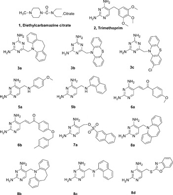
All reagents and chemicals were obtained from commercial sources (Himedia Laboratories Pvt. Ltd, Mumbai and Sigma Aldrich Chemicals Pvt. Ltd, Mumbai). The 2, 4-diaminopyrimidine and 2, 4-diamino-s-triazine derivatives (Fig. 1) were synthesized and purified in our laboratory. The structures and purity of these compounds were established using spectroscopic methods (IR, 1H-NMR, 13C-NMR, MS analysis and chromatography) (Bag et al. Reference Bag, Tawari, Degani and Queener2010a).
Structure of diethylcarbamazine citrate, trimethoprim and the synthesized test compounds used in the study.

Preparation and collection of B. malayi microfilariae and adult worms
The use of animals for this study was approved by the Institutional Animal Ethics Committee, India, which follows the norms of the Committee for the Purpose of Control and Supervision on Experiments on Animals (CPCSEA). The B. malayi life cycle was established and maintained in jirds (Meriones unguiculatus), mastomys (Mastomys natelansis) and mosquitoes (Aedes aegypti) by standard methods (Sanger et al. Reference Sanger, Lammler and Kimmig1981). Microfilariae were obtained by lavage of the peritoneal cavities of jirds with intraperitoneal filarial infection of 3 months or more in duration. Similarly, adult worms of B. malayi were recovered from the peritoneal cavities of jirds after 4–6 months of infection with infective larvae (L3). The microfilariae and adult worms were washed with RPMI 1640 medium (containing 20 μg mL−1 gentamycin, 100 μg mL−1 penicillin, 100 μg mL−1 streptomycin) plated on the sterile plastic Petri dishes and incubated at 37 °C for 1 h to remove the jird's peritoneal exudate cells. The microfilariae were collected from the Petri dishes, washed with RPMI 1640 medium and used for in vitro experiments (Ash and Riley, Reference Ash and Riley1970; Chandrashekar et al. Reference Chandrashekar, Rao, Rajasekariah and Subrahmanyam1984).
In vitro screening of compounds for antifilarial activity
In an attempt to find out whether a newer series of DHFR inhibitors were active against the B. malayi, a series of structurally diverse compounds belonging to the 2, 4-diaminopyrimidine and 2, 4-diamino-s-triazine class (Fig. 1) were evaluated using in vitro assays. Initially, all compounds were evaluated against microfilariae, at a single pre-optimized concentration (20 μg mL−1) using the standardized protocol (Sahare et al. Reference Sahare, Anandharaman, Meshram, Meshram, Gajalakshmi, Goswami and Reddy2008a; Bag et al. Reference Bag, Tawari, Sharma, Goswami, Reddy and Degani2010c). The highest concentration of DMSO used was <1% and hence 1% DMSO was taken as the control. Trimethoprim and pyrimethamine, which are well-known antifolate compounds, were used as the positive control. In a sterile 24-well culture plate (Nunc, Denmark) containing 900 μL of RPMI medium in each well, approximately 100 microfilariae in 100 μL of RPMI medium were introduced into each well for every test sample and also for the corresponding control samples (each individual sample was tested in triplicate). The plates were incubated at 37 °C for 48 h in a 5% CO2 incubator. Microfilariae motility was assessed by microscopy after incubation (using Nikon Diaphot, TMD inverted microscope). The observations were recorded as the number of non-motile microfilariae out of the total recovered in each well and is reported as a percentage. Previous studies in our laboratory had shown that a correlation exists (r=0·97) between this motility assay and the conventional MTT assay (3-[4,5-dimethylthiazol-2-yl]-2,5-diphenyltetrazolium bromide reduction assay) for actual parasite mortality (Sharma et al. Reference Sharma, Janardhanan, Gajalakshmi, Reddy and Goswami2008). Each experiment was done in triplicate to check the reproducibility.
Similar experiments were carried with adult worms. Considering the paucity of procuring adult worms, only compounds with high efficacy against microfilariae were further evaluated against adult worms. Precisely, 2 worms (1 male and 1 female) were incubated in 1 mL of the medium alone and medium containing 20 μg mL−1 of each of the compounds along with similar control worms. The plates were incubated at 37 °C, with 5% CO2, for 48 h and the motility was assessed by microscopy, wherein the adult worm viability was assessed visually by direct microscopic observations (using a Nikon Diaphot, TMD inverted microscope as indicated previously ) and the observations were scored as −, inactive or dead; +, less active; ++, moderately active; and +++, highly active. Experiments were done in triplicate to check the reproducibility.
Reversal of the activity of test compounds by folic acid and folinic acid
Freshly prepared folic acid and the microfilariae were pre-incubated with the final dose of 20 μ m for 1 h as described earlier (Sharma et al. Reference Sharma, Janardhanan, Gajalakshmi, Reddy and Goswami2008; Bag et al. Reference Bag, Tawari, Sharma, Goswami, Reddy and Degani2010c). After pre-incubation with folic acid, the test compound was added to the wells at a dose with which ∼100% loss of motility was achieved, and the 24-well culture plates were incubated in an atmosphere of 5% CO2 at 37 °C for 48 h. The antagonism between either the test compounds or the standard drugs and the folic acid was observed as the percentage reduction in loss of motility after addition of the compounds. The mean of 3 observations was calculated to obtain the result. Following the same procedure, the experiment was repeated using the adult worms. The reversal study was also done with folinic acid following the similar protocol.
Ethidium bromide–acridine orange (EB/AO) staining for detection of apoptosis
The EB/AO staining protocol was employed which is a standardized method for detection of apoptosis. This method relies on differential staining by ethidium bromide (EB) and acridine orange (AO) to record characteristic bright green to orange fluorescence in the event of apoptotic change. The staining procedure was carried out according to the reported procedure (Ribble et al. Reference Ribble, Goldstein, Norris and Shellman2005). The dye mix for the EB/AO staining consisted of 100 μg mL−1 EB and 100 μg mL−1 AO in phosphate saline buffer (PBS). Briefly, microfilariae from each well with either the test compound or staurosporine (20 μ m; as positive control for the induction of apoptosis) along with the RPMI control were transferred to 1·5 mL centrifuge tubes and pelleted by centrifugation at 100 g (REMI; RM12 C centrifuge, India) for 5 min and then washed once washed with 1 mL of cold PBS. The pelleted microfilariae was then re-suspended in 25 μL of cold PBS and 5 μL of EB/AO dye mix was added. Positively stained microfilariae (10 μL) were placed on a clean microscope slide and covered with a coverslip. The microfilariae were viewed using a Nikon LABOPHOT epifluorescence microscope with excitation filter 480/30 nm and barrier filter 535/40 nm. Pictures were taken with a Nikon camera. Tests were done in triplicate, counting a minimum of 10 microfilariae in each observation. Similarly, 2 adult worms, treated with the agents, were also tested along with the control.
Poly(adenosine diphosphate-ribose) polymerase (PARP) activity assay
To further validate apoptosis as the underlying principle behind the action of the active compounds, PARP activity in B. malayi microfilariae was determined using a commercial kit (R & D Systems Inc, Minneapolis, MN) according to the manufacturer's instruction. Briefly, 100 μL aliquots of suspension (containing about 100 microfilariae) were washed twice with 1×PBS and the pellet was suspended in 5–10 pellet volumes of cold 1×PARP buffer (provided in the kit) containing 0·4 mm PMSF, other protease inhibitors and 0·4 m NaCl. The aliquot was further lysed with 1% Triton X-100. The protein concentration of the clear supernatant was determined using the Bradford assay. Lysate (20 μg) of protein was added to each well in 96-well plates pre-coated with histone. PARP activity was determined from the incorporation of biotinylated PARP on to immobilized histone, which was measured by the addition of streptavidin-conjugated horseradish peroxide and a suitable chromogenic substrate to the incubation mixture. A standard curve for PARP enzymatic activity (A450 versus PARP units) was initially generated using 0·01, 0·05, 0·1, 0·5 and 1 unit of enzyme per well. The absorbance obtained with each test sample (microfilarial lysate) was extrapolated based on the standard curve to obtain the corresponding PARP activity. The percentage inhibition of enzymatic activity in other test samples (lysates treated with different reagents) was calculated using the control sample (microfilaria without any pre-treatment) as the 100% activity reference point. The experiment was carried out in triplicate and the mean value of percentage inhibition was calculated.
RESULTS
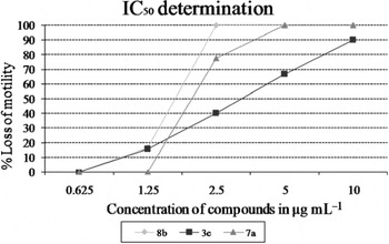
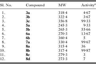
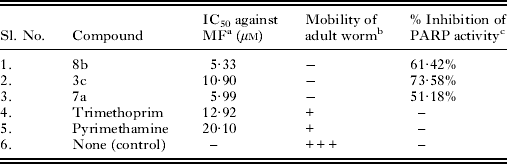
In the microscopic observation of the motility of the worms pre-incubated with 12 synthesized compounds belonging to the 2,4-diaminopyrimidine and 2,4-diamino-s-triazine class (Table 1), three molecules, 8b, 3c and 7a, showed ∼100% efficacy in terms of complete loss of motility of all the parasites. The results of the corresponding drug withdrawal through wash experiments confirmed that the drug effect was permanent and irreversible. These compounds were considered for further screening against adult worms. All 3 compounds showed better activity than trimethoprim and pyrimethamine (Table 2). The graph displaying IC50 determination for the 3 effective compounds is shown (Fig. 2).
Graphical representation for the calculation of IC50 values for each of the compounds.

Biological activity of the compounds against microfilariae
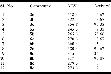
a Expressed as % loss of motility at 20 μg mL−1 after 48 h. Trimethoprim and pyrimethamine showed 100% loss of motility at this concentration. DMSO, RPMI medium (negative controls) showed activity of 3·33 and 3·0 respectively.
IC50 determination results on microfilariae and in vitro effect of active compounds on B. malayi adult worms
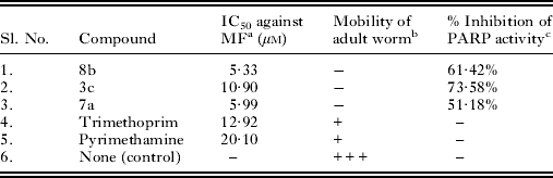
a IC50 determination against microfilariae.
b Two adult worms were incubated per well with 20 μg mL−1 compound at 37 °C, with 5% CO2 for 48 h, after which the worm viability was assessed and scored under an inverted microscope and designated as: −, inactive or dead; +, less active; +++, highly active.
c The percentage inhibition of PARP activity was calculated taking the controls RPMI alone and DMSO (vehicle control) as the baseline. The PARP activity of the positive standard staurosporine was found to be 67·82%.
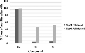
To validate the DHFR inhibition as a mechanism of action of these active compounds, folic acid and folinic acid reversal studies were carried out (Fig. 3). Out of the 3 most active compounds, the effect of 2 compounds, 3c and 7a, could be reversed up to ∼95% by folic acid and up to ∼50% by folinic acid, whereas no reversal was seen in the case of compound 8b.
Folic and folinic acid reversal studies. Microfilariae were pre-incubated with folic acid and folinic acid and the percentage loss of motility in microfilariae after 48 h of incubation with the test compound was measured.

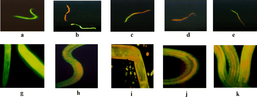
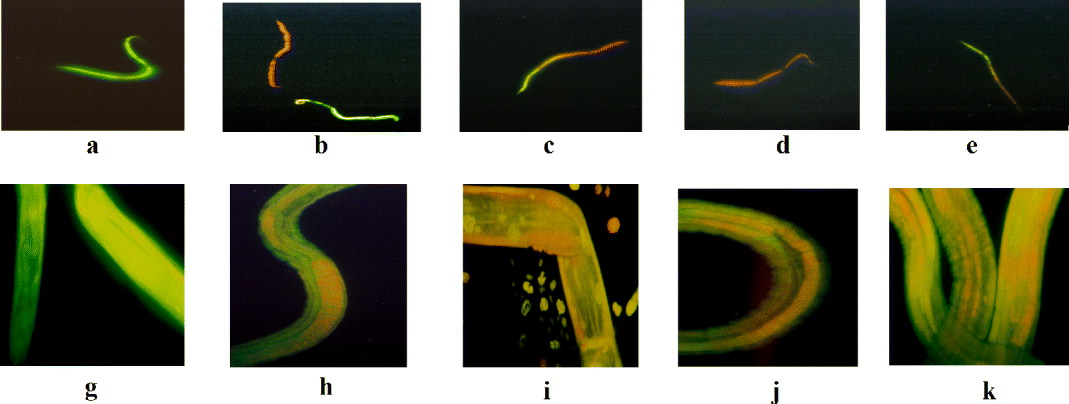
The EB/AO-stained filarial worms (microfilariae and adult worms) were observed under the Nikon LABOPHOT epifluorescence microscope and the photographs recorded (Fig. 4). Staurosporine used as a standard inducer for apoptosis, showed the presence of orange and green fluorescence indicative of apoptosis. Preliminary evidence of apoptotic damage was also observed for the test compounds in both microfilariae and adult worms but not with the negative control.
(A) Results of ethidium bromide–acridine orange (EB/AO) dual staining (10× magnified images). First row (a–e) shows results for microfilariae, a: live worms (−ve control-RPMI), b: dead worms (+ve control-staurosporine), c: compound 3c, d: compound 7a, e: compound 8b; second row (g–k) shows results for adult worms, g: live worms (−ve control-RPMI), h: dead worms (+ve control-staurosporine), i: compound 3c, j: compound 7a, k: compound 8b. The presence of orange and green fluorescence indicative of apoptosis is noticeable in the case of staurosporine and test compounds (3c, 7a and 8b) but not in the negative control. (B) Results of EB/AO dual staining (10× magnified images). First row (a–e) shows results for microfilariae, a: live worms (−ve control-RPMI), b: dead worms (+ve control-staurosporine), c: compound 3c, d: compound 7a, e: compound 8b; second row (g–k) shows results for adult worms, g: live worms (−ve control-RPMI), h: dead worms (+ve control-staurosporine), i: compound 3c, j: compound 7a, k: compound 8b. The live worms have excessive blurring effect due to bright green coloured fluorescence that appears as greyish blurring around the worms. The presence of whitish grey colouration defining the internal cell organelles and irregular sheath of worms indicates apoptosis and is noticeable in the case of staurosporine and test compounds (3c, 7a and 8b) but not in the negative control (Figure is presented in colour online.).
These 3 compounds (8b, 3c and 7a) were tested for PARP enzyme activity along with staurosporine and RPMI medium alone (as positive and negative controls respectively). Staurosporine was found to show 67·82% inhibition, whereas 61·42, 73·58 and 51·18% inhibition in PARP activity was found by compounds 8b, 3c and 7a respectively against the negative control (Table 2). These results provide robust proof of the apoptotic mechanism in the observed pharmacological effect.
DISCUSSION
In the absence of an enzyme structure for the DHFR enzyme of filarial worms, the compounds of diverse nature have been designed to probe the active site of the enzyme through systematic structural variation. Compounds 3a–c, have a bulky tricyclic substitution and the basic DHFR pharmacophore, i.e. the 2,4-diamino-substituted pyrimidine ring system is changed to a 2,4-diamino-s-triazine ring; compounds 8a–b on the other hand have pharmacophoric features of trimethoprim coupled with a bulky distal tricyclic ring system. Compounds 5a–b, 7a and 8c–d explore bridges with different steric and electronic properties; linking the 2,4-diaminopyrimidine nucleus and distal part, and lastly compounds 6a–b have a conformationally constrained bridge.
Three compounds (8b, 3c and 7a) were found to be effective in the initial in vitro screening against the microfilariae. These 3 active compounds were also found to be effective against the adult worms. These compounds have bulky tricyclic and naphthyl substitutions with an extended electronegative bridge. This suggests that the plausible hydrophobic nature of the distal binding pocket of the enzyme could be used as a clue for further development of inhibitors.
Trimethoprim and pyrimethamine showed lesser efficacy against microfilariae and failed to show any effect against the adult stage of the parasite. Theoretically, the effects of compounds acting through competitive antagonism with the substrate folic acid should significantly be reverted by addition of excess of folic acid to the test system. Such a reversal effect on certain inhibitors of folate metabolism was observed earlier (Bag et al. Reference Bag, Tawari, Sharma, Goswami, Reddy and Degani2010c). In the present study, a significant reversal effect was observed with compounds 3c and 7a, which points towards a competitive inhibition mechanism by these two compounds. However, the irreversibility of the effect of compound 8b, along with its lowest IC50 value, suggests that this compound might either be a more avid inhibitor or that its action may not be mediated by a direct competitive mode. Another approach to reverse the observed antifilarial action of these compounds could be by supplying the product of the inhibited enzyme, i.e. folinic acid. Only a weak reversal of effect was produced by compound 8b activity, as obtained using folinic acid. However, this particular effect was subjected to the ability of microfilariae and adult worms to take up folinic acid from the medium. Hence, establishing an accurate mechanism of inhibition would need further experimentation.
Impairment of the folate biosynthetic pathway has been linked with cell death, possibly by apoptosis (Huang et al. Reference Huang, Ho, Lin, Wei and Liu1999). Recently, some bio-flavonoids have actually been shown to inhibit the DHFR enzyme and induce apoptosis (Navarro-Peran et al. Reference Navarro-Peran, Cabezas-Herrera, Garcia-Canovas, Durrant, Thorneley and Rodriguez-Lopez2005). To test whether the action of the tested compounds in this study is by DHFR inhibition-mediated apoptosis, experiments were conducted to detect apoptosis by using EB/AO differential staining. Interestingly, this study displayed preliminary evidence of apoptotic damage. PARP is a key nuclear enzyme involved in DNA repair and the inhibition of PARP activity is a well-founded measure for the evaluation of cellular apoptosis (Mullen, Reference Mullen2004). During apoptosis, PARP cleavage by caspase-3 leads to reduced PARP enzyme activity, thus preventing the apoptotic cells from repairing their own DNA (Bohm, Reference Bohm2006). Hence, PARP inhibition is considered as a valid means for the detection of apoptosis. Our laboratory also reported this method for the demonstration of apoptosis in the filarial model (Singh et al. Reference Singh, Goswami, Sharma, Reddy and Dash2012). While staurosporine, the well-known apoptosis inducer, inhibited 67·82% of the PARP enzyme activity, a comparable inhibition of 61·42, 73·58 and 51·18% was shown by the compounds 8b, 3c and 7a respectively, thus pointing towards the possible role of apoptosis as the underlying mechanism of action.
Taken together, the DHFR inhibitors could emerge as promising lead molecules effective against filariasis via a possible apoptotic impact.
ACKNOWLEDGEMENTS
The authors acknowledge Mr Harshanand Patil for assistance rendered in handling of the fluorescent microscope.
FINANCIAL SUPPORT
SB would like to thank the University Grant Commission (UGC), India; NRT would like to thank the Department of Biotechnology (DBT), India, for a Fellowship; and RS would like to thank the DBT, India, for funds to support the project ‘Repository for filarial parasites and reagents’.