
I. INTRODUCTION
Over the last several decades, conventional Ni-based alloys became the most common used material for high temperature applications, such as vanes and blades in aircraft engines and industrial steam turbines. This is due to a combination of outstanding mechanical properties (creep strength, ductility) and good high temperature oxidation resistance by the two-phase microstructure. Reference Pollock and Tin1 For Ni-based alloys, the presence of γ′-phase, a stable ternary Ni3Al compound with an L12 structure, allows precipitation strengthening to provide superior high temperature strength, so they have come to dominate the modern gas turbines industry by now.
In contrast to this material, conventional Co-based alloys are utilized mostly in mechanically low loaded parts of turbines or corrosive environment, they exhibit superior corrosion resistance but inferior mechanical properties compared to common Ni-based alloys due to lack of the strengthening mechanism at high temperature. Reference Reed2,Reference Ruan, Wang, Zhao, Yang, Yang and Liu3 However, in 2006 Sato et al. discovered a new ternary intermetallic compound phase in the Co–Al–W system, Co3(Al,W), which precipitates in the matrix with an L12 structure. Reference Sato, Omori, Oikawa, Ohnuma, Kainuma and Ishida4 Therefore, this exciting discovery makes the γ/γ′-strengthening in Co-based alloys by the Co3(Al,W) phase possible, which is similar to the Ni3Al phase in Ni-based alloys and provides a new research path for the development of novel Co-based alloys.
Apart from high temperature strength, the oxidation resistance is another essential important property for this new alloy. Meanwhile, Co–Al–W alloys are now considered for the use in high temperature applications, there is a requirement to understand the oxidation process and how it might be tuned through alloying. Compared to Ni-based alloys, only little research has been done so far concerning the oxidation properties of γ′-strengthened Co-based alloys. Reference Li, Sun, Li and Hu5–Reference Liu, Ma and Sun9 Furthermore, oxidation of new γ′- strengthened Co-based alloys at high temperatures leads to the formation of a thin inner Al2O3 layer in addition to cobalt oxides, which mainly contains Co, Al, and W. Therefore, this Al2O3 layer has the potential to improve the oxidation behavior considerably. Reference Klein, Bauer, Neumeier, Göken and Virtanen10
The effects of those alloy elements on the oxidation behavior of the Co-based alloy have been studied extensively. In isothermal tests at high temperature, Klein et al. found that additions of boron (0.12 at.%) and silicon benefit the formation of inner protective Al2O3 due to enhanced selective Al-oxidation. Although Si-containing phases improve the oxidation resistance, they are also expected to decrease the mechanical properties of the alloy. Reference Klein, Killian and Virtanen11 N. Vermaak et al. studied the effect of MCrAlY coatings on the cyclic high-temperature oxidation behavior for two separate Co-based alloys and found that MCrAlY systems could be promising candidates as protective coatings for these new materials. Adherence between the coating and the substrate was maintained during thermal corrosion. In the meantime, the initial work by H-Y. Yan et al. showed that alloying was beneficial for the oxidation behavior of Co–7Al–7W alloys at 800 °C, whereby included adding Cr, Fe, and Si. Reference Yang-Tao, Xia, Yan and Zhao12–Reference Yan, Vorontsov, Coakley, Jones, Stone and Dye16 However, few studies have been carried out to examine the oxidation behavior of this new alloy system by the additions of rare earth (RE) elements (such as La, Y, Ce, and Dy). And the high temperature oxidation behavior of them has until now not been systematically investigated.
In this work, the high temperature oxidation behaviors of the novel γ/γ′-strengthened Co–Al–W and Co–Al–W–X alloys were discussed at 900 °C. In addition to oxidation kinetics curves, the morphology, crystal structure, and then chemical composition of oxide layer was characterized using scanning electron microscopy (SEM), energy dispersive X-ray (EDX) spectroscopy, and X-ray diffraction (XRD), to describe the oxidation process more deeply. Moreover, the influence of these rare alloying elements on oxidation resistance, which was expected to be beneficial, was also investigated.
II. EXPERIMENTAL PROCEDURE
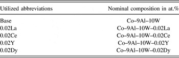
The nominal compositions of the investigated five Co-based alloys are listed in Table I. In the course of this work, only the abbreviations given in Table I are used to name the alloys. The as-cast alloy was solution heat treated in a vertical furnace at 1300 °C for 4 h in a protective Ar-atmosphere and aged for 24 h at 900 °C in air, with the purpose of obtaining the desired γ/γ′ microstructure. The generated oxide layer on the sample surface was completely removed subsequent to aging of the alloy in air. After cutting the rod samples (diameter of 7.6 mm and length of 30 mm) from the cylindrical bar by electro-discharge machining, they were cleaned and degreased, after that wet grinding and polishing was performed using SiC grinding paper (280#, 500#, 800#, and 1200#) and different diamond suspensions (up to a grain size of 1 μm), respectively. All these were performed before isothermal oxidation experiments at 900 °C in air. Flat-bottomed alumina crucibles were used to contain these samples which were oxidized in air. To hold any spallation products, lids were placed on the crucibles during the periodic specimen removal and air cooling prior to the weighing of the crucibles and specimens.
Compositions of the investigated alloys and utilized abbreviations.

Each oxidation test was performed according to the following processes: (1) placement of the specimens into the furnace, (2) oxidizing experiments in terms of the schedule, (3) removing the specimen from the furnace, (4) cooling from 900 °C to the room temperature for approximately 60 min, and (5) weighing of the specimens using a balance with a resolution of 0.10 mg.
Oxidation test was carried out for 400 h to determine the oxidation process with increasing oxidation time in a vertical tube furnace under atmospheric conditions. RE elements such as La, Ce, Dy, and Y were added to original alloy (see Table I); the work was to explore their influence on the oxidation resistance. And the results were compared. To observe the microstructure, the prepared cross-section morphology of the oxide layer was investigated by SEM. EDX spectroscopy attached was carried out to determine the chemical composition. The qualitative and quantitative analysis was determined by EDX mappings and EDX point scans, respectively. Then the crystalline phase identification in the oxide layers were detected by using XRD with Cu Kα radiation over a range of 2θ from 10° to 70°.
III. RESULTS
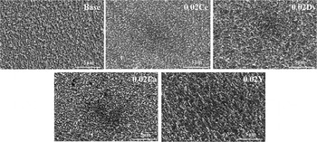
By reason of the specific heat treatment process consisting of homogenization and aging, the investigated alloy establishes the desired γ/γ′-microstructure. Reference Klein, Bauer, Neumeier, Göken and Virtanen10,Reference Klein, Killian and Virtanen11,Reference Coutsouradis, Davin and Lamberigts17 In Fig. 1, five representative SEM images of the etched metal surface were given.
Microstructures of alloys Co–9Al–10W, Co–9Al–10W–0.02Ce, Co–9Al–10W–0.02Dy, Co–9Al–10W–0.02La, and Co–9Al–10W–0.02Y in etched condition.

A. Oxidation kinetics results
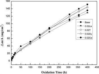
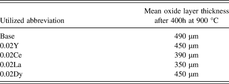
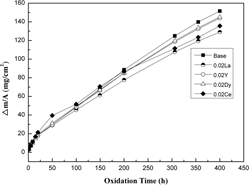
Figure 2 shows those oxidation kinetics results with representative curves of the Co-based alloys at 900 °C. The 0.02 La alloy shows the least (129.008 mg/cm2), while the base alloy shows the most (151.544 mg/cm2). From those data, we can find that oxidation at 900 °C leads to a higher mass gain per area, Δm/A, where forms a much thick oxide layer. Figure 2 displays approximately a linear growth model behavior, which generated continuous mass gains because the spallation products were retained and a ranking from best to worst as follows: 0.02La > 0.02Ce > 0.02Y > 0.02Dy > base. A lower mass gain per area exhibits better oxidation resistances for these novel alloys. A comparison of the corresponding mean oxide layer thicknesses (determined from cross-sections) was summarized in Table II. At 900 °C, oxidation kinetics was rapid and then high mass gain could be measured for all these alloys. Smaller the oxidation weight of the sample is, the thinner its average oxide film becomes. The layer thickness results correlate with the oxidation kinetics curves shown in Fig. 2.
Mass gain (Δm/A) of five Co-base alloys oxidized for 400 h at 900 °C.

Mean oxide layer thicknesses of the investigated oxidized alloys.

During the different oxidation experiments’ process, peeling and spallation occurred at the specimen surface for all alloys, these phenomena may happen during the cooling period time, which may lead to the weight of the specimen decreasing with increasing oxidation time. Reference Hebsur and Miner18 However, the total mass of these specimens increase with prolonged exposure time in this work due to the fact that the spallation products were always contained inside the crucibles.
B. Morphology and chemical composition of the oxide layers at 900 °C
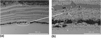
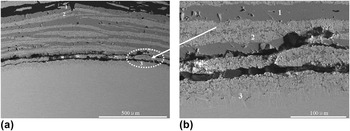
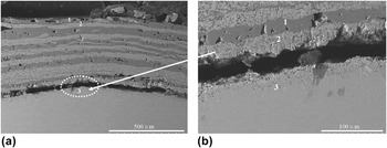
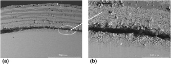
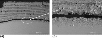
Figures 3–7 show a comparison of cross-sections SEM micrographs and partial enlarged drawings of five Co-based alloys oxidized at 900 °C in air. After long term oxidation at high temperature, a multi-layered oxide structure establishes on the investigated alloy surface, as illustrated in Figs. 3(a)–7(a). Then Figs. 3(b)–7(b) display partial enlarged details of those elliptic areas. From these diagrams, we can see that the oxide film thickness of adding the RE element sample was thinner than that of the base sample. At the same time, there exists a difference about the crack width for those five alloy systems.
(a) Back scattered electron images of the oxide scale formed on the base alloy surface after 400 h oxidation at 900 °C, (b) SEM micrographs about partial enlarged details of the red elliptic area.

(a) Back scattered electron images of the oxide scale formed on the 0.02Ce alloy surface after 400 h oxidation at 900 °C, (b) SEM micrographs about partial enlarged details of the red elliptic area.

(a) Back scattered electron images of the oxide scale formed on the 0.02Dy alloy surface after 400 h oxidation at 900 °C, (b) SEM micrographs about partial enlarged details of the red elliptic area.

(a) Back scattered electron images of the oxide scale formed on the 0.02La alloy surface after 400 h oxidation at 900 °C, (b) SEM micrographs about partial enlarged details of the red elliptic area.

(a) Back scattered electron images of the oxide scale formed on the 0.02Y alloy surface after 400 h oxidation at 900 °C, (b) SEM micrographs about partial enlarged details of the red elliptic area.

The oxide layers for all alloys show a similar morphology and comparable structure, consisting of three distinctive oxide layers. Furthermore, wide cracks can be observed near the oxide/alloy interface after long term oxidation at 900 °C. This is due to the inevitable release of thermal stresses during the cooling period after taking the samples out of the furnace.
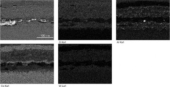
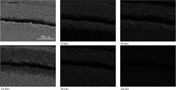
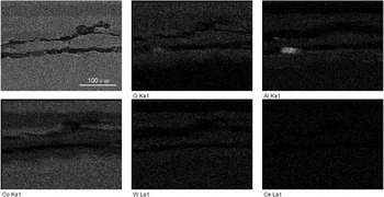
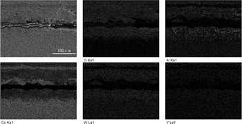
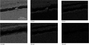
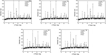
Figures 8–12 show a comparison of EDX mappings of the above corresponding partial enlarged drawing for all samples oxidized at 900 °C for 400 h. It shows the distribution of main elements and RE elements in the multilayer structure, and that the oxygen element focuses above the wide crack. Surface scanning distribution of these main elements is noticeable, but their distribution is not completely consistent. Figure 13 shows a comparison of XRD patterns of Co-based alloys oxidized at 900 °C for 400 h in air. And combined with those above EDX analysis results, it indicates the primary presence of CoO and Co3O4 phases, meanwhile accompanied with few CoWO4 and Al2O3. The strength of the primary peak is higher and five patterns show a similar tendency. The outermost dark layer (1) mainly consists of CoO and Co3O4 oxides according to EDX and XRD data, whereas the second outer light layer (2) consists of oxides containing Al, W, and Co (e.g., CoWO4). Some pores were also found in the outermost dark layer simultaneously, which suggests a vacancy formation mechanism. Then, according to EDX data, the next dark layer was similar to layer (1) and next light layer was similar to layer (2). Whereas the innermost layer (3) consist of little discontinuous oxides, no distinctive Al2O3-rich zone can be observed, a few needlelike Co3W compounds exist between the crack and alloy substrate by EDX point scanning. As a consequence of outward Al-diffusion during the oxidation process, the γ/γ′-microstructure gets instable and transforms into γ-Co and elongated Co3W precipitates. Reference Klein, Bauer, Neumeier, Göken and Virtanen10 The composition of those Co3W precipitates was determined by EDX scans. Recent studies confirm this phase transformation in the ternary Co–Al–W system. Reference Tsukamoto, Kobayashi and Takasugi19
Backscatter electron images of the oxide scale formed, and EDX mappings of the elements present, obtained in the base alloy after 400 h isothermal oxidation at 900 °C.

Backscatter electron images of the oxide scale formed, and EDX mappings of the elements present, obtained in the 0.02La alloy after 400 h isothermal oxidation at 900 °C.

Backscatter electron images of the oxide scale formed, and EDX mappings of the elements present, obtained in the 0.02Ce alloy after 400 h isothermal oxidation at 900 °C.

Backscatter electron images of the oxide scale formed, and EDX mappings of the elements present, obtained in the 0.02Y alloy after 400 h isothermal oxidation at 900 °C.

Backscatter electron images of the oxide scale formed, and EDX mappings of the elements present, obtained in the 0.02Dy alloy after 400 h isothermal oxidation at 900 °C.

Oxide product detected by XRD after oxidizing at 900 °C for 400 h in air.

Oxidation process generated the discontinuous structure at 900 °C, those formed oxide film was loose and not dense. And it did not form a continuous layer of alumina at the same time, so it was easy to cause oxygen invasion into the matrix and the antioxidant performance was degraded. But compared to the matrix, the structure of these 0.02La, 0.02Ce, 0.02Y, and 0.02Dy alloy samples were improved, and the oxide film thickness became smaller, thus their antioxidant properties show a certain degree of improvement after adding these elements.
IV. DISCUSSION
In this work, we have compared and discussed the oxidation behavior of these novel Co–9Al–10W alloys at 900 °C. Entire experiments show that alloying Y, Ce, Dy, and La into the Co–Al–W base alloys improved the oxidation resistance at 900 °C, while it has emerged a linear upward trend all the time.
At the beginning of the oxidation process, Co elements’ concentration was high, Co-oxides first developed at the alloy surface, it quickly formed continuous Co-oxide which leads to a Co-depleted zone in the matrix, and the oxidation weight increase was manifested on the dynamic curve. Moreover, it could cause the enrichment of Al and W elements in these Co-depleted areas, then they were surrounded by Co-oxides and gradually formed composite spinel phase CoWO4 or Co2AlO4 by a solid phase reaction.
According to the selective oxidation laws of multi-component alloys, Al element activity is very high, but for the early stage of oxidation, Co concentration is higher than Al, then lead to the cobalt selective oxidation and aluminum internal oxidation. Outer Co-oxides’ formation could lower the activity of oxygen between the oxide film and matrix. Therefore, for tungsten and aluminum elements in the alloy, their elements’ concentration is low while they still can generate selective oxidation at the inner side of the outer oxide film. Reference Wang, Gong, Wang, Sun, Huang and Wen20 Then the interface between the middle oxide layer (light color layer) and matrix gathered some Al oxides. Al concentration will increase gradually which is conducive to produce an Al2O3 thin layer. Reference Khalid, Hussain and Shahid21 Due to different activation energies, the affinity of Al and O is very strong, which will produce a thin Al oxide layer at the inner side in the process of diffusion. It could effectively prevent the proliferation of O and Co, lowering the oxidation rate. After the whole oxidation, the outermost has holes and defects in the oxide film, mainly by the reason of the large oxidation film stress, which has to be released by spalling and distortion. As a consequence, predominantly cobalt oxides and oxides containing Co, Al, and W were formed in the oxide film, and then the interface between the oxide film and alloy matrix (including adding specific RE elements) occurs a transformation from the γ/γ′-matrix to γ/Co3W microstructure. Due to decreasing Al content of the matrix during oxidation, the Al-depleted zone transforms into a two-phase γ/Co3W microstructure. Reference Sato, Omori, Oikawa, Ohnuma, Kainuma and Ishida4,Reference Kobayashimailto, Tsukamoto, Takasugi, Chinen, Omori, Ishida and Zaefferer22 Partial phase diagrams of the ternary Co–Al–W system illustrate this process at 900 °C. Recent studies of Tsukamoto et al. already reported the mentioned phase transformation in the ternary Co–Al–W system. Reference Wang, Gong, Wang, Sun, Huang and Wen20
Co can form different valence oxides at high temperatures. CoO has a cubic rock salt structure with a lattice constant of 4.26 Å. It is stable above 900 °C. Reference Kannan and Seehra23,Reference Brauer24 Co3O4 is another stable oxide that has a cubic spinel structure with a lattice constant of 8.15 Å. Reference Liu and Prewitt25 Under readily attainable oxygen partial pressures, it is thermodynamically favorable to form Co3O4. Reference Petitto, Marsh, Carson and Langell26 Co3O4 compound decomposition usually occurs in more than 900 °C, and CoO oxide formation is due to exceeding its decomposition temperature of Co3O4. Reference Holleman, Wiberg and Wiberg27 Thus it mainly leads to the formation of Co-oxides accompanied with few CoWO4 and Al2O3 oxides at 900 °C eventually. At final-state conditions, different oxides coexist and can be divided into different distributions. There mainly did not appear a distinct aluminum oxide layer at 900 °C, only several sporadic alumina products existed. This alumina layer has a slow growth rate, high stability, and a compact structure with few defects. Reference Li, Sun, Li and Hu5 A slower growth rate of Al2O3 was the reason for lower reaction kinetics. And it appeared as a set number of layers in the process of oxidation at 900 °C, similar to the accumulation of two layers (outside layer was a layer of cobalt oxide and inside layer was a layer of aluminum and tungsten oxide). Between the matrix and oxide film, it produced a wide crack that results in oxygen which is easy to invade into the substrate. During the serial process of in and out for the oxidation experiment at 900 °C, oxygen intrusion on the alloy surface was severe because the base alloy and oxide scales had different thermal expansion coefficients and then generated large stresses during the cooling period ultimately caused cracking and spallation on their surfaces. On re-heating, freshly uncovered regions then behaved like a new metal surface, thus resulting in an accelerated overall oxidation. Reference Smialek28 Therefore, the multilayer oxide structure and wide crack produced at the surface at 900 °C, as shown in Figs. 3–7. Knowledge of oxygen solubility and diffusivity in Co-based ternary and quaternary systems was limited.
It is likely that after joining the REs, where prevents the entry of oxygen and makes the crack become narrower. Therefore, it could make the material suitable for service under the high temperature environment. And then the addition of specific RE elements had a beneficial effect on the oxidation resistance of this alloy. Reference Young29
Added to those high temperature alloys, maybe RE elements mainly act as a purifying agent for deoxidizing and desulfurizing, thus reducing the harmful effects of oxygen and sulfur in the grain boundary. Their addition can increase the fluidity of liquid metal, which combined with the capillary action between these particles, could prompt the filling of the pore area and reduce the defects included loose organization and holes. The purifying effect makes the organization type more compact and improves the materials’ bonding strength. In addition, they may promote the dendrite formation by promoting its fusing and decreasing the gap. Then they segregate in the grain boundary as micro alloying elements, strengthening the grain boundary effect. They could increase the adhesion between the oxide film and substrate to improve their surface stability. Their atomic radius is greater than these matrix elements, which can cause the lattice distortion, lower the diffusion activation energy, increase the diffusion coefficient, result in the oxide film thickness decreasing, and significantly decrease the oxidation rate.
RE oxide generated in the process of oxidation, which reduced the stress concentration on the pore edge and maintained a stable chemical property at high temperature. Mainly because their ion radius is larger than these matrix elements, the partial concentration is higher at the grain boundary, which could effectively prevent the material from transferring, retarding the grain growth process. In addition, they make the dislocation accumulation to decrease, that reduce stress concentration and the rate of crack nucleation and propagation. As grain boundaries increase, RE oxide can increase the crack propagation resistance.
These behaviors’ existence strengthens oxidation film adhesion, and at the same time may hinder the oxidation process to continue. The addition causes the cationic vacancy of the original oxide film layer to disappear. Due to the defects, dislocation or other short diffusion path was blocked by these RE atoms, the generated dense oxide film made the metal ions spread through the oxide layer was blocked, then the diffusion rate was reduced, so as to slow down the oxidation process.
V. CONCLUSIONS
Based on the experimental results of the investigation, the following conclusions can be drawn:
-
(1) Oxidation experiments show that adding RE elements improved the oxidation resistance observably at 900 °C, especially La shows the best. All these additive elements had positive effects.
-
(2) Multilayer oxide scales were observed in all five alloys, which included three distinctive types of oxide layers. There existed the different crack width and the film thickness was greater than 350 μm. Among that the base alloy had the widest crack.
-
(3) At 900 °C the outermost dark layer mainly consists of CoO and Co3O4 oxides, the second outer light layer consists of oxides containing Al, W, and Co, the innermost layer consists of little discontinuous oxides, and no distinctive Al2O3-rich zone can be observed. Few needlelike Co3W compounds were proved to all exist between the oxide layer and alloy substrate.
-
(4) Compared with the matrix elements, the larger atomic radius of RE elements may have caused the lattice distortion and lowered the diffusion activation energy, thus resulting in decreasing the oxide film thickness at relatively high temperature and significantly decreasing the oxidation rate. And then it provides a possibility for these alloys used at high temperature fields.
ACKNOWLEDGMENTS
The authors gratefully acknowledge the financial support from the National Science Foundation of China (Grant No. 51201074). Useful discussions with Dr. Kang Wang (Northwestern Polytechnical University) are also acknowledged.