INTRODUCTION
Phytomonas serpens is a tomato parasite considered an important tool for biochemical and molecular studies in trypanosomatids (Jaskowska et al. Reference Jaskowska, Butler, Preston and Kelly2015) due to its easy cultivation in vitro and the expression of similar molecules to those described in pathogenic species to humans, including virulence-associated ones. Among these, our research group has demonstrated the presence of a 63-kDa metallopeptidase with similarity to Leishmania spp. gp63, and two cysteine-type peptidases of 38 and 40 kDa that have similar antigenic properties with the major Trypanosoma cruzi cysteine peptidase, named cruzipain (Santos et al. Reference Santos, D´Avila-Levy, Elias, Vermelho and Branquinha2007; d'Avila-Levy et al. Reference d'Avila-Levy, Altoé, Uehara, Branquinha and Santos2014). This is reflected in the strong reactivity of P. serpens antigens with sera from Chagas’ disease patients (Santos et al. Reference Santos, D´Avila-Levy, Elias, Vermelho and Branquinha2007). Both proteolytic groups participate in the adhesion process of P. serpens to the explanted salivary glands of the phytophagous insect Oncopeltus fasciatus (Santos et al. Reference Santos, D´Avila-Levy, Elias, Vermelho and Branquinha2007; d'Avila-Levy et al. Reference d'Avila-Levy, Altoé, Uehara, Branquinha and Santos2014), which is considered an experimental model to study the interaction of the phytomonad with its invertebrate host (Jaskowska et al. Reference Jaskowska, Butler, Preston and Kelly2015).
The characterization of calpains in trypanosomatids is a growing field of research (Branquinha et al. Reference Branquinha, Marinho, Sangenito, Oliveira, Gonçalves, Ennes-Vidal, D'Avila-Levy and Santos2013). Calpains are calcium-dependent cysteine peptidases that have been originally studied in humans because calpain activation seems increased during normal aging and in many pathologies; in addition, homologues were found in a wide range of organisms (Ono and Sorimachi, Reference Ono and Sorimachi2012). The use of calpain inhibitors is demonstrably beneficial in animal models of human diseases; hence, this class of proteolytic enzyme has been proposed as a potential drug target in mammalian and non-mammalian taxa (Branquinha et al. Reference Branquinha, Marinho, Sangenito, Oliveira, Gonçalves, Ennes-Vidal, D'Avila-Levy and Santos2013; Donkor, Reference Donkor2015). Synthetic reversible peptide calpain inhibitors, such as MDL28170, also known as calpain inhibitor III, which displays high membrane permeability (Rami et al. Reference Rami, Ferger and Krieglstein1997), may inhibit calpains. In this sense, our group has demonstrated that MDL28170 affected many biological aspects in T. cruzi and Leishmania amazonensis life cycles (Branquinha et al. Reference Branquinha, Marinho, Sangenito, Oliveira, Gonçalves, Ennes-Vidal, D'Avila-Levy and Santos2013): these studies have pointed at ultrastructural alterations (Branquinha et al. Reference Branquinha, Marinho, Sangenito, Oliveira, Gonçalves, Ennes-Vidal, D'Avila-Levy and Santos2013; Marinho et al. Reference Marinho, Gonçalves, Oliveira, Gonçalves, Matteoli, Seabra, Oliveira, Bellio, Oliveira, Souto-Padrón, d'Avila-Levy, Santos and Branquinha2014), fluctuations of the expression levels of calpain-like proteins (CALPs) and distinct peptidases, reduction in the adhesion to the insect midgut and in the interaction with macrophages, impairment of differentiation and viability of infective forms (Branquinha et al. Reference Branquinha, Marinho, Sangenito, Oliveira, Gonçalves, Ennes-Vidal, D'Avila-Levy and Santos2013), besides the induction of apoptosis (Marinho et al. Reference Marinho, Gonçalves, Oliveira, Gonçalves, Matteoli, Seabra, Oliveira, Bellio, Oliveira, Souto-Padrón, d'Avila-Levy, Santos and Branquinha2014). Moreover, addition of distinct calpain inhibitors to the culture medium of P. serpens led to a dose-dependent reduction in the proliferation of the parasite, being MDL28170 the most potent inhibitor (Oliveira et al. Reference Oliveira, Goncalves, Garcia-Gomes, Goncalves, Seabra, Menna-Barreto, Lopes, d'Avila-Levy, Santos and Branquinha2017).
Another relevant aspect in this field is the differential expression of components of the calpain family in trypanosomatid strains displaying resistance to different antimicrobials (Vergnes et al. Reference Vergnes, Gourbal, Girard, Sundar, Drummelsmith and Ouellette2007; Andrade et al. Reference Andrade, Murta, Chapeaurouge, Perales, Nirdé and Romanha2008; Brotherton et al. Reference Brotherton, Bourassa, Leprohon, Légaré, Poirier, Droit and Ouellette2013). Drug resistance mechanisms can be promptly studied in resistant parasites obtained in vitro under drug pressure since a reliable comparison between the resistant strain and the parental, wild-type (WT) strain from which it was generated is provided (Andrade et al. Reference Andrade, Murta, Chapeaurouge, Perales, Nirdé and Romanha2008; Brotherton et al. Reference Brotherton, Bourassa, Leprohon, Légaré, Poirier, Droit and Ouellette2013). In view of the potential use of calpain inhibitors against trypanosomatids, the aim of this study was to select a P. serpens population resistant to the calpain inhibitor MDL28170 by in vitro drug pressure and to comparatively evaluate the changes in the biology and physiology of the MDL28170-resistant parasite population, with relevance to expression of peptidases and interaction with the invertebrate host and a possible mechanism of drug resistance.
MATERIALS AND METHODS
Parasite and cultivation
Phytomonas serpens (isolate 9T), isolated from tomato (Lycopersicon esculentum), is deposited under the accession number COLPROT 189 at Protozoa Collection, Instituto Oswaldo Cruz – Fundação Oswaldo Cruz, Rio de Janeiro, Brazil. The plant flagellate was grown in Warren medium (3·7% brain heart infusion medium supplemented with folic acid 10 µg L−1 and hemin 1 mg L−1) containing 5% (v/v) heat-inactivated fetal bovine serum at 28 °C for 4 days.
Calpain inhibitors
Calbiochem (San Diego, CA, USA) supplied the three cell-permeable calpain inhibitors employed in this study: MDL28170 (Z–Val–Phe–CHO; Z = N-benzyloxycarbonyl) is a reversible peptidomimetic inhibitor, also known as calpain inhibitor III; calpain inhibitor V (Mu-Val-HPh-FMK; Mu = morpholinoureidyl; HPh = homophenylalanyl; FMK = fluoromethylketone) is an irreversible peptidomimetic inhibitor; and PD150606 (3-(4-Iodophenyl)-2-mercapto-(Z)-2-propenoic acid) is a non-competitive calpain inhibitor directed towards the calcium-binding sites of calpain. Stock solutions were prepared in dimethylsulfoxide (DMSO; Sigma-Aldrich, Saint Louis, MO, USA) at 5 mm.
Generation of MDL28170-resistant promastigotes
Phytomonas serpens WT promastigotes were initially cultured in the presence of 5 µ m MDL28170, a sub-lethal concentration of the drug corresponding to a sixth of the IC50 value (50% inhibitory concentration) to the WT cells (30·9 µ m) (Oliveira et al. Reference Oliveira, Goncalves, Garcia-Gomes, Goncalves, Seabra, Menna-Barreto, Lopes, d'Avila-Levy, Santos and Branquinha2017). A stepwise increase in drug concentration was undertaken only when the drug-exposed promastigotes had a growth rate similar to those of the WT culture. This process was applied until the final concentration of 70 µ m MDL28170 was reached. Resistant parasite cells (MDLR) were maintained under constant drug pressure by cultivating in Warren medium plus 70 µ m MDL28170 continuously. The IC50 of MDLR population was determined by culturing parasites in the presence of the calpain inhibitor in the 70–140 µ m range up to 96 h. The number of live promastigotes was evaluated under optical microscopy at 24-h intervals and the IC50 was determined after 48 h (mid-log growth phase). This value was assessed by linear regression analysis, by plotting the log number of viable promastigotes vs drug concentration by use of Origin Pro 7.5 computer software. The resistance index (the ratio of IC50 value for MDLR and WT populations) was then calculated. In order to evaluate the persistence of the resistant phenotype, MDLR population was cultured in a drug-free medium for a period of 6 months, after which the culture was again subjected to the presence of MDL28170 at final concentrations in the 10–70 µm range.
Cross-resistance to other calpain inhibitors
The determination of cross-resistance to calpain inhibitor V and PD150606 was carried out using the MDLR population described above. Promastigote forms were washed in cold phosphate-buffered saline (PBS; 150 mm NaCl, 20 mm phosphate buffer, pH 7·2) prior to resuspension in the drug-free Warren medium to yield 106 cells mL−1. Drug solutions of calpain inhibitor V and PD150606 were then added so as to obtain the 10–70 µm concentrations range. The number of live promastigotes was evaluated under optical microscopy at 24-h intervals and the IC50 values were determined after 48 h, as described above.
Parasite viability analysis
Resazurin dye/AlamarBlue® (7-hydroxy-3H-phenoxazin-3-one 10-oxide) and MTT [3-(4,5-dimethylthiazol-2-yl)-2,5-diphenyltetrazolium bromide] were employed for promastigote viability testing. Assays were performed in sterile 96-well plates using WT and MDLR P. serpens promastigotes. For the resazurin assay, 20 µL of a solution containing 0·0125% resazurin (w/v) in PBS were added to each well (containing 5 × 10⁶ cells each), and plates were incubated for 4 h at 28 °C. After incubation, cells were analysed at a microplate reader (SpectraMax spectrofluorometer, Molecular Devices) using a pair of 590 and 544 nm as emission and excitation wavelengths, respectively. For the MTT assay, both populations (107 cells well−1) were resuspended in fresh medium (200 µL) and MTT solution (5 mg mL−1 in PBS, 50 µg well−1) was added. Plates were then incubated for 3 h in the dark at 37 °C. After centrifugation at 672 g for 7 min, the supernatant was removed, the pellet was dissolved in 200 µL of DMSO and absorbance was measured in the same microplate reader at 490 nm. In both experiments, parasites were also treated with sodium azide (0·95 g L−1) for 30 min in order to obtain non-viable cells to use as a positive control.
In order to confirm cell membrane integrity, WT and MDLR P. serpens promastigotes (5 × 10⁶ cells) were washed twice in PBS and incubated with 10 µ m propidium iodide (PI), a DNA-binding vital dye, for 30 min at room temperature, protected from light. The parasites were washed in PBS and their fluorescence was quantified on a flow cytometer (FACSCalibur, BD Bioscience, USA) equipped with a 15 mW argon laser emitting at 535 nm. As a control of dead cells, promastigotes were boiled for 20 min and then processed for PI staining as described above.
Cell morphology and ultrastructural analysis
Light microscopy evaluation was performed in order to detect possible alterations on parasite morphology between WT and MDLR parasites. Parasites were stained with Giemsa and then observed in a Zeiss microscope (Axioplan, Oberkochen, Germany). By flow cytometry (FACSCalibur, BD Bioscience, USA), each experimental population was mapped by using a two-parameter histogram of forward-angle light scatter (FSC) vs side scatter (SSC) to measure two morphological parameters: cell size and granularity, respectively.
Ultrastructural analysis was also performed in WT and MDLR cultures. For scanning electron microscopy observation, promastigotes were fixed for 40 min at 25 °C with 2·5% glutaraldehyde in 0·1 m cacodylate buffer, pH 7·2. After fixation, cells were washed in cacodylate buffer and post-fixed with a solution of 1% OsO4, 0·8% potassium ferrocyanide and 5 mm CaCl2 in the same buffer for 20 min at 25 °C. Cells were dehydrated in graded series of acetone (30–100%) and then dried by the critical point method, mounted on stubs, coated with gold (20–30 nm) and observed in a Jeol JSM 6490LV scanning electron microscope (Massachusets, USA) (Oliveira et al. Reference Oliveira, Goncalves, Garcia-Gomes, Goncalves, Seabra, Menna-Barreto, Lopes, d'Avila-Levy, Santos and Branquinha2017). For transmission electron microscopy observation, cells over coverslips were fixed and post-fixed as described above, dehydrated in an ascending acetone series and embedded in PolyBed 812 resin. Ultrathin sections were stained with uranyl acetate and lead citrate and examined in Jeol JEM1011 transmission electron microscope (Tokyo, Japan) at Plataforma de Microscopia Eletrônica, IOC, FIOCRUZ (Oliveira et al. Reference Oliveira, Goncalves, Garcia-Gomes, Goncalves, Seabra, Menna-Barreto, Lopes, d'Avila-Levy, Santos and Branquinha2017).
Expression of calpain-like, gp63-like and cruzipain-like proteins
WT and MDLR P. serpens promastigotes (106 cells mL−1) were fixed for 15 min in 0·4% paraformaldehyde in PBS (pH 7·2) at room temperature, followed by extensive washing in the same buffer. Alternatively, fixed cells were permeabilized by 0·01% Triton X-100 in PBS for 15 min at room temperature and then washed twice in PBS. The fixed and permeabilized cells maintained their morphological integrity, as verified by optical microscopic observation. Cells were then incubated for 1 h at room temperature with a 1:250 dilution of the following rabbit antibodies: anti-Dm-calpain (polyclonal, raised against the 70-kDa C-terminal region of calpain from Drosophila melanogaster and kindly donated by Dr Yasufumi Emori – Department of Biophysics and Biochemistry, Faculty of Sciences, University of Tokyo, Japan) (Emori and Saigo, Reference Emori and Saigo1994); anti-CAP5·5 (monoclonal, raised against the cytoskeleton-associated protein from Trypanosoma brucei and kindly provided by Dr Keith Gull – Sir William Dunn School of Pathology, University of Oxford, England) (Hertz-Fowler et al. Reference Hertz-Fowler, Ersfeld and Gull2001); anti-CDPIIb (polyclonal, raised against Homarus americanus calpains and kindly donated by Dr Donald L. Mykles – Colorado State University, USA) (Beyette et al. Reference Beyette, Emori and Mykles1997); anti-gp63 (raised against the recombinant gp63 molecule from Leishmania mexicana and kindly provided by Dr Peter Overath – Max-Planck-Institut für Biologie, Abteilung Membranbiochemie, Germany) and anti-cruzipain (raised against cruzipain from T. cruzi and kindly provided by Dr Ana Paula C. A. Lima – Instituto de Biofísica Carlos Chagas Filho, Universidade Federal do Rio de Janeiro, Brazil). Cells were then incubated for an additional hour with a 1:100 dilution of fluorescein isothiocyanate (FITC)-labelled goat anti-rabbit IgG (Sigma-Aldrich). Finally, cells were washed three times in PBS and analysed in a flow cytometry (FACSCalibur) equipped with a 15 mW argon laser emitting at 488 nm. Non-treated cells and those treated with the secondary antibody alone were run in parallel as controls (Oliveira et al. Reference Oliveira, Goncalves, Garcia-Gomes, Goncalves, Seabra, Menna-Barreto, Lopes, d'Avila-Levy, Santos and Branquinha2017).
WT and MDLR P. serpens promastigotes (106 cells) were also fixed in paraformaldehyde and then processed as described for the flow cytometry analysis using a 1:250 dilution of the anti-gp63 antibody for the localization of gp63-like proteins by confocal fluorescence microscopy. In this set of experiments, the fixed cells maintained their plasma membrane integrity, as verified by PI labelling (described previously). Subsequently, cells were incubated with the FITC-labelled anti-IgG secondary antibody as well as with 4′,6-diamidino-2-phenylindole (DAPI) at 10 µg µL−1 for 15 min to stain nucleus and kinetoplast. Cells were then washed three times in PBS and observed in a Leica TCS SP5 confocal microscope.
Alternatively, cruzipain-like proteins were detected by Western blotting. WT and MDLR P. serpens promastigotes (2 × 10⁸ cells) were collected by centrifugation at 3000 g for 5 min at 4 °C, washed three times with cold PBS and lysed with 100 µL of a buffer containing 125 mm Tris, pH 6·8, 4% sodium dodecyl sulphate (SDS), 20% glycerol, 1 mm β-mercaptoethanol and 0·002% bromophenol blue. Immunoblot analysis was performed with total cellular extracts (equivalent to 5 × 10⁶ cells) as previously described (Oliveira et al. Reference Oliveira, Goncalves, Garcia-Gomes, Goncalves, Seabra, Menna-Barreto, Lopes, d'Avila-Levy, Santos and Branquinha2017). The secondary antibody used was peroxidase-conjugated goat anti-rabbit IgG at 1:25 000 followed by chemiluminescence immunodetection after reaction with ECL reagents. An anti-α-tubulin monoclonal antibody (Sigma-Aldrich) at 1:1000 dilution was also used as a control for sample loading in the immunoblot. The relative molecular mass of the reactive polypeptides was calculated by comparison with the mobility of SDS–PAGE standards and the densitometric analysis was performed using the ImageJ program.
Detection and measurement of calcium-dependent and calcium-independent cysteine peptidase activity
Calcium-dependent and calcium-independent cysteine peptidase activity over two fluorogenic substrates was determined using both WT and MDLR promastigotes. Whole parasite cellular extracts were obtained by repeated freeze-thawing cycles of 107 viable cells in a buffer containing 70 mm imidazole, 2 mm dithiotreitol (DTT), 1% CHAPS [3-((3-cholamidopropyl)dimethylammonio)-1-propanesulfonate], pH 7·0. Then, the cellular extract was centrifuged at 10 000 g for 30 min at 4 °C, and the supernatant immediately used to determine the protein content and the proteolytic activity. The protein concentration was determined by the method described by Lowry et al. (Reference Lowry, Rosebrough, Farr and Randall1951), using bovine serum albumin (BSA) as standard. The cleavage of the fluorogenic substrates Z–Leu–Tyr–AMC (AMC = amidomethylcoumarin) and Z–Leu–Leu–Val–Tyr–AMC (Sigma-Aldrich), commonly used to measure calpain activity (Atsma et al. Reference Atsma, Bastiaanse, Jerzewski, Van der Valk and Van der Laarse1995), was monitored continuously in a spectrofluorometer (SpectraMax Gemini XPS, Molecular Devices, CA, USA) using an excitation wavelength of 380 nm and an emission wavelength of 460 nm. A 5-mm stock solution of each fluorogenic substrate was prepared in DMSO. The reaction was started by the addition of each substrate (20 µ m) to the parasite extract (10 µg protein) in a total volume of 60 µL of a buffer containing 70 mm imidazole, 2 mm DTT, 1% CHAPS, pH 7·0, in the presence or in the absence of 100 mm calcium chloride. In this set of experiments, MDL28170 at 10 µ m, the cysteine peptidase inhibitor E-64 [trans-epoxysuccinyl L-leucylamido-(4-guanidino) butane] at 10 µ m and the calcium chelator EGTA (ethylene glycol tetraacetic acid) at 1 mm were also used. The reaction mixture was incubated at 37 °C for 1 h. The assays were controlled for self-liberation of the fluorophore over the same time interval.
In parallel, the hydrolysis of the fluorogenic substrate Z–Phe–Arg–AMC (Sigma-Aldrich), commonly used to detect T. cruzi cruzipain activity (Cazzulo et al. Reference Cazzulo, Cazzulo Franke, Martínez and Franke De Cazzulo1990), was also measured using both populations. Whole parasite cellular extracts were obtained as described above. The reaction was started by the addition of the fluorogenic substrate (20 µ m, starting from a 5-mm stock solution in DMSO) to the parasite extract (10 µg protein) in a total volume of 60 µL of 50 mm sodium phosphate buffer, pH 5·0, containing 2 mm DTT, in the absence or in the presence of 10 µ m E-64. The reaction mixture was incubated at 37 °C for 1 h. The assays were also controlled for self-liberation of the fluorophore over the same time interval.
Cysteine peptidase activities were also assayed in gelatin-containing SDS-PAGE (Heussen and Dowdle, Reference Heussen and Dowdle1980). WT and MDLR promastigotes were washed three times in PBS and then lysed by the addition of 0·1% SDS. Cells were broken in a vortex by alternating 1-min shaking and 2-min cooling intervals, followed by centrifugation at 10 000 g for 30 min at 4 °C, in order to obtain the whole-parasite cellular extracts. Samples containing 50 µg of protein of each system were resuspended in 125 mm Tris, pH 6·8, 4% SDS, 20% glycerol and 0·002% bromophenol blue. After electrophoresis at a constant voltage of 120 V at 4 °C, SDS was removed by incubation with 10 volumes of 2·5% Triton X-100 for 1 h at room temperature under constant agitation. Then, the gels were incubated at 37 °C in 50 mm sodium phosphate buffer supplemented with 2 mm DTT, pH 5·0, for 48 h, to promote the proteolysis. The molecular masses of the peptidases were estimated by comparison with the mobility of low molecular mass standards.
Phytomonas serpens – O. fasciatus salivary glands interaction
A milkweed bug (O. fasciatus) culture kit was purchased from Carolina Biological Supply Company, Burlington, North Carolina, USA. These insects originated the colony maintained at Laboratório de Bioquímica de Microrganismos (Instituto de Microbiologia Paulo de Góes, UFRJ, Brazil). Adult O. fasciatus insects (Hemiptera: Lygaeidae) were kept in plastic pitchers under a 12 h light/dark cycle at 28 °C with 70–80% relative humidity and fed with peeled and toasted sunflower seeds and distilled water (Romeiro et al. Reference Romeiro, Solé-Cava, Sousa, De Souza and Attias2000). WT and MDLR P. serpens promastigotes (107 cells) were cultured in Warren medium and then cells were washed three times in cold PBS, resuspended in PBS (5 × 10⁶ cells in 100 µL) and added to dissected salivary glands (five per group). The parasites were allowed to bind for 1 h at room temperature in PBS. After the interaction period, the salivary glands were extensively washed with PBS, individually transferred to microcentrifuge tubes containing 50 µL of PBS and homogenized (Oliveira et al. Reference Oliveira, Goncalves, Garcia-Gomes, Goncalves, Seabra, Menna-Barreto, Lopes, d'Avila-Levy, Santos and Branquinha2017). The released trypanosomatids were counted in a Neubauer chamber. The results are shown as the mean ± standard error of the mean (s.e.m.) of three experiments.
Intracellular accumulation of Rhodamine 123
The accumulation of the fluorochrome dye Rhodamine 123 (R123) is inversely related to the amount of efflux pumps present on the surface of the parasite (Forster et al. Reference Forster, Thumser, Hood and Plant2012), and it was explored in the present paper in order to identify one of the possible mechanisms involved in the resistance of MDLR cells to the calpain inhibitor MDL28170. In addition, the calcium channel blocker verapamil and the immunosuppressant cyclosporin A, known to reverse multidrug resistance (Xiong et al. Reference Xiong, Mao and Liu2015), were also used.
First, WT and MDLR promastigotes of P. serpens (5 × 10⁶ cells mL−1) in the log-phase of growth were incubated at 28 °C in Warren medium in the presence and in the absence of cyclosporin A (50 µ m) or verapamil (50 µ m) for 2 h at 28 °C. R123 (5 µg mL−1) was then added, and the cells were incubated for 30 min at 28 °C. Cells were then washed three times in PBS, resuspended with PBS and their fluorescence was quantified on a flow cytometer (FACSCalibur) equipped with a 15 mW argon laser emitting at 488 nm. In parallel, cells were mounted on 0·2 M n-propylgallate in glycerol:PBS (9:1) and the fluorescence images were acquired using appropriated filters in a Zeiss epifluorescence microscope (Axioplan 2).
Statistical analysis
All experiments were performed in triplicate, in three independent experimental sets. The data were analysed statistically by means of Student's t test using EPI-INFO 6.04 (Database and Statistics Program for Public Health) computer software. P values of 0·05 or less were considered statistically significant.
RESULTS
Selection of MDL28170-resistant promastigotes of P. serpens
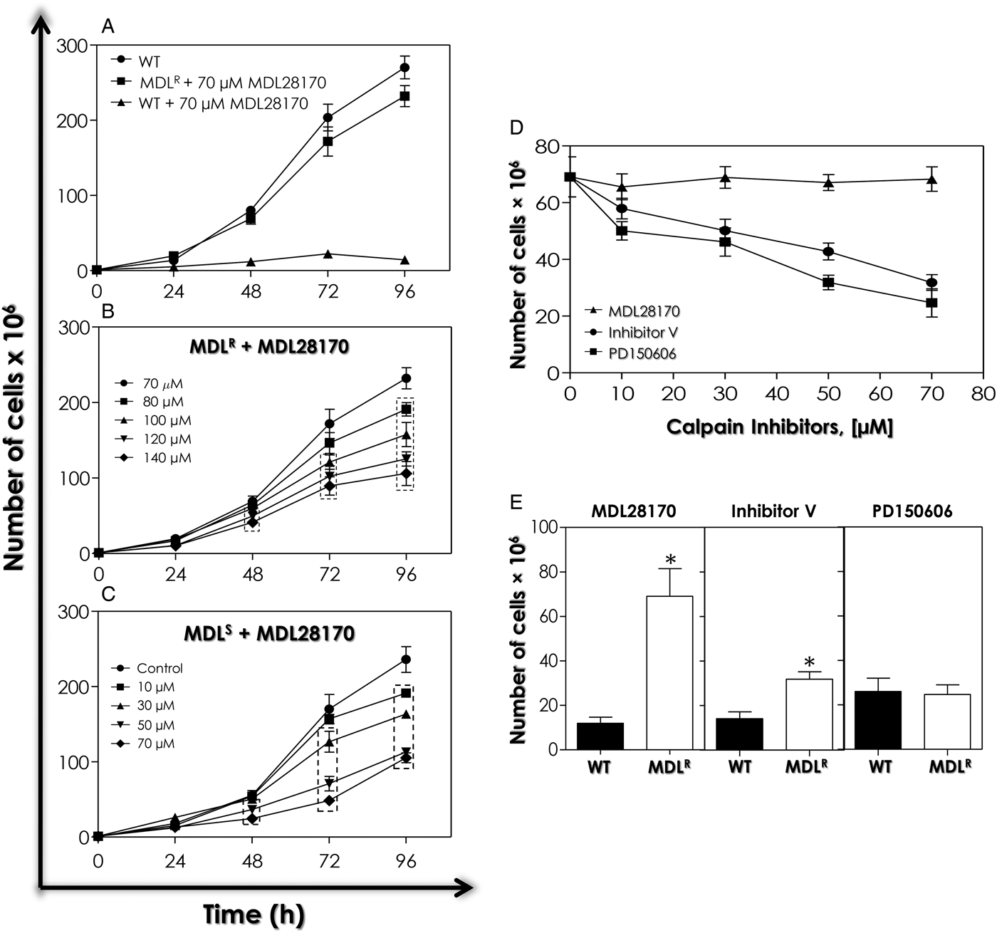
In order to generate a population of P. serpens promastigotes displaying MDL28170 resistance, WT cells were initially cultured in the presence of sub-lethal concentrations of this calpain inhibitor and a stepwise increase in its concentration was performed. After approximately 6 months, the culture was resistant to the concentration of 70 µ m MDL28170 (MDLR population). The construction of a comparative growth curve between WT and MDLR populations of P. serpens showed that MDLR cells presented a proliferation rate in the presence of 70 µ m MDL28170 comparable with the WT population (Fig. 1A). The doubling time of WT and MDLR populations were 9 h 28 min and 9 h 45 min, respectively. This drug concentration inhibited WT parasite growth by approximately 85% after 48 h (Fig. 1A). The kinetics of growth pattern of MDLR in the presence of higher concentrations of MDL28170 was then investigated, and a dose-dependent reduction in the proliferation rate was observed (Fig. 1B). The IC50 value for MDLR culture was determined as 115·4 µ m after 48 h of growth. For comparison, the IC50 value for the WT culture in the same time interval was determined as 30·9 µ m (Oliveira et al. Reference Oliveira, Goncalves, Garcia-Gomes, Goncalves, Seabra, Menna-Barreto, Lopes, d'Avila-Levy, Santos and Branquinha2017), and as such the resistance index for MDLR population was calculated as 3·7.
Growth profile and effect of calpain inhibitors on the growth rate of MDL28170-resistant (MDLR) promastigotes of P. serpens. (A) Comparative growth curve of the wild-type (WT) population, in the absence or in the presence of 70 µ m MDL28170, and MDLR cells plus 70 µ m MDL28170. (B) Effect of MDL28170 on the proliferation rate of MDLR promastigotes. (C) Evaluation of the stability of resistance of MDLR population. Cells kept for six months in the absence of the calpain inhibitor (MDLS cells) were cultured in the presence or absence (control) of MDL28170. (D) (MDLR) promastigotes were cultured in the presence of the calpain inhibitors V and PD150606 for 48 h in concentrations ranging from 10 to 70 µ m. (E) Comparison of the number of WT and MDLR cells in the presence of each calpain inhibitor at 70 µ m after 48 h of growth. Data shown represent the mean ± standard deviation of three independent experiments performed in triplicate. The dotted boxes in (B and C) highlight growth rates significantly different from control (P < 0·05). The asterisks denote statistic difference to control (P < 0·05).

In order to test the in vitro stability of the resistance phenotype, MDLR cells of P. serpens were subcultured in the drug-free Warren medium for 6 months (MDLS population), and then subjected to susceptibility assays in the presence of MDL28170. The effect of the calpain inhibitor in MDLS population was intermediary between WT and the original MDLR population: the IC50 value of MDL28170 for MDLS promastigotes was calculated as 66·7 µ m after 48 h of growth, with a reduction in the resistance index to 2·2 (Fig. 1C).
Cross-resistance to other calpain inhibitors
The in vitro susceptibility of P. serpens WT population to the calpain inhibitors V and PD150606 was previously determined and the IC50 values calculated as 32·3 and 42·8 µ m, respectively (Oliveira et al. Reference Oliveira, Goncalves, Garcia-Gomes, Goncalves, Seabra, Menna-Barreto, Lopes, d'Avila-Levy, Santos and Branquinha2017). For MDLR population, the IC50 values of the same calpain inhibitors were determined after 48 h (Fig. 1D). For calpain inhibitor V, the IC50 value was calculated as 65·9 µ m, which is approximately twice as that determined for the WT population, being suggestive of cross-resistance of MDLR population for this inhibitor. On the other hand, no cross-resistance was found to the calpain inhibitor PD150606, since the IC50 value for the MDLR culture was 49·5 µ m, which is very close to the one previously determined to the WT promastigotes. In order to further explore this result, a comparison of the proliferation rate of P. serpens WT and MDLR populations was performed in cells pre-treated with either 70 µ m MDL28170 or 70 µ m calpain inhibitor V for 48 h (Fig. 1E). It was possible to observe that proliferation was 5·8- and 2·2-fold lower in WT cells, respectively, when compared with the proliferation rate of MDLR cells cultured during the same time interval, which confirms the cross-resistance to both calpain inhibitors. In contrast, WT and MDLR promastigotes pretreated with 70 µ m PD1500606 presented a similar proliferation rate (Fig. 1E).
Cell viability
Parasite viability in MDLR population was confirmed using the resazurin and MTT assays, which showed similar levels of fluorescence and absorbance signals, respectively, to WT cells (Fig. S1A and B). This result indicated that the selection of the resistant population did not induce loss of parasite viability. Incubation with azide showed a reduction in cell viability when compared with WT and MDLR populations (Fig. S1A and B).
WT and MDLR P. serpens cells were also incubated in the presence of PI in order to compare plasma membrane integrity. Analysis by flow cytometry indicated no change in plasma membrane permeability in MDLR parasites when compared with WT promastigotes (Fig. S1C and D), in contrast to the positive control of cell death.
Morphological and ultrastructural analysis
Flow cytometry, light microscopy, scanning and transmission electron microscopy techniques were employed in order to evaluate the morphology and ultrastructure of P. serpens MDLR population. Analysis by flow cytometry showed that WT and MDLR cells presented similar cell size and granularity, as revealed by FSC and SSC parameters, respectively (Fig. S2A). Corroborating this result, both light microscopy (Fig. S2A, inset) and scanning electron microscopy (Fig. S2B) revealed no morphological differences between both populations, showing the typical P. serpens promastigote morphology, like an elongated cell body and a single, long flagellum (Fig. S2A inset and S2B), besides an anterior kinetoplast in relation to the nucleus (Fig. S2A, inset). These results are in accordance to the similar growth rate and viability of both populations (Fig. 1A, Fig. S1). MDLR promastigotes apparently presented an increased amount of small vesicles in the parasite surface (Fig. S2B).
When the ultrastructure of these parasites was compared by transmission electron microscopy, no significant changes were found between WT (Fig. S2C-a and b) and MDLR (Fig. S2C-c–e) populations: the typical morphology of nucleus, mitochondrion, kinetoplast and flagellar pocket of promastigotes was observed. However, the presence of microvesicles within the flagellar pocket of MDLR promastigotes and in the culture supernatant was detected (Fig. S2C-e, inset).
Expression of cysteine peptidases
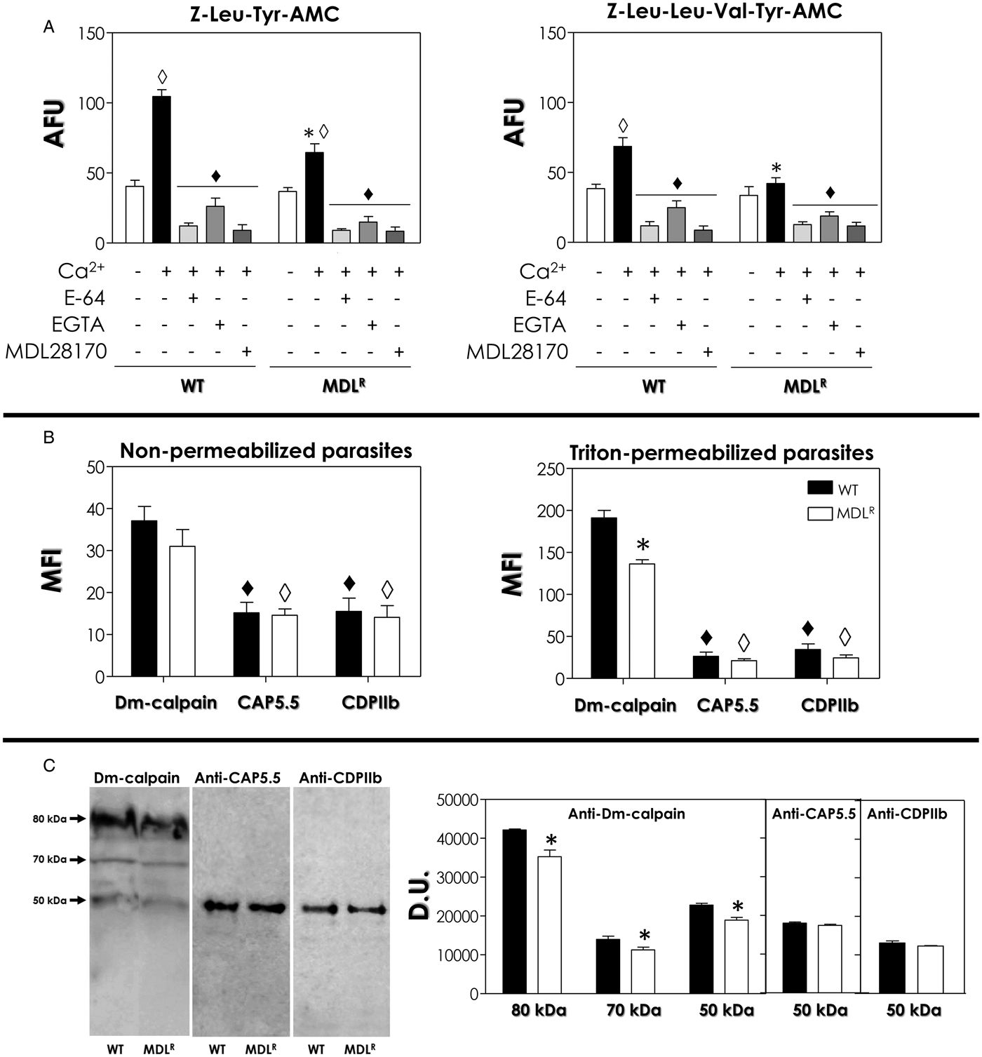
Our next goal was to evaluate the expression/activity of cysteine peptidases in both populations in order to compare the cruzipain-like proteolytic activity previously detected by our group in WT cells (Santos et al. Reference Santos, D´Avila-Levy, Elias, Vermelho and Branquinha2007) and, simultaneously, to detect calpain-like proteins whose expression could be affected in MDLR promastigotes. At first, the activity of calcium-dependent cysteine peptidases was compared using two substrates commonly used for detection of calpain activity: Z–Leu–Tyr–AMC and Z–Leu–Leu–Val–Tyr–AMC. Our data showed that the soluble extract of WT and MDLR cultures of P. serpens was able to cleave both calpain substrates at similar rates in the absence of calcium (Fig. 2A).
Detection of calcium-dependent cysteine peptidase activities (A) and calpain-like proteins (B, C) in wild-type (WT) and MDL28170-resistant (MDLR) promastigotes of P. serpens. (A) The whole cellular extract of each population was incubated with the fluorogenic substrates Z–Leu–Tyr–AMC and Z–Leu–Leu–Val–Tyr–AMC in the absence (– Ca2+) or in the presence (+ Ca2+) of calcium chloride, E-64, EGTA and MDL28170. The results were expressed as arbitrary fluorescence units (AFU). The values represent the mean ± standard deviation of three independent experiments performed in triplicate. The diamonds highlight the significant differences in each population in the presence or in the absence of calcium (white) or each modulator of calpain activity (black); the asterisks, between WT and MDLR cells in the presence of calcium (P < 0·05). (B) Fixed cells of each population, permeabilized or not, were incubated in the presence of anti-Dm-calpain, anti-CDPIIb and anti-CAP5·5 antibodies and then analysed by flow cytometry. Representative data of the analysis of 10 000 cells are shown and expressed as the mean fluorescence intensity (MFI). The asterisk highlights significantly different MFI levels between WT and MDLR promastigotes after incubation with anti-Dm-calpain antibody, while diamonds denote significant differences in MFI values between labelling with anti-CAP5·5 and anti-CDPIIb antibodies in comparison with anti-Dm-calpain antibody in WT (black diamonds) and MDLR cells (white diamonds). Labelling of permeabilized cells was significantly different from non-permeabilized cells for each of the three anti-calpain antibodies tested (P < 0·05), and consequently the scale for MFI levels in each graph was different. (C) Western blotting showing the CALPs recognized in the whole cellular extract of each population by the anti-calpain antibodies. Anti-tubulin monoclonal antibody was used as a control for sample loading in the blots, revealing a protein band of 50 kDa in similar amount in both populations (not shown). The apparent molecular masses, expressed in kilodaltons, of each detected band are shown. Right panel, the densitometric analysis of the reactive bands detected in Western blotting is expressed as densitometric units (DU). The results represent means standard deviation of three independent experiments, and the asterisks denote statistic difference between WT cells and MDLR promastigotes (P < 0·05).

When calcium was added to the reaction mixture, an increase of 158% was found for the hydrolysis of Z–Leu–Tyr–AMC in WT cells, while for MDLR soluble extract the increase was by 75% (Fig. 2A). Under these conditions, the addition of the highly selective cysteine peptidase inhibitor E-64 or the calpain inhibitor MDL 28170 inhibited the hydrolytic activity at similar levels: by 88 and 91%, respectively, in the soluble extract of WT, and by 85 and 87%, respectively, in the soluble extract of MDLR population (Fig. 2A). When the calcium chelator EGTA was included in the reaction mixture, the reduction of the substrate hydrolysis was by 74 and 78% in WT and MDLR cells, respectively (Fig. 2A). These results suggest the predominance of hydrolysis of Z–Leu–Tyr–AMC by calcium-dependent cysteine peptidases.
The hydrolysis rate for Z–Leu–Leu–Val–Tyr–AMC was also raised for WT soluble cellular extract in the presence of calcium, although at minor levels (78%), while for MDLR population no significant change in hydrolysis was detected (Fig. 2A). Similarly, E-64, EGTA and MDL28170 inhibited the hydrolytic activity on both cellular extracts in the presence of calcium: in WT population, the reduction in the hydrolysis of this substrate was 83, 64 and 88%, respectively; MDLR soluble extract presented an inhibition of 71, 57 and 73%, respectively, in the hydrolytic ability of the substrate (Fig. 2A).
A more detailed kinetics analysis confirmed the results described above: the addition of calcium to the assay buffer reduced the K m values for both substrates in the soluble extract of WT and MDLR cultures of P. serpens (Table S1). In addition, K m values were higher for Z–Leu–Tyr–AMC when compared with Z–Leu–Leu–Val–Tyr–AMC, and for MDLR population in comparison with WT cells (Table S1). E-64 and MDL28170 were also evaluated as inhibitors of calcium-dependent cysteine peptidases activity against both substrates. In our assay, the calculated apparent inhibition constants (K i values) for E-64 were found to be lower in both substrates when compared with MDL28170, which exhibited the highest K i values. The values of K i calculated for both inhibitors in the MDLR population were at least two times higher than that of WT cells (Table S2).
Previously, our group found by Western blotting analysis in WT cells that anti-Dm-calpain antibody reacted with three polypeptide bands migrating at approximately 80, 70 and 50 kDa, while a single 50-kDa polypeptide was detected with anti-CAP5·5 and anti-CDPIIb antibodies. In that work, pre-treatment of P. serpens WT cells with the IC50 value of MDL28170 led to a reduction in the expression of epitopes reactive to anti-Dm-calpain, anti-CAP5·5 and anti-CDPIIb antibodies, indicating that calpain-like molecules are potential targets of the calpain inhibitor (Oliveira et al. Reference Oliveira, Goncalves, Garcia-Gomes, Goncalves, Seabra, Menna-Barreto, Lopes, d'Avila-Levy, Santos and Branquinha2017). A search for Dm-calpain, CAP5·5 and CDPIIb homologous proteins in the streamlined genome of Phytomonas sp. isolate EM1 (Porcel et al. Reference Porcel, Denoeud, Opperdoes, Noel, Madoui, Hammarton, Field, da Silva, Couloux, Poulain, Katinka, Jabbari, Aury, Campbell, Cintron, Dickens, Docampo, Sturm, Koumandou, Fabre, Flegontov, Lukeš, Michaeli, Mottram, Szöőr, Zilberstein, Bringaud, Wincker and Dollet2014) was performed, and confirmed the presence of at least one calpain-conserved domain as well as the theoretical molecular mass consistent with the recognition by each antibody (Oliveira et al. Reference Oliveira, Goncalves, Garcia-Gomes, Goncalves, Seabra, Menna-Barreto, Lopes, d'Avila-Levy, Santos and Branquinha2017). Although the genome of P. serpens isolate 9T is available in Genbank, the genes have not been identified and no protein was predicted. Even so, in the present work a search for P. serpens sequences that could encode proteins homologous to the fragments employed to generate each antibody used in this work was performed on tBlastn. The P. serpens whole-genome shotgun sequence (WGS) AIHY01000314·1 encodes a calpain-like sequence of around 94 kDa capable of being recognized by anti-Dm-calpain and anti-CDPIIb antibodies (31 and 28% of amino acid identity, respectively). In addition, 17 homologues of the T. brucei CAP5·5 were found in P. serpens genome, eight of them with the conserved domain CysPc (pfam:Peptidase_C2) of the classical calpains. Moreover, two sequences (AIHY01001500·1 and AIHY01001547·1) presented a predicted molecular mass of 50 kDa, which supports the recognition of P. serpens 50-kDa CALP (31 and 26% identity, respectively). One of these sequences (AIHY01001500·1) is also homologous to the H. americanus CDPIIb and could be the same calpain-like molecule recognized by this antibody, displaying 31% identity. There also other ten homologous sequences in P. serpens genome that can be recognized by anti-CDPIIb antibody, although the predicted molecular mass do not support the experimental recognition by this antibody. These data encouraged us to evaluate the expression of CALPs in P. serpens MDLR promastigotes.
Flow cytometry analysis of WT and MDLR promastigotes of P. serpens was then performed using the three anti-calpain antibodies from distinct origins and different specificities in order to check for the differential expression of CALPs. In the present study, the highest MFI values were detected in the cross-reactivity with anti-Dm-calpain antibody, both at the cell surface as well as in the intracellular milieu, when compared with the binding of anti-CAP5·5 and anti-CDPIIb antibodies (Fig. 2B). The binding of the three anti-calpain antibodies was significantly enhanced when control, fixed cells were permeabilized, which indicates that CALPs cross-reactive to these antibodies are located mainly in intracellular compartments, especially for anti-Dm-calpain antibody, for which the increase was 416% for WT population and 338% for MDLR population (Fig. 2B). Furthermore, the expression of intracellular molecules cross-reactive to anti-Dm-calpain antibody was significantly lower (approximately 30%) in MDLR promastigotes when compared with WT cells, while the reactivity of the same antibody in the cell surface as well as anti-CAP5·5 and anti-CDPIIb antibodies was not altered (Fig. 2B). No significant difference in the percentage of fluorescent cells between WT and MDLR promastigotes was observed in non-permeabilized and permeabilized cells (data not shown). Interestingly, the same polypeptide bands detected previously in WT population by Western blotting (Oliveira et al. Reference Oliveira, Goncalves, Garcia-Gomes, Goncalves, Seabra, Menna-Barreto, Lopes, d'Avila-Levy, Santos and Branquinha2017) were also detected In MDLR population, but a significant reduction in the expression (in the 15–21% range) was observed only for the bands cross-reactive with anti-Dm calpain antibody, which confirmed the flow cytometry result (Fig. 2C).
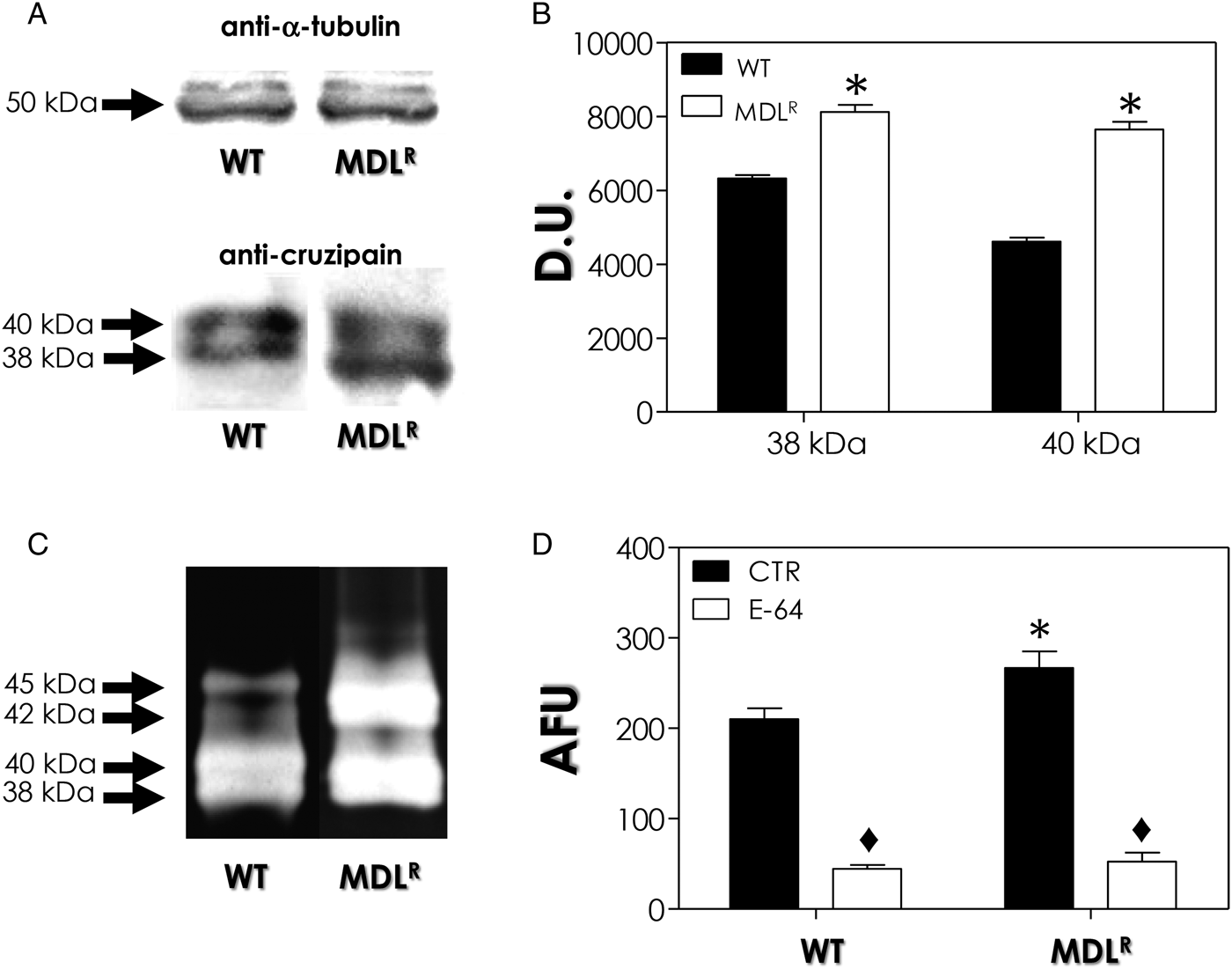
The next step of the present study was to detect papain-like cysteine peptidase activity in P. serpens WT and MDLR promastigote cells. Previous studies from our group have already identified and characterized the presence of two cell-associated cysteine peptidases at 38 and 40 kDa in P. serpens displaying cross-reactivity to the well-known cruzipain, the major cysteine peptidase from T. cruzi (Santos et al. Reference Santos, D´Avila-Levy, Elias, Vermelho and Branquinha2007; d’Ávila-Levy et al. Reference d'Avila-Levy, Altoé, Uehara, Branquinha and Santos2014). Our results from Western blotting analysis showed that MDLR cells presented a significantly increased expression of the 38 and 40-kDa proteins that cross-reacted with anti-cruzipain antibody, in comparison with the expression in WT cells, by 32 and 58%, respectively (Fig. 3A and B). The cruzipain used to produce the anti-cruzipain antibody have 59% of amino acid identity with P. serpens isolate 9T WGS AIHY01001722·1, which could encode a 44-kDa protein and also have the cruzain domain. Concomitantly, the cell-associated proteolytic profile detected by gelatin–SDS–PAGE at pH 5·0 revealed the presence of two bands migrating at the same molecular masses in both populations, besides the detection of two proteolytic activities at 42 and 45 kDa (Fig. 3C). The latter bands had their expression increased in MDLR cells (Fig. 3C). The quantification of the proteolytic activity against Z–Phe–Arg–AMC, a fluorogenic substrate commonly used to detect cysteine peptidase activity, including cruzipain (Cazzulo et al. Reference Cazzulo, Cazzulo Franke, Martínez and Franke De Cazzulo1990), was then tested. Confirming the results detected in gelatin–SDS–PAGE, MDLR population presented a significant increase (27%) in the hydrolysis of the fluorogenic substrate when compared with WT cells (Fig. 3D). The addition of the cysteine peptidase inhibitor E-64 in the reaction mixture diminished significantly the hydrolytic activity in both systems by 80% (Fig. 3D).
Differential expression of intracellular cruzipain-like molecules and papain-like cysteine peptidase activity by wild-type (WT) and MDL28170-resistant (MDLR) promastigotes of P. serpens. (A) Western blotting analysis showing the proteins recognized by the anti-cruzipain antibody. Anti-tubulin antibody was used as a control for sample loading. (B) The densitometric analysis of the reactive bands detected in Western blotting (A) is expressed as densitometric units (D.U.). (C) Peptidase profile analysis by gelatin–SDS–PAGE. The apparent molecular masses, expressed in kilodaltons, of each detected band in (A, C) are represented on the left. (D) The papain-like cysteine peptidase activity in parasites lysates was assessed by measuring the hydrolysis of Z–Phe–Arg–AMC in the absence (control, CTR) or the presence of E-64. Results are expressed as arbitrary fluorescence units (AFU). In (B, D), the values represent the mean ± standard deviation of three independent experiments performed in triplicate. The asterisks denote statistic difference between WT and MDLR promastigotes, while diamonds highlight significant differences in the hydrolytic activity in the absence or the presence of E-64 (P < 0·05).

Interaction with O. fasciatus salivary glands
Besides the evaluation of the expression of CALPs and cruzipain-like molecules in P. serpens WT and MDLR cells, we also decided to study the expression of gp63-like molecules in these parasites. The rationale behind these experiments is the previous observation by our group that P. serpens expresses metallopeptidases that are related to Leishmania spp. gp63, which displays virulence-related functions in these pathogenic parasites (Santos et al. Reference Santos, D´Avila-Levy, Elias, Vermelho and Branquinha2007). More importantly, P. serpens surface gp63-like proteins play an important role in the parasite adhesion to the salivary glands of the insect vector (Santos et al. Reference Santos, D´Avila-Levy, Elias, Vermelho and Branquinha2007).
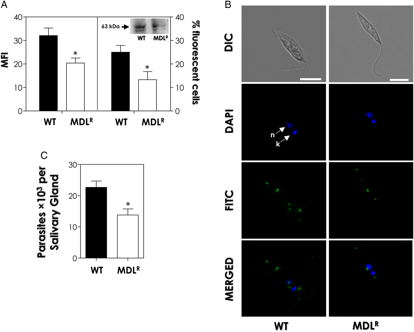
By flow cytometry, our results showed that the expression of molecules cross-reactive to anti-gp63 antibody in the surface of P. serpens MDLR cells presented a reduction of 37% in the mean fluorescence intensity (MFI) value and of 46% in the percentage of fluorescent cells when compared with WT population (Fig. 4A). As previously described by our group (Santos et al. Reference Santos, D´Avila-Levy, Elias, Vermelho and Branquinha2007), two polypeptides migrating in the 50–65 kDa range cross-react with anti-gp63 antibodies in P. serpens whole cellular extract. In the present work, Western blotting analysis showed the presence of both gp63-like protein bands; MDLR cells presented a decreased expression of these proteins, in comparison with WT cells (Fig. 4A). The most expressed gp63 sequence of L. mexicana amastigotes (used to generate the anti-gp63 employed in the present work) have at least six homologues in P. serpens isolate 9T genome: AIHY01014097·1, AIHY01014205·1, AIHY01015147·1, AIHY01014315·1, AIHY01014400·1 and AIHY01014106·1, displaying 40–42% of amino acid identity. The predicted molecular mass of each one ranges from 44 to 59 kDa.
Correlation between the expression of surface gp63-like molecules in wild-type (WT) and MDL28170-resistant (MDLR) promastigotes of P. serpens and the interaction with the explanted salivary glands of Oncopeltus fasciatus. (A) Fixed promastigotes were incubated in the presence of anti-gp63 antibody and analysed by flow cytometry. The data are expressed as the mean of fluorescence intensity (MFI) levels and as the percentage of fluorescent cells (% FC). Representative data of the analysis of 10 000 cells in experiments performed in triplicate are shown and the results were considered significantly different when P < 0·05 (*). In the inset, Western blotting showing the gp63-like proteins recognized in the whole cellular extract of each population by the anti-63 antibody. Anti-tubulin monoclonal antibody was used as a control for sample loading in the blots, revealing a protein band of 50 kDa in similar amount in both populations (not shown). The apparent molecular mass, expressed in kilodaltons, of the major band is shown. (B) Differential interference contrast microscopy (DIC) and immunofluorescence images showing the double labelling of P. serpens cells for gp63-like molecules cross-reactive to anti-gp63 antibody (FITC, green labelling) and DAPI (blue labelling). Below, the overlay of anti-gp63 and DAPI labelling. Bars: 5 µm. (C) Following interaction with each population, the salivary glands were washed and homogenized, and the released trypanosomatids were counted. The results are shown as the mean ± standard deviation of three independent experiments and the asterisk highlights significantly different values (P < 0·05).
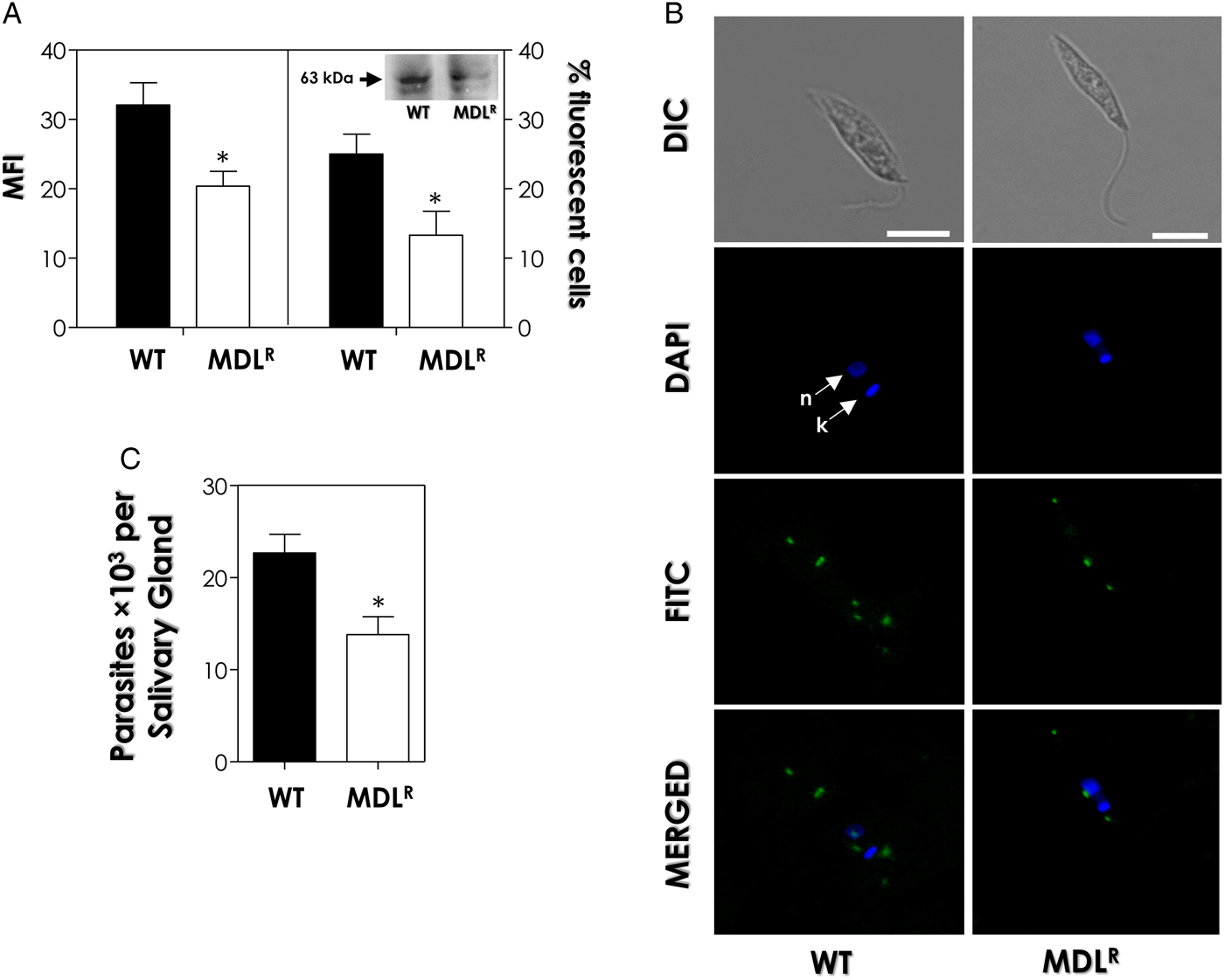
Our group has previously demonstrated the surface location of gp63-like molecules in P. serpens by agglutination assay, flow cytometry and immunofluorescence microscopy analyses (reviewed by Santos et al. Reference Santos, D´Avila-Levy, Elias, Vermelho and Branquinha2007). In the present work, immunofluorescence microscopic images confirmed the surface location of gp63-like molecules in both populations and corroborated the reduction of expression in MDLR parasites when compared with WT promastigotes (Fig. 4B). The next step was to verify whether MDL28170 resistance would influence the capacity of P. serpens to adhere to O. fasciatus explanted salivary glands: our results showed a significant reduction (38%) in the capacity of MDLR cells to adhere to the salivary glands when compared with WT population (Fig. 4C).
Possible mechanism of resistance
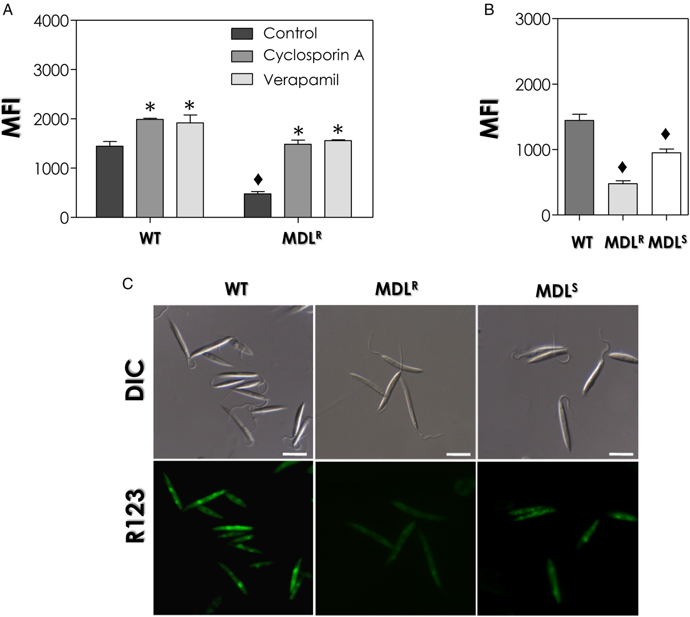
By flow cytometry, our data showed that MDLR cells presented a significant reduction (67%) in R123 accumulation in comparison with WT population (Fig. 5A). Since the efflux rate of R123 indicates the ability of the parasite to actively expel the drug from its body (Forster et al. Reference Forster, Thumser, Hood and Plant2012), it is possible to infer that efflux pumps may play a role MDL28170 resistance. In order to confirm this hypothesis, cells were pretreated with two inhibitors of ABC transporters, verapamil and cyclosporin A. First, the cytotoxicity of cyclosporin A and verapamil over the parasites was evaluated by colorimetric assay with resazurin, and the pretreatment of cells with both inhibitors up to the concentration of 50 µ m was not capable of altering the viability of the parasites, when compared with untreated control cells (data not show). The pretreatment of WT and MDLR cells with both inhibitors of efflux pumps led to a significant increase in the intracellular accumulation of R123, although with different intensities (Fig. 5A): the pretreatment of WT and MDLR cells with verapamil led to an increase of 32·8 and 210·6%, respectively, whereas cyclosporin A increased by 37·5 and 225·7%, respectively, the intracellular accumulation of R123. For MDLR cells, treatment with verapamil and cyclosporin A increased the MFI values to levels reaching the WT population (Fig. 5A).
Intracellular accumulation of rhodamine 123 (R123) in wild-type (WT) and MDL28170-resistant (MDLR) promastigotes of P. serpens. The graphs in (A, B) represent the mean of fluorescence intensity (MFI) levels of intracellular accumulation of R123 analysed by flow cytometry. (A) WT and MDLR cells were previously treated or not (control) with the ABC transporters inhibitors cyclosporin A and verapamil and subsequently incubated with R123. (B) WT and MDLR cells accumulation of R123 was compared with the MDL28170-resistant population grown in the absence of MDL28170 for 6 months (MDLS). The data presented in the graphs are representative of analysis of 10 000 cells in experiments performed in triplicate. The diamonds denote significant different values of accumulation of R123 between WT, MDLR and MDLS promastigotes, while the asterisks highlight significant different values in WT and MDLR cells pretreated or not with ABC transporters inhibitors (P < 0·05). (C) Differential interference contrast microscopy (DIC) and fluorescence microscopy of WT, MDLR and MDLS promastigotes showing the intracellular accumulation of R123. Bars: 5 µm.
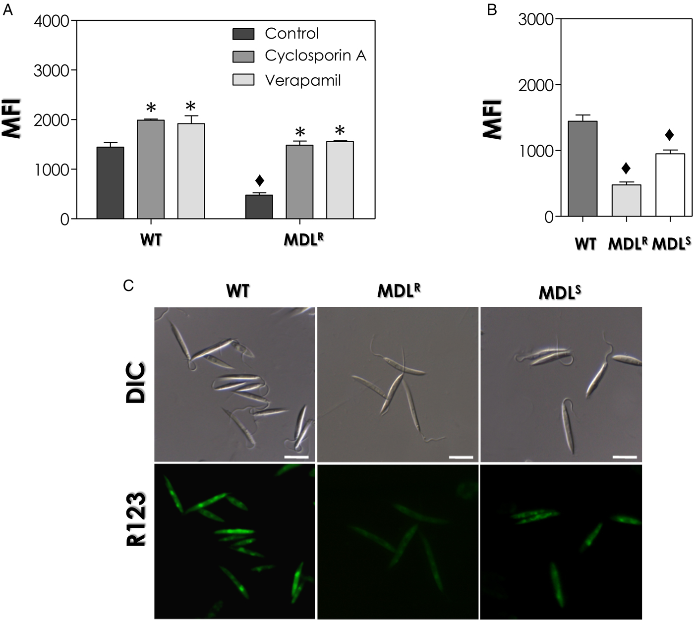
In this same set of experiments, the capacity of P. serpens WT and MDLR promastigotes to accumulate R123 was compared with MDLR cells grown in the absence of the drug for 6 months (MDLS population). By flow cytometry, our data showed that MDLS population showed a 32% reduction in the MFI value when compared with WT promastigotes and an increase of 98·4% when compared with the resistant population (Fig. 5B), which indicates a partial reversion to the WT phenotype when compared with MDLR promastigotes. In parallel to the flow cytometry analysis, cells were also observed under fluorescence microscopy, confirming the intermediary fluorescence of MDLS cells between MDLR and WT populations (Fig. 5C). Altogether, these data confirm the previous observation (Fig. 1C) that the effect of MDL28170 in the MDLS population growth curve was intermediary between WT and the original MDLR population.
DISCUSSION
There is not much information available about the roles of calpains and CALPs in trypanosomatids (Branquinha et al. Reference Branquinha, Marinho, Sangenito, Oliveira, Gonçalves, Ennes-Vidal, D'Avila-Levy and Santos2013; Ennes-Vidal et al. Reference Ennes-Vidal, Menna-Barreto, Branquinha, Santos and D'Avila-Levy2017). It is worth mentioning that whole genome analysis characterized a large family of genes encoding CALPs in T. brucei, T. cruzi and Leishmania major (Ersfeld et al. Reference Ersfeld, Barraclough and Gull2005). Considering the high number of genes and sequence diversity of calpains in these parasites (Ersfeld et al. Reference Ersfeld, Barraclough and Gull2005), it is not an easy task to completely characterize this protein family and to assess its functions (Ennes-Vidal et al. Reference Ennes-Vidal, Menna-Barreto, Branquinha, Santos and D'Avila-Levy2017). Although there is no knockout available, the RNAi of three calpain genes in T. brucei revealed their roles in parasite growth, morphology and flagellum assembly (Olego-Fernandez et al. Reference Olego-Fernandez, Vaughan, Shaw, Gull and Ginger2009; Hayes et al. Reference Hayes, Varga, Olego-Fernandez, Sunter, Ginger and Gull2014). Our group has been working with the feasible use of calpain inhibitors as leading compounds to search for new therapeutic options to treat trypanosomatid infections (Branquinha et al. Reference Branquinha, Marinho, Sangenito, Oliveira, Gonçalves, Ennes-Vidal, D'Avila-Levy and Santos2013). Mechanisms of action of MDL28170 against P. serpens so far identified (Oliveira et al. Reference Oliveira, Goncalves, Garcia-Gomes, Goncalves, Seabra, Menna-Barreto, Lopes, d'Avila-Levy, Santos and Branquinha2017) include: effects on mitochondrion and trans-Golgi network; perturbation in processing of cruzipain-like molecules; repression of the activity of calcium-dependent cysteine-peptidases and expression of CALPs; and reduction in the interaction process with O. fasciatus salivary glands. Besides investigating the mechanism of action of MDL28170, it is fundamental to investigate the possibility of resistance to this calpain inhibitor in trypanosomatids, and that was the major goal of the present study. The biochemical analysis of the drug-resistant parasites was useful in trying to delineate the drug target(s) as well as the mechanisms developed by P. serpens promastigotes to reduce the lethal effect of MDL28170.
In this work, a viable population of P. serpens resistant to the calpain inhibitor MDL28170 at 70 µ m (MDLR population) was selected by culturing promastigotes in increasing concentrations of the drug. In trypanosomatids, the drug resistance phenotype is a complex physiological process, which is often polyclonal and involves several different co-existing genotypes in a given population. The heterogeneity in both genotype and phenotype in resistant populations may be either stable, being maintained in the absence of drug pressure, or unstable, and is therefore partially or totally lost when the parasite is kept in a drug-free medium (Vanaerschot et al. Reference Vanaerschot, Huijben, Van den Broeck and Dujardin2014). In the present work, the decrease in the resistance index in P. serpens MDLS population (i.e., MDLR cells subcultured in the drug-free Warren medium for 6 months) in comparison with MDLR promastigotes indicates a partial reversion of MDL28170 susceptibility to the WT phenotype. However, it is important to note that the IC50 value of MDLS promastigotes is still significantly higher than the IC50 of WT cells, which suggests that once any degree of resistance is achieved the culture does not revert back to the WT phenotype. Based on these data, the partial reversion in the resistance to MDL28170 in P. serpens MDLS population observed in the present study may indicate the involvement of more than one molecular mechanism in the process of MDL28170 resistance, as will be discussed below. A more detailed genomic analysis may help to elucidate the possible pathways used by the parasite for protection against the effects of the drug.
Cross-resistance of P. serpens MDLR population for calpain inhibitor V was also suggested by our results due to the increase in the IC50 value for the latter, although at lower levels. Differences in the degree of cross-resistance between MDL28170 and calpain inhibitor V might be explained by differences in the chemical structure, mechanism of action or specificity for a particular calpain structure (Donkor, Reference Donkor2015). Although MDL28170 and calpain inhibitor V are both active site-directed inhibitors, they have different chemical structures that have been developed for typical mammalian calpains. Consequently, we cannot exclude the possibility that these inhibitors act on similar targets in the trypanosomatid cell as well as on different targets. On the other hand, the calpain inhibitor PD150606 is specific for calcium-binding domains present in mammalian calpains that are essential for the enzymatic activity, which may explain the absence of cross-reactivity to MDL28170 (Wang et al. Reference Wang, Nath, Posner, Raser, Buroker-Kilgore, Hajimohammadreza, Probert, Marcoux, Ye, Takano, Hatanaka, Maki, Caner, Collins, Fergus, Lee, Lunney, Hays and Yuen1996). Although amino acid residues that are critical for calcium binding in mammalian calpains are partially conserved in some kinetoplastid sequences, these calcium-binding motifs are absent in trypanosomatid CALPs (Ersfeld et al. Reference Ersfeld, Barraclough and Gull2005). Analysis of these sequences revealed the presence of conserved domains, although residues critical for catalytic activity were frequently changed (Ersfeld et al. Reference Ersfeld, Barraclough and Gull2005).
MDLR cells presented similar growth pattern and doubling time to WT promastigotes, and neither cell viability nor plasma membrane integrity were lost in the selection of the resistant population. Furthermore, no changes in the morphology and the typical ultrastructure of the parasites were visualized. These data provided supportive evidence that the experimental resistance selection was successful, adding the particular advantage that the parent WT cells and the derived MDLR population could be directly compared for phenotypic analysis. The observation of microvesicles in the flagellar pocket of MDLR promastigotes by transmission electron microscopy is suggestive of a possible drug sequestration for excretion, as observed in other drug-resistant trypanosomatids. For instance, when Engel et al. (Reference Engel, Torres, Hsieh, Doyle and McKerrow2000) generated T. cruzi epimastigotes that were 20 times more resistant to a peptidomimetic cruzipain inhibitor than the WT, the secretory pathway was up-regulated in the resistant parasites due to the release of inactive cruzipain precursors complexed to the cysteine peptidase inhibitor. In a similar result to that observed in this study, transmission electron microscopy showed a multilamellar membrane-like material in the flagellar pockets of both amphothericin B-resistant L. mexicana amastigotes and promastigotes that was absent in WT cells (Al-Mohammed et al. Reference Al-Mohammed, Chance and Bates2005). These data are suggestive of similar responses of drug-resistant cells to the presence of drugs in the flagellar pocket, the site where endocytosis and exocytosis take place in trypanosomatids (Morgan et al. Reference Morgan, Hall, Denny, Carrington and Field2002).
In the present study, differences in CALPs expression and calcium-dependent cysteine peptidase activity between WT and MDLR promastigotes were found. The reduced expression/activity of these molecules in MDLR promastigotes may allow the parasite to survive in the presence of MDL28170. Indeed, the sub-expression of the target molecules of a drug has been reported in different trypanosomatids that have developed drug resistance. Yong et al. (Reference Yong, Schmitz, Vannier-Santos, De Lima, Lalmanach, Juliano, Gauthier and Scharfstein2000) showed that T. cruzi epimastigotes (clone Dm28c) resistant to Z–(SBz)Cys–Phe–CHN2, an irreversible cysteine peptidase inhibitor, presented significantly lower cysteine peptidase activity than that observed in parental cells, and this fact was accompanied by the reduced expression of cruzipain molecules, the major epimastigote cysteine peptidase, in the resistant parasites.
In contrast to the reduced calcium-dependent cysteine peptidase activity in P. serpens MDLR promastigotes detected in this work, a higher expression of cruzipain-like molecules displaying papain-like cysteine peptidase activity was found in this population. A possible explanation to this difference may be found in T. cruzi. As previously mentioned, a T. cruzi population resistant to the cathepsin L-like cysteine peptidase inhibitor Z–(SBz)Cys–Phe–CHN2 displayed a decreased cruzipain expression and a lower rate of cysteine peptidase fluorogenic substrate hydrolysis (Yong et al. Reference Yong, Schmitz, Vannier-Santos, De Lima, Lalmanach, Juliano, Gauthier and Scharfstein2000). Moreover, marked differences were seen in the protein profiles of T. cruzi resistant cells: the decreased expression of cruzipain was paralleled by the increased expression of a 30-kDa cysteine peptidase. This proteolytic activity was refractory to inhibition by Z–(SBz)Cys–Phe–CHN2 and strongly recognized by anti-cathepsin B-like peptidase antibody, which indicated that the 30-kDa cysteine peptidase corresponds to a cathepsin B-like peptidase. This fact indicated that the deficit of metabolic functions promoted by inhibiting cruzipain was probably compensated by the increased expression of a cathepsin B-like target in T. cruzi (Yong et al. Reference Yong, Schmitz, Vannier-Santos, De Lima, Lalmanach, Juliano, Gauthier and Scharfstein2000). These results, besides the ones shown in the present work, may be correlated to the possible over-expression of distinct cysteine peptidase activities in order to compensate for the selection of cells resistant to cysteine peptidase inhibitors.
The induction of resistance may cause alterations in the expression of molecules involved in different cellular processes, as detected in this study for calpain-like and papain-like cysteine peptidases, and also virulence factors, which may affect the life cycle of the parasite. In addition, a reduced ability to replicate and/or cause disease is detected when a pathogen acquires drug resistance (Caljon et al. Reference Caljon, De Muylder, Durnez, Jennes, Vanaerschot and Dujardin2016). Several studies have shown that mutations that render pathogens resistant to drug treatment are likely to result in a loss of fitness, also defined as ‘proficiency’ (Caljon et al. Reference Caljon, De Muylder, Durnez, Jennes, Vanaerschot and Dujardin2016). In this sense, the lower levels of adhesion of MDLR cells to O. fasciatus explanted salivary glands suggest that the fitness of the parasite might be affected in this population. The lower expression of gp63-like molecules in the cell surface of MDLR promastigotes must be correlated to the reduced level of parasite adhesion to the invertebrate host, since these molecules were already described as potential adhesins expressed by the parasite to bind to salivary glands of phytophagous insects (d’Ávila-Levy et al. Reference d'Avila-Levy, Altoé, Uehara, Branquinha and Santos2014).
Resistance to chemotherapy in trypanosomatids such as Leishmania spp. and T. cruzi is often caused by changes in the expression of membrane transport proteins (Campos et al. Reference Campos, Castro-Pinto, Ribeiro, Berredo-Pinho, Gomes, da Silva Bellieny, Goulart, Echevarria and Leon2013; Yasinzai et al. Reference Yasinzai, Khan, Nadhman and Shahnaz2013). Genome analysis of Phytomonas isolates EM1 and HART1 allowed the identification of 24 and 23 gene families, respectively, as belonging to the family of ABC transporters (Porcel et al. Reference Porcel, Denoeud, Opperdoes, Noel, Madoui, Hammarton, Field, da Silva, Couloux, Poulain, Katinka, Jabbari, Aury, Campbell, Cintron, Dickens, Docampo, Sturm, Koumandou, Fabre, Flegontov, Lukeš, Michaeli, Mottram, Szöőr, Zilberstein, Bringaud, Wincker and Dollet2014). Although resistance to drugs is a complex phenomenon, the rate of efflux of the fluorescent dye R123 from the parasite cell, using flow cytometry, is an indication of the isolate's ability to efflux drugs, thus avoiding death, including trypanosomatids (Campos et al. Reference Campos, Castro-Pinto, Ribeiro, Berredo-Pinho, Gomes, da Silva Bellieny, Goulart, Echevarria and Leon2013; Rai et al. Reference Rai, Bhaskar, Goel, Nath Dwivedi, Sundar and Goyal2013). In this sense, the lowest accumulation of the fluorochrome dye R123 detected in MDLR promastigotes allowed us to assume that overexpression of efflux pumps is a possible mechanism of resistance developed by P. serpens MDLR promastigotes. However, the R123 dye accumulation assay is considered less specific with respect to the transporter subtype activity, since dyes can be substrates of different ABC protein subtypes expressed in WT cells or native tissues (Cunha et al. Reference Cunha, Burkhardt-Medicke, Wellner, Santos, Moradas-Ferreira, Luckenbach and Ferreira2017).
To investigate the phenomenon of multidrug resistance in L. amazonensis, it was demonstrated that the vinblastine-resistant phenotype was partially reverted by verapamil, which suggested that the cell line exhibited a multi-drug resistance phenotype. Confirming this assumption, R123 presented a lower intracellular accumulation in vinblastine-resistant cells due to an increased efflux (Gueiros-Filho et al. Reference Gueiros-Filho, Viola, Gomes, Farina, Lins, Bertho, Wirth and Lopes1995). A similar profile was detected in T. cruzi: strains selected in vitro for the resistance to the compounds 4-N-(2-methoxy styryl)-thiosemicarbazone (2-Meotio) and benznidazole and then treated with R123 presented lower fluorescence than that exhibited by the parental line. Moreover, when the resistant lines were treated with cyclosporin A or verapamil, the fluorescence inside the resistant lines increased significantly, indicating the participation of efflux pumps in the process and, consequently, in the elimination of R123 by the resistant parasites (Campos et al. Reference Campos, Castro-Pinto, Ribeiro, Berredo-Pinho, Gomes, da Silva Bellieny, Goulart, Echevarria and Leon2013). In the present work, the pretreatment of MDLR cells with both inhibitors of efflux pumps, verapamil and cyclosporin A, led to a significant increase in the intracellular accumulation of R123 in comparison with WT cells, which confirmed the increase in the expression of ABC transporters in the resistant population.
Another aspect that deserves consideration is the increase in R123 accumulation in MDLS cells when compared with MDLR population and its reduction in comparison with WT cells. This may indicate that the partial reversion to the WT phenotype when MDLR cells were cultured in a drug-free medium can be correlated to the reduced expression of membrane transporters. These data reinforce the previous observation that the partial reversion in the resistance to MDL28170 may indicate the involvement of more than one molecular mechanism in the process of MDL28170 resistance. Since no further decrease in resistance was observed in MDLS cells and the resistance index in this population is still significantly higher than WT cells, the biochemical modifications described in the present paper in the resistant population may be stable, while others (such as the expression of ABC transporters) may be lost or reduced during the reversion process.
In conclusion, resistance to chemotherapy in a parasite reveals its gene plasticity and capability to adaptation to different environments. The stress caused by the presence of the drug probably leads to the selection of parasites with appropriate mechanisms to evade the lethal effects caused by antiparasitic compounds (Vanaerschot et al. Reference Vanaerschot, Huijben, Van den Broeck and Dujardin2014). In the present study, biochemical changes were observed that support the existence of more than one mechanism that act in concert to provide MDL28170 resistance in P. serpens. In this sense, the decreased expression of calpain-like molecules as well as calcium-dependent cysteine peptidase activity is, probably, a mechanism employed by P. serpens MDLR cells to try to repeal the adverse effects of the continuous presence of the calpain inhibitor MDL28170. In contrast, the overexpression of cruzipain-like molecules, as well as the increase in its papain-like cysteine peptidase activity can be considered as a compensatory mechanism generated in MDLR cells for the reduced presence of calcium-dependent cysteine peptidase activity. Induction of resistance to MDL28170 also significantly reduced the expression of gp63-like molecules, which is probably associated to the lowest ability of MDLR promastigotes to interact with O. fasciatus salivary glands. This may indicate that the selection of the MDL28170-resistant population also affects the expression of molecules other than calpain-like or cysteine peptidases. However, we cannot affirm at the present moment that MDL28170 either has more than one target in trypanosomatids and may act in concert in the mechanisms cited above, or act sequentially in these processes. Additionally, the overexpression of efflux pumps in the cell surface of MDLR promastigotes is suggestive of another possible mechanism of resistance developed by the parasite. Our study yielded notions about the multiplicity of mechanisms leading to MDL28170 resistance in P. serpens promastigotes.
SUPPLEMENTARY MATERIAL
The supplementary material for this article can be found at https://doi.org/10.1017/S0031182017001561.
ACKNOWLEDGEMENTS
The authors thank Denise da Rocha de Souza, who is supported by a FAPERJ fellowship, for technical assistance.
FINANCIAL SUPPORT
This work was supported by grants from the Brazilian Agencies: Conselho Nacional de Desenvolvimento Científico e Tecnológico (CNPq), Fundação de Amparo à Pesquisa no Estado do Rio de Janeiro (FAPERJ) and Fundação Coordenação de Aperfeiçoamento de Pessoal de Nível Superior (CAPES).