CHD are the most common congenital anomalies accounting for 8/1000 live births globally. It has been suggested that folic acid and/or multivitamins, taken periconceptionally, have a role in the prevention of many other congenital anomalies. However, the results of these studies are not consistent.Reference Botto, Mulinare and Erickson 1 – Reference Shaw, O’Malley, Wasserman, Tolarova and Lammer 5 Some studies have shown a decrease in the rates of one or more defects, whereas others have not confirmed these findings.Reference Canfield, Collins and Botto 6 – Reference Botto, Khoury and Mulinare 10
It has been suggested that maternal/placental microenvironment before conception has influenced the development of the fetal organs and that cardiac defects can be explained by abnormalities of the microenvironment of the fetus. The folate pathway relates not only to purine and pyrimidine synthesis, which is important in deoxyribonucleic acid synthesis and cell proliferation, but also to the synthesis of the primary methyl donor S-adenosylmethionine, which is important in methylation reactions of cellular lipids, proteins, ribonucleic acid, and deoxyribonucleic acid. deoxyribonucleic acid methylation is critical to epigenetic regulation of gene expression.Reference Aguilera, Fernandez and Munoz 11 Epigenetic factors that predispose to CHD and placental dysfunction are suspected to be the cause of an increase in the recurrence risk of CHD after one affected child. There is increasing evidence that folate supplementation can prevent or reduce the risk and severity of CHD induced by an abnormal uterine microenvironment. In human epidemiological studies, folate doses of 10 mg/kg have been effective in preventing cardiovascular defects.Reference Czeizel 12 In mouse studies, a metabolic weight dose equivalent of 10.5 mg/kg maternal weight completely prevented cardiac defects induced during gastrulation, but a more moderate dose of 6.2 mg/kg provided only partial protection. These results suggested that a slightly higher dose of folate may be necessary for the prevention of cardiac birth defects than for neural tube defects where 1 mg/kg folate may be given for high-risk pregnancies.Reference Huhta and Linask 13
Zinc is essential for normal reproduction and development, and a severe deficit of zinc can affect the development of multiple organs, including the brain, lungs, skeleton, and heart. Heart development can be particularly sensitive to Zn deficiency. In rats, severe maternal zinc deficiency has been associated with a high incidence of fetal heart anomalies, which were speculated to result in part from a reduced expression of heart-specific genes that contain Zn-finger transcription factor binding sites in their promoter sequence.Reference Duffy, Overmann and Keen 14 In a rat model, Lopez et alReference Lopez, Keen and Lanoue 15 also proposed that maternal zinc deficiency is associated with an increased risk for fetal heart malformations.
However, progress in the primary prevention of heart defects has been slow. If vitamins have a role in prevention, it is reasonable to expect lower maternal blood levels of these vitamins in pregnancies affected by congenital malformations other than neural tube defects. It is very difficult to test this hypothesis because blood samples can rarely be obtained from pregnant women before they begin taking prenatal vitamins. Therefore, we planned to investigate the serum micronutrient levels in mother–infant pairs with CHD compared with ones without CHD.
Subjects and methods
Study participants
This prospective case–control study was conducted in a tertiary neonatal intensive care unit of Turkey between May 2013 and May 2015. The babies born in our hospital or transferred to our neonatal intensive care unit in the neonatal period were included. In the study group, the babies born at 35–42 weeks of gestational age and diagnosed as CHD before 1 month of age were included. The diagnosis of the CHD was made on clinical ground, typical findings on chest radiograph, and electrocardiogram. The diagnosis was confirmed by echocardiography or catheterisation. Gestational week, postnatal age, and sex-matched healthy newborns were assigned as controls. The mothers of the subjects were also included. In CHD infants, serum samples were taken within the 1st days of the admission (<30 days of life). The samples from the controls were taken on similar postnatal ages to the study subjects. Mothers’ serum samples were taken simultaneously.
The study was approved by the local Institutional Review Board and Ethical Committee. An informed verbal and written consent was obtained from the accompanying parent (s).
Maternal and infant characteristics were recorded to previously prepared forms. Micronutrient (folic acid, homocysteine, zinc, vitamin A, vitamin D, and vitamin B12) levels were detected in all subjects.
Laboratory methods
Serum samples were separated after collection of blood specimens and forwarded to the laboratory where they were consecutively numbered at arrival and stored at −80°C until tested.
Vitamin B12 and folate measurements were performed by chemiluminescence immunoassay method using an immunoassay autoanalyser (ADVIA Centaur XP; Siemens Diagnostics, Tarrytown, NY, United States of America). Serum homocysteine and vitamin A levels were determined by high-pressure liquid chromatography methods. Plasma zinc was evaluated by energy-dispersive X-ray fluorescence spectrometry (Shimadzu AA-680; Shimadzu Corp., Kyoto, Japan). Levels of vitamin D were measured by using Vitamin D-Ria-CT kits (BioSource Europe SA Rue de I’Industrie, 8, B-1400 Nivelles, Belgium). Cases and controls were randomly mixed in every assay, and operators were not aware of the status of any samples. An ongoing quality control system within the laboratory ensured long-term performance of the assays within established limits over the time scale of analysis of the two groups.
Statistics
The data were entered into an SPSS spread sheet and analysed with SPSS version 16.0 (Chicago, IL, United States of America). A p value of <0.05 was accepted as statistically significant. The Shapiro–Wilk test was used to test the distribution of variables. Continuous variables were expressed as mean ± SD and range (min–max values). Categorical variables were denoted as numbers and percentages. Student’s t-test and χ 2-test were used to compare the normally distributed continuous and categorical variables of the two groups, respectively. Pearson’s correlation test was used to analyse relations between maternal and neonatal micronutrient levels. Multivariate analysis was used to test the effect of micronutrient levels on CHD. Two-tailed p values <0.05 were accepted to be statistically significant.
Results
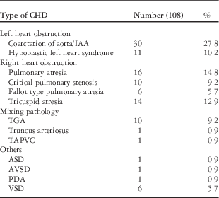
A total of 211 subjects (study group: n=108 and control group n=103) were included. Table 1 shows the types of CHD in study patients. Most of them had right-sided CHD.
The types of CHD s in study patients.

ASD=atrial septal defect; AVSD=atrioventricular septal defect; IAA=interrupted aortic arch; PDA=patent ductus arteriosus; TAPVC=total anomalous pulmonary venous connection; TGA=transposition of great arteries; VSD=ventricular septal defect
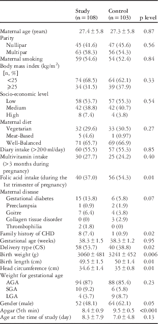
The mean gestational age and birth weight of all subjects were 38.3±1.4 weeks and 3149±475 g, respectively. Study patients and controls did not differ in terms of socio-demographic characteristics including maternal age, smoking, and socio-economic level. The rate of overweight (body mass index >25 kg/m2) women was similar in groups.
In the CHD group, 13 (12.4%) had prenatal diagnosis. Among them, 8 (61.5%) underwent C/S, and 5 (38.4%) underwent vaginal delivery (p=0.60). Caesarean rate was higher and birth size was lower in newborns with CHD compared with the control ones. The rate of folic acid supplementation during the 1st trimester was lower in the study group. Family history for CHD was higher in the study group (Table 2).
Characteristics of the subjects by groups.

AGA=appropriate for gestational age; LGA=large for gestational age; SGA=Small for gestational age
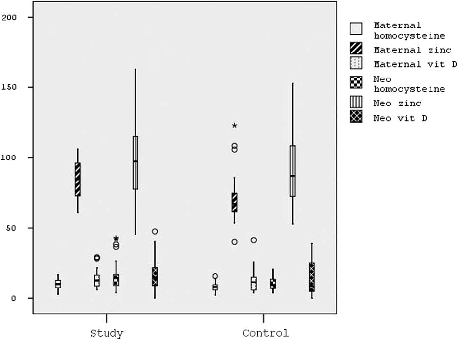
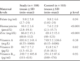
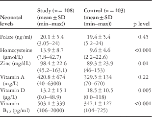
Table 3 and 4 show maternal and neonatal micronutrient levels. For both maternal and neonatal data, homocysteine and zinc levels were higher and vitamin D levels were lower in the study group compared with the control group (Fig 1). Neonatal serum vitamin B12 levels were higher in the study group, although the maternal levels were similar in groups. Among all subjects, none of the mothers had low folic acid levels (<2 ng/ml). However, 15 newborns (study group; n=3, control group; n=12) had low folic acid levels (<14 ng/ml) for age. Other micronutrient levels did not differ between the groups.
Maternal and neonatal data; homocysteine, zinc, and vitamin D levels by groups.* shows extreme values.

Maternal levels of micronutrients in all study subjects by groups.

Neonatal levels of micronutrients in all study subjects by groups.

There were positive correlations between maternal and neonatal levels of folic acid (p<0.001, r=0.56), homocysteine (p<0.001, r=0.51), zinc (p=0.04, r=0.29), vitamin D (p<0.001, r=0.77), and vitamin A (p=0.01, r=0.33). However, there was no correlation between maternal and neonatal vitamin B12 levels (p=0.15, r=0.24).
In multivariate analysis, when maternal age, multivitamin use, maternal education and socio-economic levels, maternal chronicle diseases, maternal homocysteine, zinc, vitamin D, and folate levels, and gender of the babies were assigned as independent variables, only maternal high zinc levels were associated with CHD in infants (p=0.02, OR: 0.9, 95% CI 0.8–0.9).
The results did not change when analysed for truncal anomalies including truncus arteriosus, tetralogy of Fallot, and d-transposition of great arteries. Maternal and neonatal homocysteine and zinc levels were higher and vitamin D levels were lower in this subgroup compared with healthy newborns.
Discussion
The exact aetiology of CHDs is unknown, but there is a tendency for addressing genetic and environmental factors such as advanced maternal and paternal age at conception, higher maternal body mass index, higher frequency of maternal medical conditions during pregnancy, higher prenatal exposure to medications, and micronutrient deficieny. This study investigated the levels of maternal and neonatal micronutrient levels in newborn patients with CHD. Two main studies examined the role of multivitamins containing folic acid in reducing the risk of all birth defects excluding neural tube defects and both suggested a significant reduction with prenatal vitamin supplementation.Reference Botto, Olney and Erickson 2 , Reference Czeizel, Vereczkey and Szabó 4 Botto et alReference Botto, Olney and Erickson 2 stated that mothers who reported periconceptional multivitamin use had a 43% lower risk of having infants with CHD including conotruncal defects. Czeizel et alReference Czeizel, Vereczkey and Szabó 4 reported a significant decrease in the prevalence of cases with ventricular septal defect, tetralogy of Fallot, d-transposition of great arteries, and secundum atrial septal defect in infants born to mothers who had taken high doses of folic acid during the critical period of CHD development. In the current study, we found that both maternal and neonatal homocysteine and zinc levels were higher, and vitamin D levels were lower in the newborns with CHD. In another study, which looked at severe CHDs in Canada before and after fortification of grain with folic acid, it was found that there was a significant decrease in birth prevalence of both conotruncal and non-conotruncal defects after fortification.Reference Aguilera, Fernandez and Munoz 11 We obtained the same results when analysed for both all CHS and this specific subgroup (conotruncal anomalies). Interestingly, although the rate of folic acid supplementation during the 1st trimester was lower in the study group, there were no differences between the maternal and neonatal folic acid levels in groups. The reference ranges of folic acid have defined as 2–20 ng/ml for adults and 14–51 ng/ml for infants.Reference Lonescu-Ittu, Marelli and Mackie 16 In Çelik et al’sReference Çelik, Aygün and Gülten 17 study, folic acid levels were measured in 141 low-birth-weight infants and their mothers. They found its neonatal levels as 21.2±12.2 ng/ml and maternal (puerperal women) levels as 12.3±7.5 ng/ml. The authors have noted that none of the mothers had low folic acid levels. In our study, serum folic acid levels were <14 ng/ml in 15 newborns, among all subjects. However, both maternal and neonatal lower folic acid levels were not associated with CHD.
Folate is a natural form of vitamin B9 and found in a variety of plant and animal food sources. The natural folate sources such as cereals, broccoli, beans, nuts, and seeds can definitely help to increase the folic acid levels in the body. As Turkey is a country of agricultural origin, most of the mothers might take necessary folic acid from these products in our study. The vegetarian ratio was high (~30%) in both study and control groups. We speculated that they were not consuming meat owing to poverty.
Folate and B12 status are important determinants of plasma homocysteine. This may explain the normal levels of folic acid levels in all mothers despite the difference in folic acid supplementation ratios between the groups. It was reported that raised total homocysteine levels in maternal blood and amniotic fluid were associated with an increased risk of neural tube defect pregnancies.Reference Mills, McPartlin and Kirke 18 , Reference Steegers-Theunissen, Boers and Blom 19 Molloy et al’s research groupReference Molloy, Kirke and Brody 20 also found that neural tube defect pregnancies were associated with low maternal levels of B12 as independent of folate status. In another study, Kapusta et alReference Kapusta, Haagmans and Steegers 21 measured folate, vitamin B6, and vitamin B12 levels in 27 mothers of children with CHDs. They showed that plasma homocysteine concentrations were significantly higher in the study group as compared with control subjects and concluded that the maternal hyperhomocysteinaemia may be a risk factor for CHDs. They also stated that the fact that women with hyperhomocysteinaemia are at increased risk for bearing children with all forms of CHDs suggested that mechanisms other than neural crest malfunction may be involved. Our data indicated that both maternal and neonatal homocysteine levels were higher in the study group. However, both maternal and neonatal folate levels did not differ between the groups. Although the maternal B12 levels were similar in groups, neonatal serum B12 levels were higher in the study group. Similarly, some micronutrient levels were higher in infants than those of their mothers. This may be explained by in vivo metabolism of these micronutrients. Zinc, which is an intracellular ion, is essential for the activity of many enzymes such as angiotensin-converting enzyme and the matrix metalloproteinases. The rapid effect of zinc deprivation in rats and the teratogenic effect of even transitory deficiency, as well as interactions between dietary zinc and other factors, had led Hurley and SwenertonReference Hurley and Swenerton 22 to investigate that zinc deficiency might be a factor in congenital abnormalities in newborns. Warkany and PeteringReference Warkany and Petering 23 also made this suggestion. Sever and EmmanuelReference Sever and Emanuel 24 claimed that maternal zinc deficiency was related to congenital malformations of the central nervous system. Moghimi et alReference Moghimi, Ashrafzadeh and Rassi 25 conducted a case–control study in Iran involving mothers of 80 neonates with congenital anomalies. They also found an association between congenital malformation in newborns and maternal zinc deficiency. In another study from Turkey, the authors showed that high maternal copper levels and low zinc levels have increased the risk of neural tube defect.Reference Zeyrek, Soran and Cakmak 26
On the other hand, toxic metals and trace elements can be transported from mother to fetus via blood circulation, and their excessive levels may accumulate in the fetus and cause harm. It was speculated that conotruncal defects might occur as deoxyribonucleic acid methylation mechanisms are impaired by cumulative effects of heavy metals and trace elements during fetal development, and fetal exposure to these inorganic substances may occur through the mothers who are living in a polluted environment.Reference Turker, Ergen and Karakoç 27
In a study from Turkey, the authors measured the concentrations of lead, cadmium, iron, zinc, and copper in 1st-day meconium specimens that were collected from 60 newborns with conotruncal heart defects and 72 healthy newborns. They found that mean concentrations of lead, cadmium, iron, zinc, and copper were significantly higher in newborns with conotruncal heart defects (p=0.001 for each).Reference Kundak, Pektas and Zenciroglu 28 Similarly, we found that both maternal and neonatal zinc levels were higher in the CHD group. It was noted that only maternal high zinc levels were associated with CHD in children.
Vitamin D plays an important role in metabolism of calcium–phosphorus and regulates bone mineralisation. Vitamin D deficiency is associated with increased risk of malignancies, diabetes, cardiovascular disease, infections, and neuromusculoskeletal and autoimmune disorders.Reference Holick 29 There is accumulating evidence that vitamin D plays a role in the central nervous system. The studies conducted by Daglar et alReference Daglar, Tokmak and Kirbas 30 and Nasri et alReference Nasri, Ben Fradj and Feki 31 showed an association between a decreased vitamin D level in pregnant women and the risk of fetal neural tube defects. In literature, there are data on maternal vitamin D level and the risk of CHD in infants. In our study, we observed that maternal serum vitamin D levels were lower in the study group.
The power of the present study is limited because of the small number of study participants. We were unaware of the maternal micronutrient levels before conception. Additionally, we could not measure exact micronutrient intake by foods. Nevertheless, this is the first study investigating both maternal and neonatal serum micronutrient levels in patients with CHD. We found that serum maternal and neonatal homocysteine and zinc levels were higher but vitamin D levels were lower in these patients. Large-scale, prospective studies are needed to clarify the role of micronutrients in the pathogenesis of CHDs.
Acknowledgements
The authors participated in the design, execution, and analysis of the paper and approved the final version, and state that the article has neither been published nor submitted elsewhere.
Financial Support
This research received no specific grant from any funding agency, commercial or not-for-profit sectors.
Conflicts of Interest
None.
Ethical Standards
The study was approved by the local Institutional Review Board and Ethical Committee.