INTRODUCTION
Erythrocytic necrosis viruses (ENVs) are associated with viral erythrocytic necrosis (VEN), a condition affecting marine and anadromous fish (CABI, 2006). Viral erythrocytic necrosis has been reported in teleosts from the north-east and north-west Atlantic and from the northern and southern Pacific (Smail & Munro, Reference Smail, Munro and Roberts2001; CABI, 2006). The double stranded DNA iridoviruses involved are generally large (up to 360 nm in diameter) and icosahedral, affecting mature and immature erythrocytic cells (Smail & Munro, Reference Smail, Munro and Roberts2001). Recognized primarily by the appearance of acidophilic, intracytoplasmic inclusion bodies in Giemsa-stained erythrocytes, VEN may be also manifest as intraerythrocytic haloes, granules and comet-tails, and involve nuclear destruction, appearance of cell ghosts, cell lysis and general changes in the blood picture, especially erythroblastosis (Johnston & Davies, Reference Johnston and Davies1973; Eiras et al., Reference Eiras, Costa, Biscoito and Davies1996; Davies & Merrett, Reference Davies and Merrett1998, Reference Davies and Merrett2000; Hershberger et al., Reference Hershberger, Hart, Gregg, Elder and Winton2006).
Recently, a VEN-like infection was observed in peripheral blood smears from an immature blackbar triggerfish, Rhinecanthus aculeatus (L.) (Teleostei: Balistidae), captured at Lizard Island, Great Barrier Reef (GBR). To our knowledge, records of iridovirus infections from Australian teleosts have been of epizootic haematopoietic necrosis viruses (see Hyatt et al., Reference Hyatt, Gould, Zupanovic, Cunningham, Hengstberger, Whittington, Kattenbelt and Coupar2000; Pallister et al., Reference Pallister, Gould, Harrison, Hyatt, Jancovitch and Helne2007), rather than ENVs. This short paper therefore considers the appearance of the infection at Lizard Island, its decline with time, and its possible mode of transmission.
MATERIALS AND METHODS
The juvenile triggerfish (8.7 cm total length (TL)) was collected from a patch reef, off Lizard Island, in November 2005, as part of a larger study of the blood parasites of fish in the region (see Smit et al., Reference Smit, Grutter, Adlard and Davies2006). The triggerfish, like other fish examined, was herded into a barrier net, captured with a hand net and transferred to a catch bag. It was then transported to Lizard Island Research Station and examined for ectoparasites. Subsequently, the fish was held in a 1000 l aquarium at 29–31°C, with continuous running seawater pumped directly from the reef, provided with shelter, and fed daily. It was then examined for infections following removal of blood from the caudal vein (see Smit et al., Reference Smit, Grutter, Adlard and Davies2006); the size of the fish meant that only sufficient blood for three thin smears was taken for initial screening. While the triggerfish recovered in fresh seawater, two blood smears on clean glass slides were methanol-fixed, Giemsa-stained and examined with a Nikon Eclipse 80i microscope. Images were captured by digital camera (Nikon DS-5M) and measured with a Nikon NIS 2.10 image analysis system, calibrated to a stage micrometer. The remaining blood smear was methanol fixed, stained with phosphate buffered (pH 6.0), 0.01% (w/v) aqueous acridine orange (AO) and observed with a triple-filter fluorescence facility (excitation wavelengths 410–585 nm) on a similar Nikon system.
In the broader study at Lizard Island, most fish were released to the wild following examination (see Smit et al., Reference Smit, Grutter, Adlard and Davies2006), but the young triggerfish was retained in the laboratory on the island because of its unusual blood infection. Its blood picture was then monitored using the screening methods above, intermittently, from November 2005–December 2007 inclusive.
Blood-feeding, ectoparasitic, juvenile gnathiids (Isopoda: Gnathiidae) (N = 20), present on the captured triggerfish in November 2005, were squashed between two glass slides up to 4 days after feeding to repletion. These preparations were fixed, stained and examined as for blood films.
RESULTS
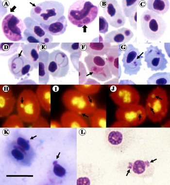
In the November 2005 blood films, ~18% of mature erythrocytes were affected by the condition resembling VEN (Figure 1A–G); erythroblasts were present (~4–8% of total red cells), but these were free from visible infection. Blood films also contained a haemogregarine (Apicomplexa: Adeleorina: Haemogregarinidae) identified as Haemogregarina balistapi (see Smit et al., Reference Smit, Grutter, Adlard and Davies2006), but this protist was confined to leukocytes in these blood films (Figure 1A).
Mature erythrocytes with VEN-like bodies had smooth or crenated outlines (Figure 1A–G), measured 10.3±0.8 µm long by 6.9±0.5 µm wide (N = 25), and were thus no larger, or smaller, than uninfected R. aculeatus erythrocytes. The inclusion bodies were single, or less commonly paired within the erythrocyte cytoplasm (Figure 1A–G), and usually at a distance from the nucleus. They were often round in outline, stained deep magenta with Giemsa and the smallest measured 1.3±0.2 µm, range 0.5–1.7 µm (N = 25) in diameter. Some bodies were larger with a distinct central granule surrounded by an indistinct cloud of eosinophilic material up to 2.2±0.4 µm across, range 2.0–3.3 µm (N = 25) (Figure 1C), while others were associated with fine, eosinophilic haloes, 4.8±0.5 µm across, range 4.1–5.2 µm (N = 25), sometimes encircling the host cell nucleus (Figure 1D, E, G). Less frequently, distinct, pink-stained, comet-tails up to 5.9 µm long (N = 3) (Figure 1F) were observed. The nuclei of infected erythrocytes were sometimes irregular in outline (Figure 1A), although some, apparently uninfected, erythrocytes also had irregular nuclei. Nuclear displacement was uncommon except where haloes or comet-tails were well defined (Figure 1D–F). No evidence was seen of nuclear destruction, cell lysis or ghost cell formation. The VEN-like bodies fluoresced green following AO staining, indicating their DNA content (Figure 1H–J).
Giemsa (A–G) and acridine orange (H–J) stained blood-film preparations from Rhinecanthus aculeatus; Giemsa-stained squashes of juvenile gnathiid isopods from R. aculeatus at 24 hours (K) and 4 days (L) following blood feeding. (A–C) Viral erythrocytic necrosis (VEN)-like bodies within erythrocytes, one within a host cell with an irregular nucleus (light arrow), and haemogregarines within small lymphocytes (heavy arrows); (D, E) infected cells with fine haloes (arrow in D) showing host nucleus displacement; (F) VEN-like body associated with comet-tail-like staining (arrow); (G) crenated erythrocytes with two VEN-like bodies each and fine haloes (arrow); (H–J) fluorescence of single and paired VEN-like bodies (arrows) in host erythrocytes; (K, L) digesting erythrocytes containing single (K) and paired (L) VEN-like bodies (arrows). Bar = 10 µm.

When the triggerfish was monitored over time (26 months), the infection fell from ~18% of erythrocytes in November 2005, to 0.5% by January 2006, 0.4% by May 2006, and persisted at this low level (0.4%) until the end of December 2007, but erythroblast levels did not change perceptibly (~4%) during this period; in December 2007 the triggerfish finally died in captivity, having attained 10.2 cm TL. Haemogregarine infections were also maintained at low levels over this period.
Giemsa-stained squashes of 2/20 juvenile gnathiid isopods contained infected erythrocytes, 24 hours–4 days after the isopods had fed on the triggerfish in November 2005. Despite the advancing stages of digestion, eosinophilic, single and paired VEN-like bodies could be seen in many erythrocytes, although less clearly than in blood films (Figure 1K, L).
DISCUSSION
Common teleost hosts for VEN infections include small intertidal fish, cod, herring, mullet, flatfish, and salmonids (Smail & Munro, Reference Smail, Munro and Roberts2001; CABI, 2006). Given its appearance by light microscopy and its green fluorescence with AO, the infection reported here also resembles VEN. Unequivocal demonstration of icosahedral viral particles within the erythrocyte cytoplasm would have confirmed this, but attempts to gain good transmission electron microscopy (TEM) images from the limited Lizard Island material were unsuccessful. The poorly preserved particles observed by TEM were of similar size (~300 nm across) and grouping to those of VEN infections (data not shown), and their ultrastructure did not resemble any part of a normal erythrocyte, or of an apicomplexan, such as the haemogregarine present in blood films. We continue to monitor fish at Lizard Island to gain samples suitable for future TEM studies; nevertheless, despite lack of convincing TEM material currently, the experience of one of us (A.J.D.) of VEN and VEN-like infections in the northern hemisphere (see Johnston & Davies, Reference Johnston and Davies1973; Eiras et al., Reference Eiras, Costa, Biscoito and Davies1996; Davies & Merrett, Reference Davies and Merrett1998, Reference Davies and Merrett2000), suggests a strong similarity between these and the triggerfish condition. Thus, this study likely represents the first record of a VEN-like infection from Australia and probably from a coral-reef fish.
Outbreaks and persistence of VEN infections in confined fish have been reported previously (see Hershberger et al., Reference Hershberger, Hart, Gregg, Elder and Winton2006). In the current case, infection apparently declined over 7 months, and then formed a stable chronic condition affecting fewer erythrocytes for a further 19 months. No VEN-like infections were observed during our earlier studies of fish at Lizard Island (see Smit et al., Reference Smit, Grutter, Adlard and Davies2006), especially among triggerfish of the family Balistidae. Subsequently, we have identified this infection, with confidence, but at a very low level (<0.1% of erythrocytes infected, unpublished data), in just one other R. aculeatus of a large population of triggerfish sampled regularly around Lizard Island.
Juvenile gnathiids are voracious blood feeders, especially on fish of the GBR; they are eaten particularly by cleaner fish (Grutter, Reference Grutter2002) and we suspect that they transmit fish haemogregarines (see Smit et al., Reference Smit, Grutter, Adlard and Davies2006). Reports of water-borne and vertical transmission of VEN exist, as well as transfer by injection (see CABI, 2006). Detection of VEN-like bodies within the digesting blood meal of gnathiids was an interesting finding, perhaps indicating that these isopods act as viral vectors on the GBR, in a manner similar to the fish louse, Argulus foliaceus L. and the leech Piscicola geometra L., which function as mechanical vectors of spring viraemia of carp virus (Ahne, Reference Ahne1985). Two of us (A.J.D. and N.J.S.) have a long experience of examining gnathiid blood meals (see Davies & Smit, Reference Davies and Smit2001; Smit et al., Reference Smit, Grutter, Adlard and Davies2006), but we have never detected VEN-like bodies within digesting erythrocytes, except in this instance.
ACKNOWLEDGEMENTS
The authors are grateful to P. Hershberger, Western Fisheries Research Center, Washington, USA, and S. Corbeil and M. Crane, both of the CSIRO, Australia, for helpful advice. Technical assistance was kindly provided by R. Giddens and G. Sappal, Kingston University, UK. Lizard Island Research Station staff generously maintained the fish in captivity. The study was funded by the Australian Research Council (to A.S.G., N.J.S. and A.J.D.) and an Ian Potter Doctoral Fellowship (to L.C.).