Introduction
Lingual thyroid is an uncommon condition, with a prevalence of between 1 in 10 000 and 1 in 100 000.Reference Akyol and Ozcan1 It occurs when the thyroid anlage fails to descend from the foramen caecum to the neck. It is more common in females than in males, with a ratio of between 4:1 and 7:1.Reference Akyol and Ozcan1 Lingual thyroid often presents during puberty, pregnancy or times of stress, when it enlarges. In 70 per cent of patients, this is the only thyroid tissue present. Thus, unless the mass is symptomatic, it is often observed until it causes obstructive symptoms, bleeds, or there is suspicion of malignant change.
Surgical access to the tongue base area is difficult; thus, non-surgical management of lingual thyroid has been used, such as thyroxine suppression and radioiodine treatment. However, the effects of such treatments are not consistent, and they do not address the issues of obstruction and bleeding from the lingual thyroid.Reference Kang2, Reference Iglesias, Olmos-Garcia, Riva and Diez3
The mainstay of treatment of a symptomatic lingual thyroid is surgical resection via an open or transoral approach. Traditionally, large lesions have been resected via an open approach, either using a suprahyoid pharyngotomy, lateral pharyngotomy or lip-splitting transmandibular approach. With advances in technology and understanding of surgical anatomy, transoral resection is now an option even for large lesions.
We report our experience in transoral robotic resection of a large lingual thyroid in a female patient who presented with obstructive symptoms.
Case report
A 17-year-old female adolescent with no previous medical history of note presented to the Singapore General Hospital ENT clinic in May 2011 after incidentally noticing a mass at the back of her tongue. Except for occasional sensations of a lump in the throat, she was otherwise asymptomatic, with no dysphagia, upper airway obstruction or bleeding from the mass. She was clinically euthyroid.
Clinical examination revealed a well-circumscribed, 2.5 cm mass at the tongue base with intact, smooth overlying mucosa. The tongue was freely mobile, with no deficits of the lingual or hypoglossal nerve. Fibre-optic nasopharyngoscopy showed that the mass was bulging into the vallecula, with partial occlusion of the airway (Figure 1).
Endoscopic view showing the lingual thyroid causing partial airway obstruction.

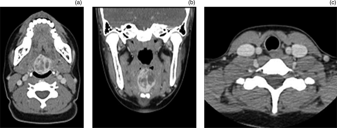
Computed tomography (CT) scanning of the neck with contrast revealed a hyperdense, midline lesion at the base of the tongue, measuring 30 × 27 mm, with no thyroid gland in the usual position (Figure 2). A diagnosis of lingual thyroid was made based on the CT findings and the clinical picture.
(a) Axial computed tomography (CT) scan of the lingual thyroid; (b) coronal CT scan of the lingual thyroid; and (c) axial CT scan of the neck showing no thyroid in the normal position.
Thyroid function testing showed subclinical hypothyroidism, with an elevated thyroid-stimulating hormone level (15 MU/l) and a normal free thyroxine level (9.3 pmol/l).
As the patient was asymptomatic, we commenced her on levothyroxine replacement (100 µg once daily) and continued to observe her.
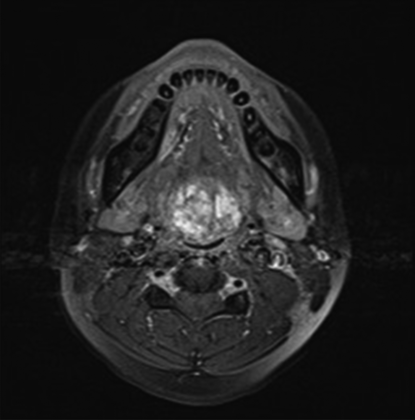
Four months later, she became symptomatic, complaining of intermittent choking episodes whilst eating, and difficulty sleeping supine. Her tonsils were not enlarged. Magnetic resonance imaging (MRI) of the head and neck showed that the mass had increased in size to 31 × 35 × 24 mm, with features of degeneration (Figure 3).
Axial, T2-weighted magnetic resonance imaging scan showing the lingual thyroid and resultant airway obstruction.
In view of the symptoms and increase in size of the mass, surgical excision was advised, and the patient underwent transoral robotic resection of the lingual thyroid in March 2012.
Under general anaesthesia, the patient was placed in a supine position and intubated transnasally with an armoured endotracheal tube. The neck was extended with a shoulder roll and then prepared and draped in a sterile fashion. Next, a 2/0 silk stitch was placed in the midline of the oral tongue to retract it anteriorly in the midline whilst positioning a Feyh-Kastenbauer-Weinstein-O'Malley retractor. A Simon mandible blade was also used.
After achieving satisfactory exposure, the da Vinci Si HD robotic system (Intuitive Surgical, Sunnyvale, California, USA) was docked with a 12 mm 30° up endoscope in the centre, 5 mm Maryland dissector on the left of the patient, and 5 mm monopolar cautery on the right (Figure 4). An incision was first made anterior to the mass, taking a small cuff of muscle around the mass. This allowed retraction of the mass without tearing the thyroid tissue. After the mass was released anteriorly, lateral incisions were made and the tongue blades repositioned deeper into the tongue base as dissection progressed. Dissection was carried out along a broad front, with careful lateral dissection in the vicinity of the lingual artery. The lingual artery was not encountered in this case. The dissection continued towards the level of the hyoid bone, and the mass was released after a final incision along the vallecula. Haemostasis was achieved with monopolar cautery (Figure 5). The larynx was examined to ensure there was no oedema, before anaesthesia was reversed and the patient extubated. The total surgical time was 85 minutes.
Endoscopic, intra-oral view showing the lingual thyroid (centre) together with the robotic surgical instruments: Maryland dissector (top left), monopolar diathermy (top right) and straight suction (mid-left and mid-right).
Endoscopic, intraoral view showing the lingual thyroid tongue base site following resection.
The resected specimen measured 4 × 3 × 3.5 cm. Histological examination was consistent with thyroid tissue with no malignant changes.
The patient was kept in a high-dependency unit for overnight observation. On the first post-operative day, she was started on oral feeding, and was taking a soft diet well by the fourth post-operative day, when she was discharged.
On initial follow up two weeks post-operatively, the patient complained of numbness of the anterior one-third of the tongue. This had resolved by the fourth post-operative week. The patient was continued on thyroxine replacement. She reported that her obstructive symptoms had resolved.
Discussion
We report a case of transoral lingual thyroid resection using a robotic system.
Traditionally, the tongue base may be accessed via the following open approaches: (1) transoral midline glossotomy, (2) transcervical approach via a lateral or suprahyoid pharyngotomy, and (3) transmandibular approach with a lip-splitting incision. The disadvantages of these approaches include prolonged recovery time, swallowing dysfunction and need for a feeding tube, risk of pharyngocutaneous fistula, longer hospitalisation, and an external scar. Minimally invasive transoral approaches avoid these problems. Dziegielewski et al. reported five cases of lingual thyroid resected transorally with CO2 laser or electrocautery; all five patients had good swallowing function post-operatively.Reference Dziegielewski, Chau, Seikaly, Allegretto, Barber and Harris4 Several published reports of lingual thyroids resected transorally state that patients were discharged without the need for tube feeding.Reference Orbelo, Ekbom and Thompson5–Reference Burkart, Richter, Rutter and Myer7
Several authors have described minimally invasive transoral approaches, using either a rigid endoscope or an operating microscope with suspension laryngoscopy. Suspension microlaryngoscopy may be suitable for smaller lesions, but would be technically difficult for larger lesions. Thus far, the majority of lingual thyroids reported to be resected transorally have been small, ranging from 0.5 to 3 cm.Reference Orbelo, Ekbom and Thompson5–Reference Burkart, Richter, Rutter and Myer7
Following O'Malley and Weinstein's first reported use of transoral robotic surgery for oropharyngeal and base of tongue cancers, its use has broadened to include benign lesions and neoplasms.Reference O'Malley, Weinstein, Snyder and Hockstein8 In the present patient, the transoral approach was chosen in order to avoid the greater morbidity associated with open approaches. Transoral resection with a robotic system has the following advantages over other minimally invasive approaches: (1) a good, wide-angle view of the whole surgical field; (2) the facility to allow the surgeon to drive the endoscope; and (3) greater dexterity (due to the robotic instruments). These advantages enable easier, faster and safer surgery, especially for larger lesions which extend posteriorly to the vallecula. A literature review identified only one previous report of this approach, in a case involving a smaller lesion.Reference May, Newman and Padhya9
• Lingual thyroid is an uncommon condition
• Open surgical approaches have traditionally been used for removal
• Safe use of transoral robotic resection is presented
• Understanding intralingual neurovascular anatomy is essential for safety
Surgeons accustomed to the open approach may find transoral resection of the lingual thyroid difficult as regards understanding the anatomy ‘from the inside out’, and visualising it in three dimensions. Understanding the relationship of the lingual artery to the mass is of the utmost importance. Injury to both lingual arteries could potentially result in necrosis of the tongue. Cadaveric studies have found the lingual artery to lie approximately 2.2 to 2.7 cm inferior and 1.3 to 1.6 cm lateral to the foramen caecum.Reference Lauretano, Li, Caradonna, Khosta and Fried10 However, in the present case this was not a useful reference point, as the mass had distorted the usual anatomical relationship. The key steps in avoiding intra-operative injury to the lingual artery are: (1) to understand the course of the lingual artery; (2) to correlate this with the pre-operative imaging; and (3) to pursue careful, broad dissection during the resection itself. In the presented case, we preferred MRI of the tongue to CT scans, as the former gives better information on soft tissue definition and anatomical relationships of the mass. In the present case, the lingual artery and the dorsal lingual branch were not encountered as they had been displaced laterally by the mass.
Conclusion
It is surgically feasible and safe to undertake transoral robotic resection of a large lingual thyroid. This approach may allow faster recovery and reduced hospitalisation. Understanding intralingual neurovascular anatomy is important for safe surgery.