Introduction
Neurosurgical and radiotherapeutical approaches are well established in the treatment of pituitary adenoma and craniopharyngioma.Reference Clark, Cage, Aranda, Parsa, Auguste and Gupta 1 Surgery is considered the treatment of choice for these tumours. The extent of resection is an important factor that determines long-term local control. Gross total resection is often not possible in these tumours due to the closeness of critical structures like optic pathways, hypothalamus, brain stem and cavernous sinus.Reference Müller 2 Radiotherapy improves the local control after a subtotal excision and is used as adjuvant treatment after surgery or in case of recurrence. Since these tumours have a good chance of prolonged local control with surgery and radiotherapy, it is vital to reduce the treatment related normal tissue damage. Modern techniques of radiotherapy such as stereotactic radiosurgery (SRS) or fractionated stereotactic radiotherapy (SRT) enables the delivery of high dose of radiation to the tumour, while not exceeding the tolerance of nearby organs at risk (OARs).Reference Harrabi, Adeberg and Welzel 3 Non-coplanar static conformal field (SCF), static conformal arc (SCA) and dynamic conformal arc (DCA) radiotherapy are the three methods of linear accelerator-based SRT techniques.Reference Colombo, Benedetti and Pozza 4 – Reference Perks, Jalali and Cosgrove 8 The SCF plan consists of six to ten non-coplanar static fields; each field is individually shaped to the beams eye view (BEV) projection of the target using micro multileaf collimator (MLC). In SCA plan, the field is shaped to BEV projection of the target at the beginning of the arc, and the shape of the field aperture remains constant during an arc. In DCA plan, the shape of the micro MLC is automatically adjusted to the projected shape of the target in the BEV for every 10° increment from the start angle till the end of the arc.
The aim of our study was to do a dosimetric comparison of the three SRT techniques. We undertook this study to decide whether we have to change our institutional protocol for SRT from SCF technique to DCA technique due to dosimetric advantage. We also wanted to see whether changing our institutional protocol from SCF to DCA technique will decrease the overall treatment time, thereby increasing the throughput of our department.
Materials and Methods
We included all the patients with pituitary adenoma or craniopharyngioma, treated in our centre with SRT from April 2014 to August 2015. There were 12 patients with pituitary adenoma and eight patients with craniopharyngioma. The study was accepted by our Institutional Review Board scientific and ethics committee. CT scan was done for patients with Gill Thomas CosmanReference Gill, Thomas, Warrington and Brada 9 stereotactic localising frame along with Brown-Robert-WellsReference Heilbrun, Roberts, Apuzzo, Wells and Sabshin 10 localiser system. The gross tumour volume (GTV) and the OARs were contoured on axial CT fused with axial T1 gadolinium magnetic resonance images. Patients’ planning CT datasets, with 3 mm slice thickness from vertex to lower border of second cervical vertebra was retrieved from the iPlan RT V.4.5.3, treatment planning system (TPS) (Brain LAB AG, Feldkirchen, Germany) for computation of treatment plans. The outlining and planning for all patients were done by the authors. The GTV was contoured by consultant radiation oncologists with more than ten years of experience and was verified by the senior neurosurgeon who operated on the patient at the time of initial treatment. The planning target volume (PTV) and OARs were contoured by the radiation oncologist. The plans were generated by medical physicist. The dosimetric analysis was done by the team of radiation oncologists and medical physicists. For ease of dosimetric analysis, a uniform dose (54 Gy in 30 fractions) was prescribed to all PTVs irrespective of the diagnosis. Brain LAB, iPlan RT version 4.5.3 (Brain LAB AG) TPS uses the pencil beam algorithm with tissue inhomogeneity correction to calculate the dose distribution. The grid size for calculation was 2-mm pixel. The same dose requirements for PTV coverage and OAR sparing were set for all the three techniques.
For SCF plan single isocenter with three to seven non-coplanar beams were used and beam shaping was done by 3 mm micro MLCs. The collimator angle was always optimised in order to irradiate the smallest possible area for each beam. SCA and DCA were planned with three to six arcs in a non-coplanar fashion with single isocenter. Dosimetric analysis was performed using dose volume histograms and 2D dose displays. Conformity index (CI), homogeneity index (HI) and quality of target coverage were used to compare the rival plans. Analysis of plans was also performed using comparison of dose received by OARs and the volume of brain receiving 5, 10, 20 and 40 Gy.
The Radiation Therapy Oncology Group (RTOG) CI was calculated based on the formula.Reference Shaw, Kline and Gillin 11
 $${\rm CI}\left( {{\rm RTOG}} \right)\,{\equals}\,{{{\rm V}_{{{\rm RI}}} } \over {{\rm TV}}}$$
where VRI is the volume of the reference isodose and TV is the target volume.The concept of HIReference Shaw, Kline and Gillin 11 was developed to analyse the spatial dose distribution in each section of the treatment plan. HI was described as per the RTOG
 $${\rm HI}_{{{\rm RTOG}}} \,{\equals}\,{{I_{{{\rm max}}} } \over {{\rm RI}}}$$
where I max is the maximum dose received by the target, RI the reference isodose.
Quality of coverage was assessed based on RTOG criteria. If the 90% of the prescription isodose line completely covers the PTV, it is considered per protocol. If the minimum dose to PTV is between 80% and 89·9% of the prescription dose, it is considered as minor deviation and if the minimum dose to PTV is less than 80% of the prescription dose, it is considered as major deviation.Reference Shaw, Kline and Gillin 11
For the OARs, maximum dose was compared across three plans. The volume of brain receiving 5, 10, 20 and 40 Gy was compared across three plans for all the patient datasets. This could help in selection of a better SRT technique with regard to spillage of lower dose.
The time taken for treatment delivery was measured for all the three techniques for one patient plan for single fraction of radiation (1·8 Gy). The measurement was done using water phantom on the machine and time was calculated from starting of the treatment till completion. The time required for set up of the patient has been assumed to be constant.
Data entry was done in Epidata version 3.1 and was analysed using SPSS 17.0. For continuous data, the descriptive statistics such as mean, standard deviation, median, minimum and maximum was represented. For categorical data, the number of patient datasets and percentage was presented. Based on the normality of the data, the parametric analysis of variance test or non-parametric Kruskal–Wallis test was used to compare the three SRT treatment techniques.
Results
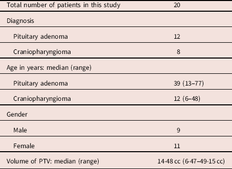
The demographic details of patients are listed in Table 1.
The demographic details of patients are as follows

Abbreviation: PTV, planning target volume.
Volumes of PTV and reference isodose
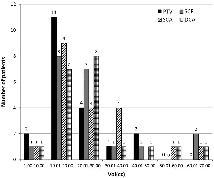
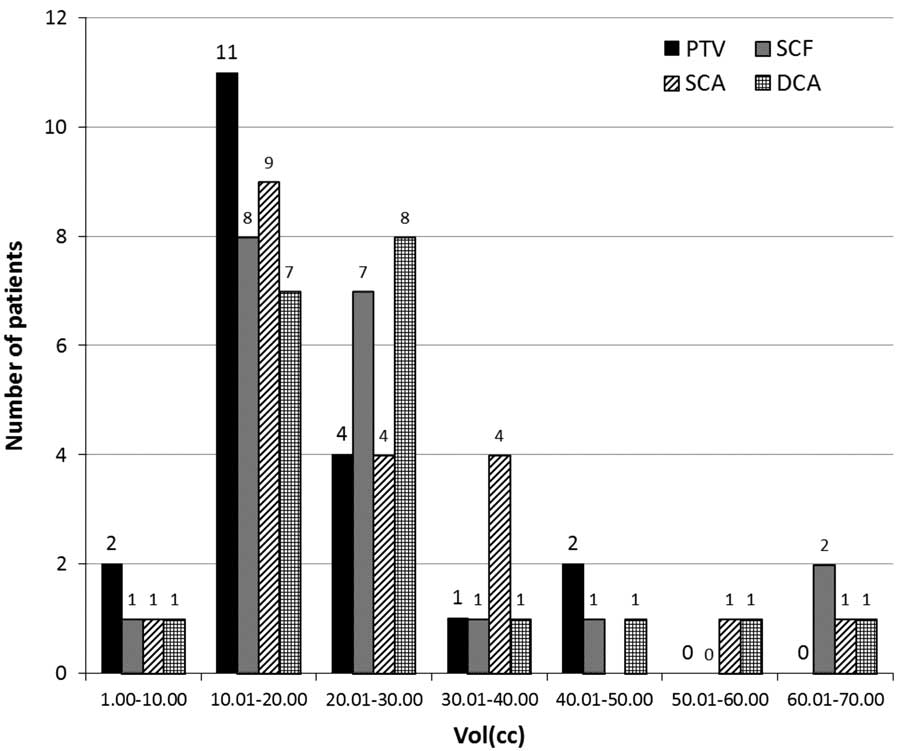
The PTV ranged from 6·47 cc to 49·15 cc, with a median PTV of 14·48 cc. A total of 13 patients had PTV<20 cc and 7 had >20 cc volume. Average volume of the reference isodose (95% isodose) was around 25 cc in all the three plans. The values are presented in Figure 1.
Planning target volume and reference isodose volumes for SCF, SCA and DCA techniques. Abbreviations: PTV, planning target volume; SCF, static conformal field; SCA, static conformal arc; DCA, dynamic conformal arc.
CI
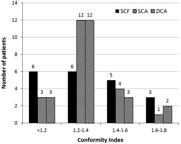
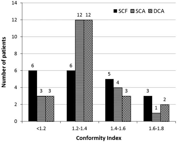
RTOG CI was within acceptable limits (between 1 and 2) for all the techniques across all patient datasets. Mean conformity for SCF, SCA and DCA was 1·34, 1·35 and 1·34, respectively. The difference in the conformity indices among the three plans was not significant (p=0·951). In SCF plan, 75% of the plans had CI of <1·5, whereas in the case of SCA and DCA plans 95% and 90% had CI of <1·5 (Figure 2).
Conformity index for three different SRT techniques. Abbreviations: SCF, static conformal field; SCA, static conformal arc; DCA, dynamic conformal arc.
HI
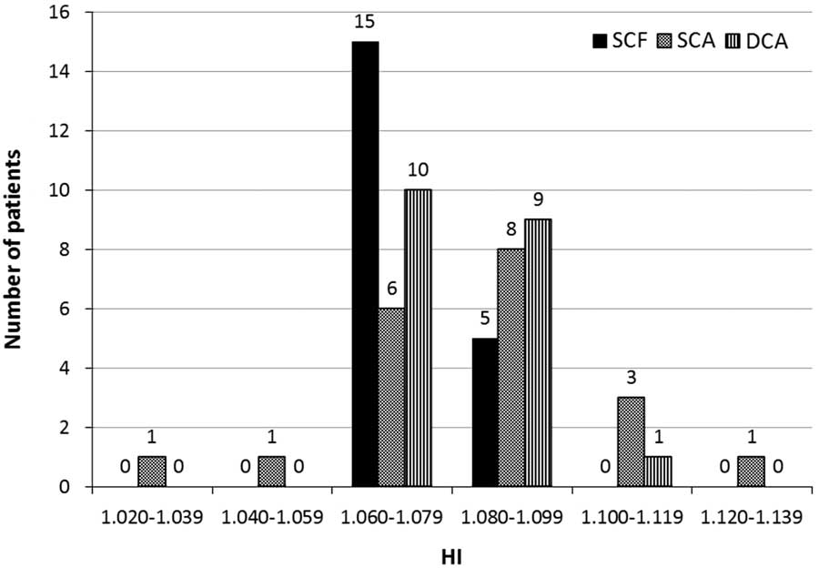
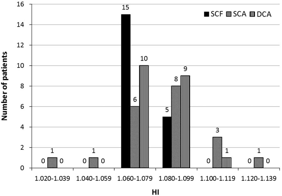
Mean HI was 1·07±0·01; 1·08±0·02 and 1·08±0·02 in SCF, SCA and DCA plans, respectively. The difference in HI was not statistically significant across the plans. HI is presented in Figure 3.
Homogeneity index for three different SRT techniques. Abbreviations: SCF, static conformal field; SCA, static conformal arc; DCA, dynamic conformal arc.
Quality of coverage
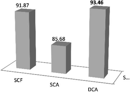
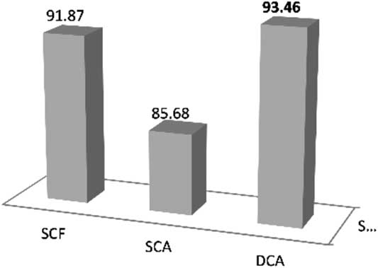
The quality of coverage was best in DCA plans. 80% of the DCA plans had quality of coverage per protocol compared with 55% of SCF and 20% of SCA plans. This difference in quality of coverage in DCA plans was statistically significant compared with SCA plans (p<0·001). There was no major deviation in quality of coverage in DCA plans whereas one SCF plan and two SCA plans showed major deviation in quality of coverage as per RTOG protocol. The quality of coverage showed minor deviation as per RTOG protocol for four DCA plans, eight SCF plans and 14 SCA plans (Figure 4).
Mean quality of target coverage of the three SRT plans. Abbreviations: SCF, static conformal field; SCA, static conformal arc; DCA, dynamic conformal arc.
Doses to OARs
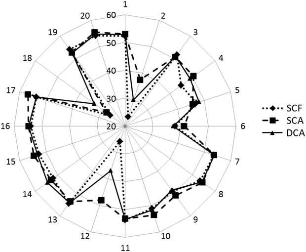
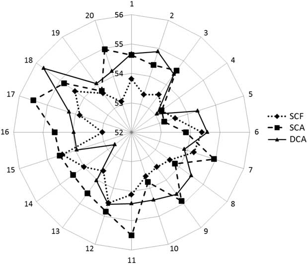
The SCF technique delivered the minimum dose to the brain stem with two plans exceeding 54 Gy while in SCA and DCA technique, five plans each exceeded 54 Gy. Highest dose to brain stem, 56·82 Gy was found in one of the SCA plans (Figure 5). The average dose to brain stem was minimal in SCF plans and it was statistically significant (p=0·019).
Radar diagram showing the maximum dose to the brain stem from 20 to 56 Gy for three SRT techniques for 20 patients’ datasets. Abbreviations: SCF, static conformal field; SCA, static conformal arc; DCA, dynamic conformal arc.

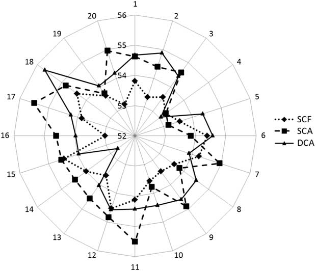
The maximum point dose received by optic chiasm ranged from 52·99 to 54·81 Gy in SCF, 53·16 to 55·51 Gy in SCA and 52·59 to 55·69 Gy in DCA plans. The SCF technique delivered the least dose to optic chiasm compared with SCA and DCA plans. None of the plans exceeded 55 Gy to optic chiasm in SCF technique, whereas the maximum dose to optic chiasm exceeded 55 Gy in two plans in SCA and one plan in DCA technique. Dose to optic chiasm is represented in radar diagram in Figure 6. The maximum dose to optic nerves ranged from 2·96 to 52·84 Gy in 20 patient datasets for the three techniques.
Radar diagram showing dose to the optic chiasm in 20 datasets for three SRT techniques (SCF, SCA and DCA). Abbreviations: SCF, static conformal field; SCA, static conformal arc; DCA, dynamic conformal arc.
Volume of brain receiving low dose radiation
The mean volume of the brain receiving 5 Gy was lower in SCF technique compared with SCA and DCA techniques, whereas volume of brain receiving 20 Gy (V20) and 40 Gy (V40) was more in SCF technique as compared with SCA and DCA techniques (p=0·008). The volume of brain receiving 5, 10, 20 and 40 Gy doses were comparable in SCA and DCA techniques.
Time taken for treatment delivery
The time taken for treatment delivery using water phantom on the machine was measured for all the three techniques for one patient plan for single fraction of radiation (1·8 Gy). The time required for set up of the patient has been assumed to be constant. In SCF plan, the time taken for single fraction radiation delivery from starting of radiation till completion was 7 minutes and 40 seconds, whereas time for both SCA and DCA plans was 8 minutes 20 seconds (difference of 40 seconds was found).
Discussion
Pituitary adenoma and craniopharyngioma are benign tumours, anatomically located over the sellar region and are closely associated with optic apparatus anteriorly and brain stem posteriorly. Stereotactic fractionated radiotherapy delivers highly precise and accurate radiation resulting in reduction in the dose to the OARs.Reference Colombo, Benedetti and Pozza 4 – Reference Bourland and McCollough 6 In this study, dosimetric comparison of the three linear accelerator-based SRT techniques was done on the CT dataset of 20 patients who were treated for pituitary adenoma or craniopharyngioma. The reference isodose volume (95% isodose) concurs well with PTV in all the three techniques.
CI estimates the dose conformity to the target and also indirectly estimates the dose to the surrounding normal organs. In this study, the CI for all the datasets in three techniques was <2, without any violation of RTOG criteria.Reference Shaw, Kline and Gillin 11 The mean conformity indices were slightly better in SCF 1·34 (±0·19) and DCA 1·34 (±0·16) than in SCA technique 1·35 (±0·13), but this was not statistically significant (p=0·720). Similar study was performed by AmmerReference Ammar, Eldebawy, Maarouf, Khalil and Zaghloul 12 evaluating the peripheral dose and CI for three SRT techniques for the intracranial targets for ten patients, found mean CI of 1·34 (±0·13) for arcs and 1·4 (±0·09) for conformal fields. The results are similar in the present study. In case of SCF, the angle of the gantry is manually modified or use of field in field approach gives more conformal dose distribution. Similar degree of freedom in DCA or SCA may not be possible for individual fields. The change in the shape of MLCs for every 10° gantry rotation would probably support the slightly better CI in DCA compared with SCA in the present study.
Dosimetric comparison of the SRT techniques with IMRT plans for the four simulated targets of the cranial tumours by S D SharmaReference Sharma, Jalali, Phurailatpam and Gupta 13 showed that CI was better in SCF (mean CI=0·71) and in DCA (mean CI=0·72) plans than in SCA plans (mean CI of 0·67). Study by WiggenraadReference Wiggenraad, Petoukhova, Versluis and van Santvoort 14 comparing IMRT with DCA technique for intracranial tumours, reported a mean CI ranging from 1·14 to 1·38 for DCA technique, which was similar to our study.
Mean HI was 1·07 (±0·01), 1·08 (±0·02) and 1·08 (±0·02) in SCF, SCA and DCA plans, respectively. In all the plans the HI was within the RTOG protocol. The HI was comparatively better in the SCF technique.
Cardinale et al.Reference Cardinale, Benedict, Wu, Zwicker, Gaballa and Mohan 15 compared three linac-based SRT techniques for three intracranial test targets having ellipsoid, hemisphere and irregular shape and found that CI and HI were higher in arc technique compared with 3D conformal technique. But in our study, we found that there was no significant difference in CI and HI among the three techniques.
WiggenraadReference Ammar, Eldebawy, Maarouf, Khalil and Zaghloul 12 comparing IMRT with DCA technique for intracranial tumours reported a mean HI ranging from 1·15 to 1·306 for DCA techniques, concluding that DCA technique is more preferred for intracranial targets irrespective of shape and size of the targets.
Mean quality of coverage was within the acceptable limits of RTOG (>90%) in SCF and DCA plans, but for SCA plans, the mean quality of coverage was 85·6%, which was a minor deviation (80–89%) as per RTOG protocol. This difference of quality of coverage among the three techniques was statistically significant (p<0·001). Our mean quality of coverage was 93·46 for DCA technique which was slightly inferior compared with Wiggenraad et al.Reference Wiggenraad, Petoukhova, Versluis and van Santvoort 14 This was probably because we included only sellar and suprasellar tumours which are in close proximity to OARS compared with Wiggenraad et al., which included all intracranial tumours.
The average D max to brain stem was 47·27, 50·07 and 48·88 Gy in SCF, SCA and DCA plans, respectively (p=0·019). SCF plan was efficient in achieving the dose constraint consistently for 18/20 patients, whereas DCA and SCA plans were able to achieve the dose constraint in 15/20 patients. The dose to brain stem was better controlled in SCF technique because individual selection of beams minimising the entry and exit dose to brain stem was possible in SCF technique. The SCF technique was more efficient in reducing the dose to the optic chiasm as compared with the arc techniques due to the better beam selection as stated above. WiggenraadReference Wiggenraad, Petoukhova, Versluis and van Santvoort 14 in the study comparing IMRT with DCA for intracranial tumours, expressed that sparing the optic system is a challenge when the target is close to it and said that the dose up to 56 Gy was acceptable with standard fractionation. The doses to the optic nerves were well below the constraints in all the cases, none of the plans had optic nerve dose >54 Gy. Again SCF technique was better in sparing the optic nerves than both the arc techniques.
In both arc techniques, the volume of the brain receiving the low dose (5 Gy) were high compared with the SCF technique which was statistically significant (p=0·001). This was because the number of fields will be higher (30–40, depending on the number and length of arc) in arc technique. The small dose from each of these fields is converging to the target resulting in high volume of brain receiving low dose.
Hamilton et al.Reference Hamilton, Kuchnir and Sweeney 16 compared the SCF technique with arc therapy in multiple intracranial targets, and found that the volume of normal tissue receiving lower doses (10% of prescription dose) increases with number of fields in the SCF technique and with number of arcs in arc technique, the difference was around 1·6 times more in arc technique compared with static fields. Also they have reported volume of the normal tissue receiving high dose (>90% of the prescribed dose) was similar in arc technique and static field technique with 8–12 fields. In our study, we noticed that the volume of brain receiving low dose of radiation was more in the arc techniques compared with static field technique. In the present study, it was also found that the volume of brain receiving intermediate (10, 20 Gy) and high dose (40 Gy) was higher in SCF technique (p=0·008).
We calculated the treatment time for all the three techniques for one patient and the monitor units required to deliver single fraction of radiation was compared for the three techniques across one dataset. The median monitor units required to deliver single fraction RT were 266·5, 265 and 260·5 for SCF, SCA and DCA techniques, respectively. Time required in completing single fraction RT was lesser (7 minutes 40 seconds) in SCF plan compared with SCA and DCA techniques (8 minute 40 seconds). Similar study on monitor units was performed by Solberg et al.Reference Solberg, Boedeker, Fogg, Selch and DeSalles 17 and concluded that dynamic arc technique was simple and the treatment time and monitor units were comparable with SCF and DCA techniques.
Limitations of this study
This work has been purely a dosimetric study and no attempt has been made to evaluate the clinical benefit. We were unable to find a significant dosimetric difference between SCF, SCA and DCA techniques in delivering SRT for pituitary adenoma and craniopharyngioma, probably due to small sample size.
Conclusions
The linac-based SRT techniques SCF, SCA and DCA are efficient in delivering highly conformal and homogenous dose to the target in pituitary adenoma and craniopharyngioma. Among these three techniques, SCF and DCA had acceptable quality of coverage. The dose received by OARs was least in the SCF technique. We did not change our institutional protocol from SCF to DCA technique in SRT.
Acknowledgements
None.
Financial Support
None.
Conflicts of Interest
None.