Introduction
Since its introduction in the 1970s, laryngotracheal reconstruction with autologous cartilage graft has become the standard augmentative airway procedure.Reference Koempel and Cotton1 The principle of the procedure is enhancement of airway calibre with graft interposition while maintaining the structural integrity of the laryngotracheal complex. Various types of reconstruction were designed depending on the underlying pathology, patient age and associated co-morbidities.Reference Gustafson, Hartley, Liu, Link, Chadwell and Koebbe2,Reference Younis, Lazar and Bustillo3
Despite advances in open and endoscopic airway reconstruction surgery, subglottic stenosis, posterior glottic stenosis, congenital bilateral abductor paralysis and congenital laryngeal webs continue to present as significant and challenging pathologies to surgeons. Patients with these conditions often undergo multiple surgical procedures and are often dependent on tracheostomy for extended periods of time. Dissemination and advances in open airway reconstruction techniques over the last decades have improved the ability to avoid tracheostomy in many cases.Reference Cotton4–Reference Yamamoto, Monnier, Holtz and Jaquet11 Worldwide spread of these surgical techniques has allowed them to be successfully performed in many centres rather than in only a few specialised centres. As a result, the number of laryngotracheal reconstruction procedures performed around the world is increasing. Children who were perceived as inoperable cases in our institution are now considered acceptable surgical candidates. Therefore, documentation of the outcomes of the procedure, rate of post-operative complications and possibility of revision surgery is an increasing necessity. The present study aimed to show five-year experience with laryngotracheal reconstruction at our tertiary referral centre including variation in the surgical technique, outcomes and complications.
Materials and methods
This was a retrospective study at a tertiary referral centre. The medical records for laryngotracheal reconstruction cases carried out in Department of Otorhinolaryngology over a five-year period from 2016 to 2020 were reviewed. Ethics committee approval was obtained (number: R.20.05.842) with consent taken from parents of the included patients.
Candidates included children who underwent laryngotracheal reconstruction by costal cartilage graft as a treatment for four underlying pathologies: congenital laryngeal web, subglottic stenosis, isolated posterior glottic stenosis and congenital bilateral abductor paralysis.
Patients with combined posterior glottic and high grade subglottic stenosis requiring extended partial cricotracheal resection, those with incomplete data and patients with history of previous open airway surgery other than tracheostomy were excluded from the study.
Reporting of this study was performed in accordance with the International Consensus Statement for laryngotracheal reconstruction surgery.Reference Balakrishnan, Sidell, Bauman, Bellia-Munzon, Boesch and Bromwich12
Pre-operative assessment
Pre-operative demographic and clinical details were evaluated. Office-based fibre-optic endoscopic evaluation was performed for all cases. Accurate dynamic laryngotracheal assessment in the operating room was performed and then laryngotracheal reconstruction surgery was planned at a later date. Retrograde evaluation through the stoma was added for patients with a pre-existing tracheostomy. In unstable patients presenting with respiratory distress, emergency evaluation and tracheostomy were performed. The site of tracheostomy was high if a single stage laryngotracheal reconstruction was planned and low if a double stage laryngotracheal reconstruction was planned. The following specific points of assessment were documented depending on the underlying pathology:
Congenital laryngeal web
Grading of laryngeal webs was performed according to Cohen's classification.Reference Cohen13 To be included in the study, the web had to be thick with a cartilaginous subglottic component. The patient's voice was classified into five grades using the voice grading system described by de Trey et al.Reference De Trey, Lambercy, Monnier and Sandu14 These were: normal voice, mild dysphonia, moderate dysphonia, severe dysphonia and aphonia. In addition, subjective analysis of patients’ voice samples was performed using Praat speech analysis software.
Subglottic stenosis
Computed tomography (CT) evaluation of the laryngotracheal complex was performed for all cases with virtual bronchoscopic reconstruction when possible. Under general anaesthesia, the site of airway narrowing was assessed with regard to grade and length. Grading was classified according to the Myer–Cotton classification,Reference Cotton15 and length of stenotic segment was determined by inserting the endoscope into the upper and lower margins of the stenosis, when possible, and making markings on the endoscope. In a tracheostomised patient, the endoscope is inserted through the stoma to count the number of uninvolved, healthy tracheal rings from the stoma down to the carina. Retrograde endoscopy through the tracheostomy was performed in order to assess the number of normal tracheal rings, if any, from the lower margin of stenosis to the tracheostoma.
Posterior glottic stenosis and congenital bilateral abductor paralysis
Congenital bilateral abductor paralysis, a neurogenic cause, is differentiated from posterior glottic stenosis by placing a laryngeal Lindholm self-retaining retractor at the level of the false vocal folds and then opening it. The interarytenoid distance is restored to its normal size in congenital bilateral abductor paralysis, whereas it remains narrow in posterior glottic stenosis with scar tissue visible in the interarytenoid region. Severity of posterior glottic stenosis was assessed using the classification of Bogdasarian and Olson.Reference Bogdasarian and Olson16 The ‘gold standard’ for management of congenital bilateral abductor paralysis is posterior cordectomy with partial arytenoidectomy, but bearing in mind the nature of demanding physical activity of a growing child, more space is sometimes needed. Thus, a posterior graft can be indicated either as a primary treatment or as a salvage treatment. Both cases of congenital bilateral abductor paralysis in the present study had a history of failed unilateral posterior cordectomy with partial arytenoidectomy.
Surgical technique
All involved cases were treated by laryngotracheal reconstruction using costal cartilage graft either as double or single stage laryngotracheal reconstruction. In single stage laryngotracheal reconstruction, grafting was limited to only the anterior tracheal wall, and the tracheostomy, if present, was closed in the same setting. In double stage laryngotracheal reconstruction, grafts were placed in both the anterior and posterior walls of the airway or in the posterior wall only with associated tracheostomy. All cases of congenital laryngeal web, congenital bilateral abductor paralysis and posterior glottic stenosis underwent a double stage laryngotracheal reconstruction. In cases of subglottic stenosis, the choice of double or single stage surgery was made depending on the tracheostomy status and whether only anterior or both anterior and posterior grafting was needed.
After adjustment of the patient in the neck-extended position, a spiral cuffed endotracheal tube was inserted either into the tracheostomy (in tracheostomised patients) or into a laryngeal inlet (in non-tracheostomised patients) for respiratory management. Incision at the right sub-mammary crease, at the level of the fifth and sixth costal cartilages, was utilised to harvest 4–6 cm of costal cartilage graft with its non-pleural side of the perichondrium. Next, saline was used to fill the wound, and a Valsalva manoeuvre was performed to test for an air leak. A rubber drain was left in the wound for a couple of days to guard against possibility of haematoma.
In tracheostomised patients with planned single stage laryngotracheal reconstruction, a skin apron incision was associated with skeletonisation of the stoma whereas in double stage laryngotracheal reconstruction, the stoma was separate from the incision. In non-tracheostomised patients with planned double stage laryngotracheal reconstruction, the procedure began with a low tracheostomy. After midline incision of the anterior cervical muscles and thyroid gland, exposure of the laryngotracheal framework was extended up to the upper margin of the thyroid cartilage superiorly and down to the lower end of the tracheostomy aperture inferiorly.
Anterior cricoid split with costal cartilage graft expansion
The laryngofissure is initiated by a caudo-cranial incision of the anterior tracheal wall, cricoid cartilage and thyroid cartilage stopping just below the anterior commissure, leaving the upper margin of the thyroid cartilage intact. In cases of glottic web surgery, combined transoral plus open approaches were preferred. This was achieved by dividing the web endoscopically with extreme care in order to stick to the midline. Then, an age-appropriate stent was placed with its upper end reaching the ventricle. In single stage laryngotracheal reconstruction, the endotracheal tube was used for stenting. Two to three 3.0 Prolene® sutures were placed in the ventricle, cricoid and trachea for stent fixation to reduce friction-induced granulation tissue. To close the laryngofissure and achieve a sharp-angle anterior commissure, the vocal folds were sutured meticulously using 4.0 Vicryl® sutures at the peak of the thyroid cartilage.
Next, anterior graft was carved and placed for subglottic expansion with the perichondrium facing the lumen (8 to 12 mm wide, 4 to 5 mm thick, 4 to 6 cm long). An important point is to keep a sufficient distance between the caudal end of the graft and the stoma, otherwise graft infection or suprastomal collapse will be inevitable. Another alternative was to close the stoma with the lower part of the graft and relocate the tracheostomy distally if a double stage laryngotracheal reconstruction is the plan. Simple suturing was preferred over mattress suturing because the more cartilage punctures that are performed, the more chondrocytes might be killed.
The stent was left in place for 4 to 6 weeks and drains were usually maintained for 48 hours. After stent removal, the cavity was usually filled with antibiotic-steroid ointment, delivered through a small endotracheal tube, to eliminate the granulations. Another control endoscopy was usually performed two weeks later before decannulation. In single stage laryngotracheal reconstruction with endotracheal tube stenting, the patients were maintained under sedation for two weeks in the paediatric intensive care unit after surgery, followed by extubation in an operating room. Corticosteroids were started 48 hours before extubation and continued 24 hours afterwards.
Posterior cricoid split with costal cartilage graft expansion
Complete laryngofissure involving the upper margin of thyroid cartilage was performed before full incision and dilatation of the cricoid lamina was performed. Then, a costal cartilage graft (8–10 mm wide, 2–3 mm thick and 1–2 cm long) was placed in the posterior incision followed by suturing of the costal and cricoid cartilages together with 4–0 polydioxanone sutures. At least one suture at each posterior graft corner was needed to fix it to the incised lamina. Additional sutures were passed between the caudal end of the graft and proximal membranous trachea and between the proximal end of the graft and interarytenoid mucosa. If the posterior graft had sufficient shoulders to be sandwiched deep to the incised cricoid lamina, it was left without suturing and only supported by the stent. After stenting, thyroid cartilage was approximated using 3–0 Prolene sutures to re-establish the anterior commissure, and the laryngofissure was stitched. In congenital bilateral abductor paralysis, it is crucial to design a small posterior graft (not wider than 4–5 mm) to guard against unnecessary over-expansion of the posterior commissure with subsequent aspiration.
Anterior and posterior cricoid split with costal cartilage graft expansion
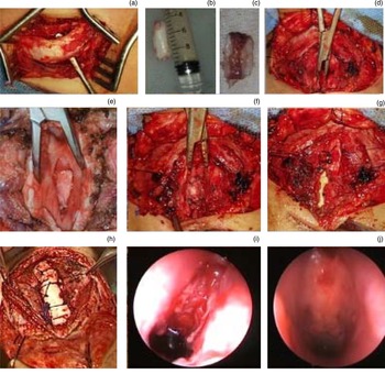
A combination of both techniques was performed (Figure 1). Peri-operatively, all patients were maintained on intravenous antibiotics. The tracheostomy was surgically closed once the child had successfully tested for decannulation.
Demonstration of the main steps of anterior and posterior laryngotracheal reconstruction by costal cartilage grafting. (a) Skeletonisation of the rib graft with the overlying perichondrium. (b) Cartilage graft length, which usually measures 4–6 cm. Note the absence of perichondrial coverage as the perichondrium on the pleural side is left in place to guard against pleural injury. (c) The luminal side of the graft with its perichondrium and carved edges to fit into the laryngofissure. (d) Posterior cricoid split. (e) Posterior cartilage graft in place between the edges of the split cricoid lamina. (f) The posterior graft is stabilised in its position by four 4–0 Vicryl sutures. (g) Stent (Foley's catheter) in place with closure of the upper end of the laryngofissure. (h) Anterior cartilage graft. The overlying Prolene suture knots are those used for stent fixation. (i) Endoscopic view immediately after stent removal. (j) Endoscopic view two weeks after stent removal showing completely taken graft.

Post-operative assessment and outcome measures
The following outcome measures were recorded: (1) adjunctive surgical procedures including granulation tissue removal, excision of suprastomal granuloma and need for cordectomy or supraglottoplasty; (2) whether or not revision reconstruction or resection was subsequently required; (3) decannulation rate; (4) voice outcome in cases of glottic web; and (5) complications.
Statistical analysis
Data were analysed using SPSS® (version 24) statistical analysis software. Normality of data was tested with the Shapiro test. Qualitative data were described using number and percentage. Association between categorical variables was tested using the Monte Carlo method. Continuous variables were presented as mean and standard deviation for parametric data and median for non-parametric data. Results were considered significant when the p-value was equal to or less than 0.05.
Results
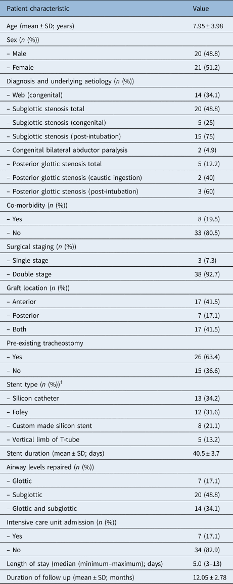
Forty-one laryngotracheal reconstructions (3 single stage laryngotracheal reconstructions and 38 double stage laryngotracheal reconstructions) were evaluated during the present study. Mean age at operation time was 7.95 ± 3.98 years. In 14 patients, the underlying pathology was congenital laryngeal web, 20 patients were diagnosed with subglottic stenosis, 2 patients with congenital bilateral abductor paralysis and 5 patients had posterior glottic stenosis. Apart from the 26 patients (63.4 per cent) who had already presented with tracheostomy, 12 patients needed tracheostomy at the time of laryngotracheal reconstruction and 3 patients remained non-tracheostomised. Eight patients (19.5 per cent) had associated co-morbidities, three patients had cardiac problems (one with ventricular septal defect, two with patent ductus arteriosus and one with tetralogy of Fallot), four patients had associated neurological problems (2 cases of cerebral palsy and 2 cases of epilepsy) and one patient had paraplegia after a motor car accident.
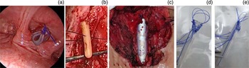
All cases of laryngeal web and 3 cases of subglottic stenosis (17, 41.5 per cent) had isolated anterior cartilage grafting, contrary to congenital bilateral abductor paralysis and posterior glottic stenosis cases where isolated posterior graft was the chosen technique. Combined anterior and posterior cartilage expansion was performed in 17 cases of subglottic stenosis. All patients with double stage laryngotracheal reconstruction had airway stenting for an average duration of 40.5 ± 3.7 days. Because of unavailability of LT-MoldTM at our institutionReference Monnier17, a custom-made silicon stent was used in 8 patients (21.1 per cent), a vertical limb of T-tube was used in 5 patients (13.2 per cent), part of a silicon catheter was used in 13 patients (34.2 per cent) and part of Foley's catheter was used in 12 patients (31.6 per cent) (Figure 2). Average duration of hospital stay ranged from 3 to 13 days, and 7 patients (17.1 per cent) required post-operative care in the paediatric intensive care unit (Table 1).
Different types of stents used in the present study. (a) Part of silicon catheter. The transfixing suture was usually added at the upper end of the stent to give a nearly triangular anterior end to fit in the anterior commissure and also to avoid aspiration. (b) Part of Foley's catheter. (c) Vertical part of T-tube. (d) Frontal and (e) lateral views of the custom-made silicon stent.

Demographic and clinical characteristics in the studied group*

*n = 41; †n = 38. SD = standard deviation
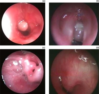
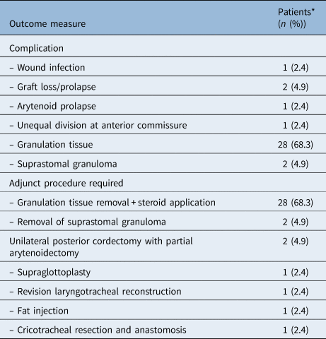
Regarding adjunctive procedures, surgical removal of granulation tissue was needed with steroid ointment application in 28 cases. In addition, a case of subglottic stenosis with single stage laryngotracheal reconstruction required a revision laryngotracheal reconstruction with stenting for graft prolapse. Another case, with grade IV subglottic stenosis, showed restenosis with loss of the anterior graft, which was scheduled for partial cricotracheal resection. Unequal division at the anterior commissure was documented in one of the earliest cases of glottic web surgery. This was managed by fat injection into the traumatised fold. Two cases required surgical excision of suprastomal granuloma; one of them was excised using coblation. Also, a case of subglottic stenosis needed supraglottoplasty for post-cricoid expansion arytenoid prolapse (Figure 3). Unilateral posterior cordectomy with partial arytenoidectomy using carbon dioxide (CO2) laser was performed in two cases for further airway widening. Neither recurrent laryngeal nerve palsy nor pneumothorax was reported, but two patients had subcutaneous emphysema at the site of graft harvest, which resolved with conservative treatment (Table 2).
Complications encountered post-laryngotracheal reconstruction. (a) Suprastomal granuloma. (b) Subglottic central granuloma. (c) Bilateral arytenoid prolapse after posterior glottis expansion. (d) Endoscopic view of prolapsed anterior wall graft causing marked narrowing of the airway.

Post-operative complications and outcome measures

*n = 41
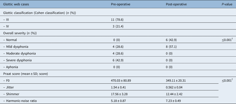
Endoscopic examination of glottic web cases showed Cohen's classification type 4 in 3 patients (21.4 per cent) and type 3 in 11 patients (78.6 per cent). Overall severity of voice affection and voice parameters, analysed by Praat software, were improved significantly post-operatively. Normal voice level was achieved in 6 cases (42.9 per cent), 8 cases (57.1 per cent) demonstrated mild dysphonia, and none of the cases had persistent moderate or severe dysphonia (Table 3).
Classification and subjective and objective assessment of severity of glottic web cases*

*n = 14; †Statistically significant results. SD = standard deviation
Myer–Cotton staging of subglottic stenosis cases showed that 7 patients (35.0 per cent) had grade II subglottic stenosis, 12 patients (60.0 per cent) had grade III subglottic stenosis and only one patient (5 per cent) had grade IV subglottic stenosis. Three patients had associated unilateral vocal fold paralysis. Length of stenotic segment ranged from 1 cm in 6 patients to 3 cm in 4 patients, and 10 patients had a stenosis of 2 cm in length. According to Olson classification, 3 patients (60.0 per cent) with posterior glottic stenosis had grade II stenosis, one patient (20.0 per cent) had a grade III stenosis and one patient (20.0 per cent) had grade IV stenosis.
The mean post-operative follow up was 12.05 ± 2.78 months. After inclusion of the revision laryngotracheal reconstruction case, 38 out of 39 tracheostomised patients were decannulated at the end of the present study. The remaining case was successfully decannulated after partial cricotracheal resection.
Discussion
Reconstruction of the laryngotracheal framework with costal cartilage graft was introduced by FearonReference Fearon18 and has become the standard treatment for many paediatric airway problems. Laryngotracheal reconstruction was primarily introduced as a double stage laryngotracheal reconstruction with anterior or posterior grafting, tracheostomy and stenting for long-term stabilisation of the graft with good decannulation rates.Reference Bailey19–Reference Ochi, Evans and Bailey21 Later on, two modifications were introduced, single stage laryngotracheal reconstruction,Reference Cotton, Myer, O'Connor and Smith22 for avoidance of tracheostomy and stenting, and partial cricotracheal resection,Reference Monnier, Savary and Chapuis23 which was later considered to be the standard procedure for high grade subglottic stenosis.
Although single stage laryngotracheal reconstruction and double stage laryngotracheal reconstruction have similar success rates,Reference Padia, Sjogren, Smith, Muntz, Stoddard and Meier24 double stage laryngotracheal reconstruction is usually adopted for complicated cases that have associated co-morbidities, cases requiring stenting (such as for laryngeal webs), or in cases of posterior glottic stenosis and congenital bilateral abductor paralysis, where stabilisation of the posterior graft is needed.Reference Kopelovich, Wine, Rutter, Mitchell and Prager9,Reference Bajaj, Cochrane, Jephson, Wyatt, Bailey and Albert25–Reference Wyatt and Hartley30 Single stage laryngotracheal reconstruction is reserved for low grade subglottic stenosis with anterior grafting only. This partly explains the dominance of double stage laryngotracheal reconstruction in the current study. In many centres, the standard treatment for high grade subglottic stenosis is partial cricotracheal resection rather than laryngotracheal reconstruction.Reference Kopelovich, Wine, Rutter, Mitchell and Prager9,Reference Bajaj, Cochrane, Jephson, Wyatt, Bailey and Albert25,Reference Agrawal, Black and Morrison31 In the present study, although the case with grade IV subglottic stenosis needed a subsequent partial cricotracheal resection for restenosis, laryngotracheal reconstruction helped to shorten the stenotic segment allowing for easier and shorter segment resection.
Advanced congenital glottic web, type 3 and 4, usually manifests early in life with severe symptoms that prompt a tracheostomy. Similar to previous reports,Reference De Trey, Lambercy, Monnier and Sandu14 57.1 per cent of patients in the present study required tracheostomy early in life. Laryngotracheal reconstruction is a successful treatment for severe congenital glottis web cases as described by several studies,Reference Bajaj, Cochrane, Jephson, Wyatt, Bailey and Albert25,Reference de Alarcon and Rutter32–Reference Rizzi, Thorne, Zur and Jacobs34 with similar results achieved in our series and successful decannulation in all patients. Success of laryngotracheal reconstruction in these cases is determined by two outcomes: decannulation and voice. To maximally improve voice results, division of the web is preferably performed endoscopically either by cold instruments or by CO2 laser. This is completed at the beginning of the surgery, as described by de Trey et al.,Reference De Trey, Lambercy, Monnier and Sandu14 to avoid unequal division of the anterior commissure with subsequent scarring. This complication was encountered once at the beginning of the series before standardisation of endoscopic assistance (Figure 4). The remaining patients had a nice triangular shape of the anterior commissure with an adequate airway. Voice assessment in paediatric patients is challenging. Post-operative voice quality was found to be poorer in cases with higher risk of anterior commissure blunting, such as in patients with severe stenosis and revision cases.Reference De Trey, Lambercy, Monnier and Sandu14
Endoscopic-assisted glottic web cutting. (a) Endoscopic view of grade IV glottic web. (b) View after cutting the glottis web demonstrated in (a) without endoscopic visualisation, showing traumatised right vocal fold resulting from unequal cut at anterior commissure. (c) Follow-up view of (a) showing atrophic anterior end of the right vocal fold. (d) Endoscopic view of grade III glottic web. (e) Endoscopic-assisted cutting of web shown in (d) (neck view). (f) Follow-up endoscopic view of (d) showing sharp anterior commissure with taken subglottic anterior graft.

Duration of stenting is still a matter of debate. Yamamoto et al.Reference Yamamoto, Monnier, Holtz and Jaquet11 recommended long-term stenting for better graft stabilisation, despite its drawbacks of granulation tissue and tracheostomy dependence. Monnier invented the LT-Mold and reported its advantages of softness, adaptation to the contour of the inner larynx and minimal mucosal trauma with less granulation tissue formation.Reference Monnier17,Reference Monnier28 Because of its unavailability, different stents were used in this series. Among them, custom-made silicon stents had the least incidence of associated granulation tissue.
Laryngotracheal reconstruction treatment requires careful attention to its consequences. When complete failure occurs, revision laryngotracheal reconstruction or partial cricotracheal resection are possible solutions. In our series, the factors associated with failure of primary surgery were high-grade subglottic stenosis and graft collapse. This is in accordance with a previous systematic review of laryngotracheal reconstruction.Reference Agrawal, Black and Morrison31 Apart from stent-associated granulations and granulomas, arytenoid prolapse is a potential complication of laryngotracheal reconstruction.Reference de Alarcon and Rutter32,Reference Rizzi, Thorne, Zur and Jacobs34 There are limited data in the literature on its risk factors; however, destabilisation of the arytenoid cartilage by large posterior graft or inadequate cutting of interarytenoid muscle can lead to this complication.Reference Li, Black, White, Zalzal and Preciado33 Hart et al. found an incidence of arytenoid prolapse of 5.7 per cent in his laryngotracheal reconstruction patients.Reference Hart, Richter, Cotton and Rutter35 The patient with arytenoid prolapse in the current study underwent supraglottoplasty with resection of collapsed supraglottic tissue by coblation.
The obvious limitation of the present study is its retrospective nature. It may be of future interest to investigate correlation between refinements in the technical details of the procedure and surgical outcomes.
• Costal cartilage laryngotracheal reconstruction is the principle augmentative procedure for many paediatric airway pathologies
• Documentation of this procedure worldwide among tertiary referral centres is mandatory to serve as baseline data for future comparative and meta-analysis studies
• Procedure staging (double or single), type of stent, possible complications and how to manage them are highlighted in the present study
Conclusion
Favourable functional outcomes were achieved with costal cartilage laryngotracheal reconstruction at our tertiary referral centre with a good decannulation rate that was comparable to large known paediatric laryngotracheal reconstruction centres worldwide.
Competing interests
None declared