Introduction
Nowadays, endoscopic sinus surgery is a well established approach for the management of sinonasal disease. The endoscopic sinus surgeon should be well aware of all anatomical features of the paranasal sinuses, and surgery should strictly adhere to the principle of identification of anatomical landmarks and adjacent vulnerable structures. Such anatomical orientation is the most important factor directly affecting endoscopic sinus surgery outcomes and complication rates. Therefore, a detailed knowledge of paranasal sinus anatomy is essential.
The roof of the ethmoidal labyrinth is formed by the fovea ethmoidalis, which is an extension of the orbital plate of the frontal bone. Medially, the fovea ethmoidalis attaches to the lateral lamella of the cribriform plate, which is a very thin bony structure offering low resistance to perforation during surgical manoeuvres. According to the literature, iatrogenic lesions of the skull base occur predominantly in the lateral lamella of the cribriform plate. The site where the anterior ethmoidal artery penetrates the cranial fossa is particularly notable, as this is the thinnest, least pressure-resistant region of the whole skull base.Reference Stammberger and Kennedy1–Reference Ohnishi and Yanagisawa3
Several cadaveric studies have described the anatomy of the ethmoid roof.Reference Ohnishi and Yanagisawa3 Keros,Reference Keros4 in a classic study, proposed a three-category classification based upon the position of the cribriform plate in relation to the ethmoid roof, and this classification has clinical relevance for sinus surgery. In the type I olfactory fossa, the olfactory sulcus is 1–3 mm deep, the corresponding lateral lamella is short and there is a significant portion of frontal bone protecting the ethmoid roof, making the roof thick and the sinus less dangerous to operate within. In the type II olfactory fossa, the olfactory sulcus is 3–7 mm deep, and the corresponding lateral lamella forms a considerable portion of the medial ethmoid roof. In the type III olfactory fossa, the olfactory sulcus is 7–16 mm deep and the ethmoid roof lies at a significant level above the cribriform plate. The thin lateral lamella forms a larger component of the ethmoid roof, which is not protected by the thick frontal bone. Therefore, the Keros type III olfactory fossa is the most vulnerable type, and is considered to pose a major risk for iatrogenic trauma of the cribriform plate.Reference Basak, Karaman, Akdilli, Mutlu, Odabasi and Erpek5
Although many studies have stressed the relevance of radiological evaluation of the ethmoid roof, and the importance of such evaluation in preventing endoscopic sinus surgery complications,Reference Stankiewicz6, Reference Kainz and Stammberger7 normative data on the height and slope of this region are somewhat limited.Reference Arslan, Aydinlioglu, Bozkurt and Egeli8, Reference Zacharek, Han, Allen, Weissman and Hwang9 Moreover, very few studies have reported the distribution of ethmoidal height measurements with respect to the various Keros olfactory fossa types.Reference Keast, Yelavich, Dawes and Lyons10
The present radioanatomical study aimed to establish normative data for some of the most important dimensions of the olfactory fossa, cribriform plate lateral lamella and ethmoid roof.
Materials and methods
Subjects included 300 adult patients who underwent high resolution, multislice, 1-mm bone window computed tomography (CT) imaging of the nose and paranasal sinuses at Alexandria University Hospitals for investigation of possible or evident sinusitis. A Toshiba Aquilion 16-slice CT machine was used (kVp (kilo voltage peak) = 120, MAs (Milli-Ampere seconds) = 120, rotation time = 1 second, FOV (field of view) = 240 mm, slice thickness = 0.5 mm, reconstruction interval = 0.3 mm, pitch = 0.7; Toshiba, Aquilion 16, Tokyo, Japan).
Subjects whose CT images showed any alteration of paranasal sinuses anatomy due to surgery or facial trauma were excluded from the study, as were those in whom sinus disease obscured the structures we wished to study.
The medical ethics committee of Alexandria Medical School approved the study.
An e-film workstation with Merge Efilm software (version 2.0.0; build 37) was used for morphological evaluation of the CT scans. This software provided tools to establish the necessary distance and angle measurements.
Measurements were recorded at two points: anterior and posterior. The anterior point was the junction between the posterior wall of the frontal sinus and the cribriform plate. The posterior point was the junction between the posterior edge of the cribriform plate and the anterior edge of the planum sphenoidale. These points were chosen because they corresponded to the extreme anterior and posterior ends of the cribriform plate, and could be easily defined with reasonable accuracy and reproducibility.
Each scan was examined by three assessors, and average measurements subsequently calculated.
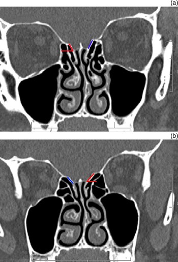
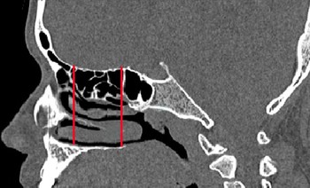
The following measurements were recorded: (1) depth of the olfactory fossa on both sides (Figure 1); (2) length of the lateral lamella and its angulation with the horizontal plane, on both sides (Figure 2); and (3) height of the ethmoid roof at the anterior and posterior points, in the sagittal plane (Figure 3).
Measurement of olfactory fossa depth: (a) anterior; (b) posterior.

Measurement of lateral lamella length (straight line) and angulation with the horizontal plane (angled line).

Measurement of ethmoid roof height at the anterior point and posterior point.

The Statistical Package for the Social Sciences software was used for statistical analysis. Student's t-test was used for comparison of means. Pearson's correlation coefficient was used to assess the correlation (linear dependence) between the variables.
Results
We analysed the CT scans of 300 adult patients: 150 men and 150 women. The mean (standard deviation) age of the patients was 39.3 (9) years for men and 36.8 (6) years for women.
Olfactory fossa depth
Table I shows the distribution of olfactory fossa depth according to the Keros classification system.
Keros olfactory fossa type by side and sex

The commonest Keros olfactory fossa type in men was type II, on both sides (66.7 per cent); in women, it was type I, on both sides (53 per cent). There were statistically significant differences between the depth of the olfactory fossae of men and women, both on the right side (t = 2.039, p = 0.04) and on the left (t = 2.118, p = 0.039). A difference between both sides of 3 mm or more was present in 11 per cent of men and 2 per cent of women.
Lateral lamella length
Table II shows lateral lamella length measurements in men and women.
Lateral lamella length by side and sex

Ant = anterior; post = posterior
The length of the lateral lamella was significantly less in women than in men (t = 2.146, p = 0.040). In both sexes, there were statistically significant differences between the lateral lamella length on the right and left sides (for men, t = 2.132 and p = 0.035; for women, t = 2.118 and p = 0.039). The length of the lateral lamella was greater anteriorly than posteriorly, in both men and women.
Lateral lamella angulation
Measurements for the angulation of the lateral lamella with the horizontal plane are shown in Table III.
Lateral lamella angulation by side and sex

Ant = anterior; post = posterior
There were significant differences between lateral lamella angulation in men and women (for the right side, r = 2.202 and p = 0.039; for the left side, r = 2.312 and p = 0.032). In addition, there were statistically significant differences between lateral lamella angulation in men and women (for the right side, r = 1.723 and p = 0.083; for the left side, r = 1.234 and p = 0.156).
Ethmoid roof height
Table IV shows ethmoid roof height measurements.
Ethmoid roof height by side and sex

Ant = anterior; post = posterior; SD = standard deviation
There were statistically significant differences between the right and left sides in both men and women (for the right side, r = 2.111 and p = 0.049; for the left side, r = 2.212 and p = 0.042). There were also significant differences in average ethmoid roof heights between men and women (for the right side, r = 2.214, p = 0.042; for the left side, r = 2.252 and p = 0.039).
Correlation between dimensions
There was a statistically significant association between lateral lamella angulation and olfactory fossa depth (r = 0.231, p = 0.020). However, there was no statistically significant association between ethmoid roof height and olfactory fossa depth (r = 0.161, p = 0.072).
Discussion
Computed tomography has contributed greatly to the diagnosis and evaluation of sinonasal diseases, and also to the characterisation of important anatomical landmarks. This fact has been documented in several studiesReference Ohnishi, Tachibana, Kaneko and Esaki2, Reference Ohnishi and Yanagisawa3 highlighting the relevance and precision of radiographic evaluation of the ethmoid roof, and its validity for the provision of safe ‘roadmaps’ for surgeons.
The present study provides a detailed quantitative analysis of the ethmoid roof, olfactory fossa and cribriform plate lateral lamella. With the help of computer-aided, multislice CT analysis, we were able to measure reliably the olfactory fossa depth, the lateral lamella length and angulation with the horizontal plane, and the ethmoid roof height in the sagittal plane.
Studies of olfactory fossa depth have demonstrated divergences in the distribution of Keros olfactory fossa types. Basak et al. Reference Basak, Akdilli and Karaman11 evaluated 64 children and found a higher frequency of Keros type II (53 per cent), followed by type III (38 per cent) and type I (9 per cent). Jang et al. Reference Jang, Park and Kin12 in a study of 205 predominantly adult patients, observed that Keros type II was most frequent (69.5 per cent), followed by type I (53.8 per cent) (in this series, no Keros type III olfactory fossae were found). Anderhuber et al. Reference Anderhuber, Walch and Fock13 studied children aged between zero and 14 years, and found the following incidences of Keros olfactory fossa types: 14.2 per cent for type I, 70.6 per cent for type II and 15.2 per cent for type III. Souza et al. Reference Souza, Souza, Idagawa, Wolosker and Ajzen14 studied predominantly adult patients, and also found that the Keros type II olfactory fossa was most frequent (73.3 per cent), followed by type I (26.2 per cent) and type III (0.5 per cent).
The present study found that men and women displayed different distributions of Keros olfactory fossa types. Type II was more common in men, while type I was commoner in women. Keros type III was present in 1.4 per cent of men but absent in women. In our subjects, 11 per cent of men and 2 per cent of women had different Keros types on both sides. This considerable inter-subject anatomical variation should alert the surgeon to the need for careful review of the patient's anatomy on pre-operative scans, prior to operation.
The present study showed that the depth of the olfactory fossa depends upon the length and angulation of the lateral lamella. This agrees with the findings of Souza et al. Reference Souza, Souza, Idagawa, Wolosker and Ajzen14 who observed that, in most cases, ethmoid roof asymmetry was related to the angulation of the lateral lamella. The length of the lateral lamella and its angulation with the horizontal plane were less in females than males. Consequently, the olfactory fossa was shallower in females than in males, as shown by the predominance of the Keros type I configuration among females.
The present study found significant differences between the length and angulation of the lateral lamella, comparing both sides of the head. This agrees with other studies demonstrating that the cribriform plate lateral lamella is symmetrical in less than 50 per cent of individuals.Reference Ohnishi and Yanagisawa15, Reference Grevers16 Another important finding of the present study was a decrease of the lateral lamella length as we traced it posteriorly. This is in keeping with the general posterior flattening of the ethmoid roof, described previously and documented in the present study as well as that of Souza et al. Reference Souza, Souza, Idagawa, Wolosker and Ajzen14
• This study aimed to establish normative measurements for the olfactory fossa depth, the length and angulation of the cribriform plate lateral lamella, and the ethmoid roof height, in adult males and females
• The cribriform plate lateral lamella was significantly shorter and less oblique in men than in women
• The ethmoid roof was lower in women than in men, and there was no statistically significant association between the ethmoid roof height and the olfactory fossa depth
In the present study, the height of the ethmoid roof was significantly less in women than men. However, there was no statistically significant association between the height of the ethmoid roof and the depth of the olfactory fossa. In agreement with common belief, there were significant differences between the right and left sides in both men and women. These findings agree with those of Dessi et al. Reference Dessi, Moulin, Triglia, Zanaret and Cannoni17 who reported that the ethmoid roof was asymmetrical in approximately 10 per cent of cases. Likewise, Lobvitz et al. Reference Lobwitz, Terk, Jacob and Holiday18 reported ethmoid roof asymmetry in 57 per cent of their patients. In a more recent study, Solares et al. Reference Solares, Walter, Batra and Citardi19 reported asymmetry of the ethmoid roof in all subjects, and attributed their findings to the use of computer-aided CT scan analysis which allowed more precise measurement of the ethmoid roof. These previous studies, together with the present one, should alert surgeons to the frequency of ethmoid roof asymmetry, which should be taken into consideration in order to avoid iatrogenic injury.
Conclusion
The current study presents a precise, quantitative analysis of the olfactory fossa and ethmoid roof position and configuration, and reports sex-related differences as well as individual asymmetry. This information may be useful during pre-operative evaluation of CT images, as well as intra-operatively.