Introduction
Parasites are common and widespread in most ecosystems (Hudson et al., Reference Hudson, Dobson and Newborn1998; Hudson et al., Reference Hudson, Dobson and Lafferty2006; Lafferty et al., Reference Lafferty, Allesina, Arim, Briggs, De Leo, Dobson, Dunne, Johnson, Kuris, Marcogliese, Martinez, Memmott, Marquet, McLaughlin, Mordecai, Pascual, Poulin and Thieltges2008). A growing body of literature suggests that the impacts of parasites on their hosts are substantial and diverse (Kuris et al., Reference Kuris, Hechinger, Shaw, Whitney, Aguirre-Macedo, Boch, Dobson, Dunham, Fredensborg, Huspeni, Lorda, Mababa, Mancini, Mora, Pickering, Talhouk, Torchin and Lafferty2008; Tompkins et al., Reference Tompkins, Dunn, Smith and Telfer2011). In particular, pathogens such as fungi (e.g., Batrachochytrium dendrobatidis, Batrachochytrium salamandrivorans) and iridoviruses (e.g., ranavirus) have been implicated in amphibian declines and extinctions (Berger et al., Reference Berger, Speare, Daszak, Green, Cunningham, Goggin, Slocombe, Ragan, Hyatt, McDonald, Hines, Lips, Marantelli and Parkes1998; Daszak et al., Reference Daszak, Berger, Cunningham, Hyatt, Green and Speare1999; Blaustein and Kiesecker, Reference Blaustein and Kiesecker2002; Lips et al., Reference Lips, Brem, Brenes, Reeve, Alford, Voyles, Carey, Livo, Pessier and Collins2006; Skerratt et al., Reference Skerratt, Berger, Speare, Cashins, McDonald and Phillott2007; Martel et al., Reference Martel, Spitzen-van der Sluijs, Blooi, Bert, Ducatelle, Fisher, Woeltjes, Bosman, Chiers, Bossuyt and Pasmans2013). However, less is known about macroparasites and their role in dictating amphibian health (Koprivnikar et al., Reference Koprivnikar, Marcogliese, Rohr, Orlofske, Raffel and Johnson2012).
One host species that has been the subject of numerous parasitological studies is the cane toad, Rhinella marina (Selechnik et al., Reference Selechnik, Rollins, Brown, Kelehear and Shine2017). This large, toxic amphibian is a widespread invasive species and studies with a parasite focus have centred on identifying useful biocontrol agents to curtail the spread of the toad (Speare, Reference Speare1990; Kelehear et al., Reference Kelehear, Webb and Shine2009; Kelehear et al., Reference Kelehear, Brown and Shine2011). The Rhinella marina species complex (previously Bufo marinus) contains 11 species: Rhinella achavalli, Rhinella arenarum, Rhinella cerradensis, Rhinella horribilis, Rhinella icterica, Rhinella jimi, Rhinella marina, Rhinella poeppigii, Rhinella rubescens, Rhinella schneideri, Rhinella veredas (Pramuk, Reference Pramuk2006; Vallinoto et al., Reference Vallinoto, Sequeira, Sodré, Bernardi, Sampaio and Schneider2010; Acevedo et al., Reference Acevedo, Lampo and Cipriani2016). Until recently, the native range of R. marina was considered to span from South Texas, USA contiguously through to the southern edge of the Amazon in Brazil (Zug and Zug, Reference Zug and Zug1979). However, accruing molecular evidence indicates that this species should be split into two, one species east and one west of the Andes (Slade and Moritz, Reference Slade and Moritz1998; Vallinoto et al., Reference Vallinoto, Sequeira, Sodré, Bernardi, Sampaio and Schneider2010). Recently, Acevedo et al. (Reference Acevedo, Lampo and Cipriani2016) provided both molecular and morphological support for this split, such that toads west of the Andes are now classified as R. horribilis and those to the east retain the name R. marina (Acevedo et al., Reference Acevedo, Lampo and Cipriani2016). Rhinella horribilis is the only species in the complex that occurs in Central America; the remaining 10 species are restricted to South America. In light of the recent taxonomic shift, it has become apparent that we know little about R. horribilis in Central America. Most existing research has been conducted on R. marina in their invasive range (Lever, Reference Lever2001; Shine, Reference Shine2010) – animals that were sourced from native populations in South America for a widespread introduction to the Caribbean, Florida, Pacific Islands, Australia and a myriad of other localities (Easteal, Reference Easteal1981). In particular, little is known about the parasites of R. horribilis and the influence of these parasites on their host. The few studies that exist are parasite surveys and do not experimentally examine the impacts of parasites on the toad.
Parasitic nematodes of the genus Rhabdias infect the lungs of reptiles and amphibians (Anderson, Reference Anderson2000; Kuzmin, Reference Kuzmin2013) where they feed on blood and reduce growth, performance and/or survival in some hosts (Goater, Reference Goater1992; Goater et al., Reference Goater, Semlitsch and Bernasconi1993; Kelehear et al., Reference Kelehear, Webb and Shine2009, Reference Kelehear, Brown and Shine2011; Marr et al., Reference Marr, Johnson, Hara and McGarrity2010; Pizzatto and Shine, Reference Pizzatto and Shine2011b, Reference Pizzatto and Shine2012; Moretti et al., Reference Moretti, Titon, Madelaire, Arruda, Alvarez and Gomes2017; Finnerty et al., Reference Finnerty, Shine and Brown2018) but not others (Pizzatto and Shine, Reference Pizzatto and Shine2011a; Nelson et al., Reference Nelson, Brown, Dubey and Shine2015). They have a direct life cycle that includes a parasitic blood-feeding stage in the lungs, and free-living larval stages in the soil (Anderson, Reference Anderson2000; Kuzmin, Reference Kuzmin2013). Rhabdias spp. infect R. marina across their native range (Kuzmin et al., Reference Kuzmin, Tkach and Brooks2007; Dubey and Shine, Reference Dubey and Shine2008; Bursey and Brooks, Reference Bursey and Brooks2010; Kuzmin et al., Reference Kuzmin, du Preez and Junker2015) and one species (Rhabdias pseudosphaerocephala) has been introduced to Australia (Dubey and Shine, Reference Dubey and Shine2008) where it negatively impacts toad growth, running performance and survival (Kelehear et al., Reference Kelehear, Webb and Shine2009; Kelehear et al., Reference Kelehear, Brown and Shine2011). In Central America, R. horribilis hosts at least four species of Rhabdias (Rhabdias alabialis; Rhabdias americanus; Rhabdias fuelleborni; R. pseudosphaerocephala), with prevalence of infection fluctuating broadly from 2.6 to 66% (Espinoza-Jiménez et al., Reference Espinoza-Jiménez, García-Prieto, Osorio-Sarabia and León-Règagnon2007; Espinola-Novelo and Guillen-Hernandez, Reference Espinola-Novelo and Guillen-Hernandez2008; Bursey and Brooks, Reference Bursey and Brooks2010; Ruiz-Torres et al., Reference Ruiz-Torres, García-Prieto, Lagunas-Calvo, Violante-González and Osorio-Sarabia2017).
Throughout Central and South America ~19 species of Rhabdias are known from anuran hosts, yet there is no published information on Rhabdias in amphibians of Panama (Online Resource ESM1); the majority of research that has been done on Rhabdias infecting R. horribilis involves parasite surveys conducted in Mexico, Costa Rica and Nicaragua (Espinoza-Jiménez et al., Reference Espinoza-Jiménez, García-Prieto, Osorio-Sarabia and León-Règagnon2007; Espinola-Novelo and Guillen-Hernandez, Reference Espinola-Novelo and Guillen-Hernandez2008; Bursey and Brooks, Reference Bursey and Brooks2010; Ruiz-Torres et al., Reference Ruiz-Torres, García-Prieto, Lagunas-Calvo, Violante-González and Osorio-Sarabia2017). A recent study from Brazil using sequencing of three mitochondrial and nuclear DNA loci, identified two new species of Rhabdias and five morphospecies, suggesting that diversity within the genus is high (Müller et al., Reference Müller, Morais, Costa-Silva, Aguiar, Ávila and da Silva2018). The Isthmus of Panama connects North and South America and is a hot spot for amphibian diversity (Brooks et al., Reference Brooks, Mittermeier, Mittermeier, de Fonseca, Rylands, Konstant, Flick, Pilgrim, Oldfield, Magin and Hilton-Taylor2002). Over recent years, the amphibian fauna on the isthmus has been adversely impacted by the spread of the pathogenic chytrid fungus (B. dendrobatidis), with rapid declines and extinctions reported for numerous species (Lips, Reference Lips1999; Crawford et al., Reference Crawford, Lips and Bermingham2010). Bolstering our knowledge of the parasite fauna impacting anurans in this vulnerable region will allow insight into those factors governing population health. We conducted field comparisons to determine the prevalence, intensity and diversity of Rhabdias infections in R. horribilis in Panama. We also conducted laboratory experiments where we exposed parasite-naïve R. horribilis toadlets to Rhabdias to determine the effect on growth, survival and locomotion.
Materials and methods
Rhabdias field comparisons
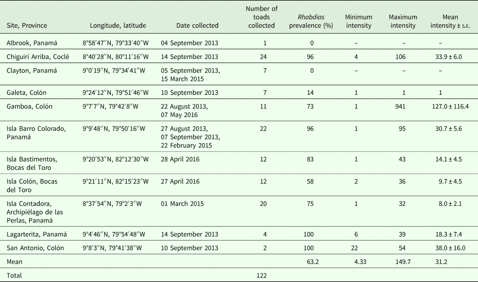
We collected 122 sub-adult and adult R. horribilis (mean snout-urostyle length ± 1 s.e. = 105.38 ± 1.63 mm, min = 52.2 mm, max = 156.0 mm) from 11 sites (four island and seven mainland) across four provinces (Bocas del Toro, Coclé, Colón, and Panamá) in the Republic of Panamá during August 2013–April 2016 (Table 1). Since levels of parasitism can vary with host sex we attempted to collect a relatively even sex ratio (59 females, 63 males). We euthanized toads by immersing them in a clove oil solution (Mitchell et al., Reference Mitchell, Riggs, Singleton, Diaz-Figueroa and Hale2009), measured their snout-urostyle length to the nearest 0.1 mm using calipers, and removed both of their lungs and examined them for Rhabdias. We counted all adult Rhabdias and preserved them in 70% ethanol for molecular and morphological analyses (see below). We report prevalence as the percentage of toads infected per site and intensity as the number of adult worms in an infected toad (Bush et al., Reference Bush, Lafferty, Lotz and Shostak1997).
Collection localities and dates (August 2013–April 2016) for lungworms (Rhabdias) surveyed in toads (Rhinella horribilis) in Panama

Prevalence = the percentage of toads infected at a site. Intensity = the number of adult Rhabdias per infected toad.
Toad breeding and husbandry
We collected two breeding pairs (source adults for Clutch 1 and 2) from Isla Barro Colorado in the Panama Canal (Table 1) and one pair (source adults for Clutch 3) from Chiguirí Arriba (Table 1) over the period 27 August 2013 to 14 September 2013 to source parasite-naïve toad egg clutches. We brought the breeding pairs to the Smithsonian Tropical Research Institute (Naos Marine Laboratory [Naos]: 8°54′58″N, 79°31′57″W) and housed them in slanted plastic enclosures with continual access to fresh water. We fed them crickets twice per week and cleaned their enclosures at least every fourth day.
We followed the same breeding and tadpole husbandry methodology used in Kelehear et al. (Reference Kelehear, Webb and Shine2009). We stimulated spawning by administering subcutaneous injections of the gonadotropin-releasing hormone agonist: leuprorelin acetate (Lucrin, Abbott Australasia, Kurnell, Australia) diluted at 1:20 with amphibian Ringer's solution. We injected each female with 0.8 mL of the solution 1 hour prior to dusk and each male with 0.5 mL of the solution on dusk and placed pairs into 114 L opaque plastic containers with 10 L of spring water situated outdoors at Naos. The following morning, we collected the fertilized egg clutches, transferred them to 10 L of aerated spring water and changed half the water every day for 3 days. When tadpoles were 5-days-old, we split each clutch (keeping clutches separate) between 3–6 large (134–220 L) glass or fiberglass aquaria (n = 13 aquaria in total) and maintained them at a density of one tadpole per 400 mL of aerated aged tap water. We performed half water changes as necessary. Aquaria were maintained outdoors beneath a roof, under natural climatic conditions (24.7–31.1°C, 70–99% relative humidity) at Naos. We fed tadpoles frozen lettuce ad libitum and checked aquaria daily for metamorphs.
The first metamorphs (Gosner Stage 46: Gosner, Reference Gosner1960) emerged 22 (Clutch 1 and 3) and 47 (Clutch 2) days after egg deposition. We weighed metamorphs to 0.0001 g (Adventurer SL AS214, Ohaus Corporation, Pine Brook, New Jersey, USA) when tail resorption was complete (mass at metamorphosis) and assigned them alternately to the control or treatment group (see below) according to their date of metamorphosis. We housed metamorphs individually in clear plastic enclosures (65 × 119 × 198 mm) with a moist sandy substrate and a rock as a dry retreat. All containers and substrates were autoclaved prior to use to ensure sterility. Enclosures were maintained on shelving outdoors beneath a roof (as for the tadpole aquaria). We fed metamorphs 10 pinhead crickets dusted in multivitamins (Reptivite and Rep-Cal) every third day.
Experimental infections
Of the 122 toads dissected for Rhabdias surveys, five (three from Largarterita, one from San Antonio, one from Gamboa; Table 1) were used as a source of infective Rhabdias larvae (L3) for experimental infections. These five toads were infected with a mean of 15 worms (range: 6–22, pooled total: 75 worms). We placed all 75 adult Rhabdias into Petri dishes of distilled water (keeping worms separate for each of the five toads) for several hours to collect their eggs. We then placed the worms into 70% ethanol for morphological examination and DNA extraction (see below). Following this methodology, we aimed to determine the identity of the Rhabdias used in experimental infections (see below), as it is impossible to distinguish between Rhabdias species at the L3 stage (used in infections).
Molecular characterization of Rhabdias
We extracted DNA from 30 of the 75 adult Rhabdias removed from the lungs of the five adult toads (four–nine Rhabdias from each adult) and used as a parasite source for the experimental infections (see above) and from three toadlets (four Rhabdias) that were experimentally infected (see below). We extracted DNA in Lysis Buffer containing 200 mm NaCl, 200 mm Tris-HcL (pH 8.0), 1% β-mercaptoethanol, and 800 µg mL−1 proteinase K (Holterman et al., Reference Holterman, van der Wurff, van den Elsen, van Megen, Bongers, Holovachov, Bakker and Helder2006). We incubated nematode tissue in 25 µL sH2O and 25 µL Lysis buffer for 1:30 h at 65°C with shaking, followed by 5 min at 99°C to deactivate the proteinase K. We amplified the cytochrome b locus using primers Rhcytb-F and Rhcytb-R (Dubey and Shine, Reference Dubey and Shine2008) in 25 µL reactions containing 2.5 µL 10X buffer, 2 mm MgCl2, 50 µmol each dNTP, 0.2 µmol each primer, 2% DMSO, 1 unit Platinum Taq polymerase, and 1–3 µL DNA extract. Amplification conditions included denaturation at 94°C for 3 min, followed by 35 cycles of 94°C for 45 s, 50°C for 45 s, and 72°C for 90 s, with a final extension cycle at 72°C for 7 min. We gel-purified amplified bands using GELase enzyme (Epicentre) and sequenced samples in both directions on an ABI 3500XL automatic sequencer. We cleaned and aligned sequences using Sequencher 5.1 (GeneCodes) and compared the sequences with others on GenBank using BLAST (NCBI). Genetic distances between sequences were calculated using MEGA 5.2.2.
Morphological characterization of Rhabdias
Prior to molecular analyses (see above), we relaxed nematodes in distilled water, preserved them in 70% ethanol and examined their morphology (within a few hours of preservation) beneath a dissecting scope. Specifically, we examined the positioning of the vulva and looked for the presence of phasmids on the tail. In addition, since lip morphology is a key feature for distinguishing between Rhabdias species (Kuzmin et al., Reference Kuzmin, Tkach and Snyder2003) we used scanning electron microscopy to examine the lips of three of the Rhabdias used as a source for the experimental infections and six of the Rhabdias recovered from experimentally infected toadlets (see below). For further information see Online Resource ESM2.
Rhabdias culture and exposure to toadlets
We placed the Rhabdias eggs and gut contents of the five dissected toads into separate Petri dishes and moistened them with distilled water. We allowed the Rhabdias eggs to develop through to the L3 stage at room temperature (23.3–26.5°C). We added 30 L3 to 48 sterile plastic Petri dishes (14 × 55 mm) lined with filter paper (Grade 2: Whatman International, Brentford, Middlesex) and moistened with distilled water. We kept L3 sourced from each adult toad separate such that each treatment (exposed) toadlet received parasites from only one adult toad; 4–15 toadlets were exposed to the larvae sourced from each of the five toads. We placed 48 treatment (exposed) toadlets from three clutches (Clutch 1: n = 9; Clutch 2: n = 3; Clutch 3: n = 36) into the individual Petri dishes with the 30 L3. After 18 h, we removed the toadlets from the dishes and counted all remaining L3 to calculate the number of L3 that had penetrated each toadlet. We repeated this protocol, except the addition of 30 L3 to the dishes, for 50 control (unexposed) toadlets (Clutch 1: n = 11; Clutch 2: n = 4; Clutch 3: n = 35). All exposures to L3 were conducted indoors at 23.3–26.5°C after which toadlets were returned to their outdoor enclosures for the duration of the experiment (20 days) and housed and fed as above.
Toadlet performance
We weighed toadlets (to the nearest 0.0001 g) at the time of treatment and 15 days post-treatment (p.t.) to calculate mean daily growth rates. At 15 days p.t. we measured the locomotor performance of surviving toadlets (control: n = 38; infected: n = 36) on a square racetrack constructed from plastic guttering (10 cm wide, 8.5 cm high, 440 cm total length taken at mid-point of guttering). Since toadlets are diurnal we conducted trials between 1030 and 1630 h at 27.8–33.4°C (mean ± 1 s.e. = 30.9 ± 0.12°C) and 63–99% relative humidity (mean ± 1 s.e. = 78.2 ± 0.88%) outdoors beneath a roof at Naos. During each locomotor trial, one of us tapped the toadlet on the urostyle with a small artist's paintbrush whenever it stopped running; trials concluded when five taps failed to induce movement and the toadlet was considered to be exhausted. We used the fastest 25 cm of the first 50 cm travelled as a measure of initial sprint speed. We used the average speed over the entire trial as a measure of sustained speed. We used total distance travelled and total time spent running (up until the point of exhaustion) as endurance measures.
Toadlet mortality and necropsy
We checked all toadlets daily; any dead toadlets were placed into Petri dishes and dissected to count larval and/or adult Rhabdias. At the conclusion of our experiments (20 days p.t.), we euthanized (via immersion in a clove oil solution) all surviving toads and examined them immediately for parasites. We removed all parasites and flushed the toadlet's body with distilled water, left it for 24 h, and then re-examined it for any previously undetected larvae. We extracted DNA from four of the recovered Rhabdias and processed six for s.e.m. photography.
Data analyses
We log-transformed initial sprint speed, sustained sprint speed, total distance travelled and total run time to meet assumptions of normality. Since temperature and humidity have a significant impact on ectotherm performance (Losos, Reference Losos1990; Niewiarowski et al., Reference Niewiarowski, Lopez, Ge, Hagan and Dhinojwala2008; Biro et al., Reference Biro, Beckmann and Stamps2010; Seebacher et al., Reference Seebacher, Tallis and James2014; Halsey et al., Reference Halsey, Matthews, Rezende, Chauvaud and Robson2015) we included these factors as covariates in the locomotor trial analyses. We explored relationships between Rhabdias prevalence and host sex and size using chi-squared analyses. We analyzed correlations between Rhabdias intensity and host size and sex using a two-way analysis of variance (ANOVA). We analyzed data on locomotor performance and growth using multivariate ANOVA. We investigated the relationship between the number of L3 that penetrated a toadlet and the number of blood-feeding Rhabdias in the toadlet's lungs at dissection using ANOVA (unexposed control toads excluded). For our performance data on growth and locomotion we conducted full factorial MANOVAs checking for interactions and removing the highest order non-significant interactions. Preliminary analyses revealed that each of the three clutches had similar relationships between the treatment and the response variables and thus, the clutch was excluded as a factor from final analyses (see Kelehear et al., Reference Kelehear, Webb and Shine2009). We analyzed data on toadlet survival using a χ2 analysis. We performed all analyses in JMP Pro 9.0.0.
Results
Rhabdias field comparisons
We found Rhabdias infecting R. horribilis at nine of 11 sites in Panama (mean prevalence of 63% per site; Table 1). Mean infection intensity was 31 worms per host, with the heaviest infection comprised 941 worms (Table 1). Prevalence of infection did not differ with toad sex (χ2 = 3.48, df = 2, P = 0.18) but varied with toad body size (chi-sq = 5.21, df = 1, P = 0.02): larger toads were more likely to be infected with Rhabdias than smaller toads. There was no influence of host sex (F 1,87 = 0.013, P = 0.91) or size (F 1,87 = 0.87, P = 0.35) on the intensity of Rhabdias infections (uninfected toads and one outlier with 941 Rhabdias excluded).
Molecular and morphological characterization of Rhabdias
Molecular characterization of Rhabdias
We sequenced 30 nematodes from the lungs of five adult toads and four nematodes from the lungs of four toadlets infected in the experiment. Four unique haplotypes were identified, which had four variable positions (GenBank Accession No. MG205601-MG205604; Table 2; Online Resource ESM3). All haplotypes were closely related, with a mean divergence of 0.003 ± 0.002 (Kimura 2 parameter). Haplotype 1 was found in two adult toads and one experimental toadlet. Haplotype 2 was found in a single adult toad. Haplotype 3 was found in all five adult toads and represented 53% of the nematodes sequenced in adults and 75% of the nematodes from the toadlets. Haplotype 4 was found in two adult toads. Four out of the five adults had co-infections with multiple haplotypes of Rhabdias, with 2 or 3 nematode haplotypes found in the same adult toad. Co-infections showed no obvious patterns of occurrence: haplotypes co-occurred within a host in multiple combinations (i.e., Haplotype 1 co-occurred with Haplotype 2 and Haplotype 3; Haplotype 2 co-occurred with Haplotype 1 and 3; Haplotype 3 co-occurred with all other haplotypes; Haplotype 4 co-occurred with Haplotype 3). BLAST results suggest that these haplotypes are closely related to R. pseudosphaerocephala (97% identity), found in R. marina from Brazil (Online Resource ESM3; Dubey and Shine, Reference Dubey and Shine2008).
Molecular results showing the number of Rhabdias lineages in each of five wild-caught adult Rhinella horribilis (n = 4–9 nematodes sequenced from each toad, n = 30 nematodes in total) from three geographic localities (Gamboa, Largarterita and San Antonio) and three experimentally infected R. horribilis toadlets (n = 4 nematodes in total)

Morphological characterization of Rhabdias
Morphological measurements showed more variation than our molecular results despite our small sample sizes. The vulva was positioned slightly pre-equatorial (47.1–48.7% of body length from anterior end; n = 2) in Haplotype 1 specimens, but in Haplotype 2 worms, ranged from pre-equatorial to equatorial (30–50% of body length from anterior end; n = 2). The positioning of the vulva was very inconsistent for Haplotype 3 specimens, ranging from 46.3 to 84.2% of body length from the anterior end (n = 14), and in Haplotype 4 ranged from 50 to 75% of body length from the anterior end (n = 6). Haplotype 4 also displayed well-developed phasmids mid-distance along the tail. Of the nine worms we processed for s.e.m. photography, we were only able to obtain satisfactory photos of the lips of three worms for which we had molecular data: lips of a Haplotype 1 individual showed morphology consistent with Rhabdias fuelleborni (n = 1) while that of Haplotype 3 most closely resembled that of R. pseudosphaerocephala (n = 2) (Kuzmin et al., Reference Kuzmin, Tkach and Brooks2007; Online Resource ESM4). We were unable to obtain satisfactory s.e.m. photos of the lips of Haplotypes 2 and 4.
Experimental infections
Infection success
On average, 21 (± 1 s.e. = 0.85) of the 30 L3 penetrated each exposed toadlet (min = 9, max = 30). Of the 48 exposed toads, 45 postmortems were conducted either immediately after euthanasia or shortly after death (the remaining three toadlets died during the night and were too decomposed to accurately dissect). Twenty of the 45 treatment toadlets had blood-feeding Rhabdias in their lungs at dissection (mean ± 1 s.e. = 3.7 ± 0.49, min = 1, max = 8). For one of these toadlets, only 7 days had passed since exposure, indicating that the L3 is able to migrate to the lungs and begin feeding on blood rapidly, even in small hosts (this toadlet weighed only 0.057 g). There was no relationship between the number of L3 that penetrated a toadlet and the number of blood-feeding Rhabdias in the toadlet's lungs at dissection (F 1,43 = 0.87; P = 0.36). No Rhabdias were recovered from any control (unexposed) toadlets.
Growth
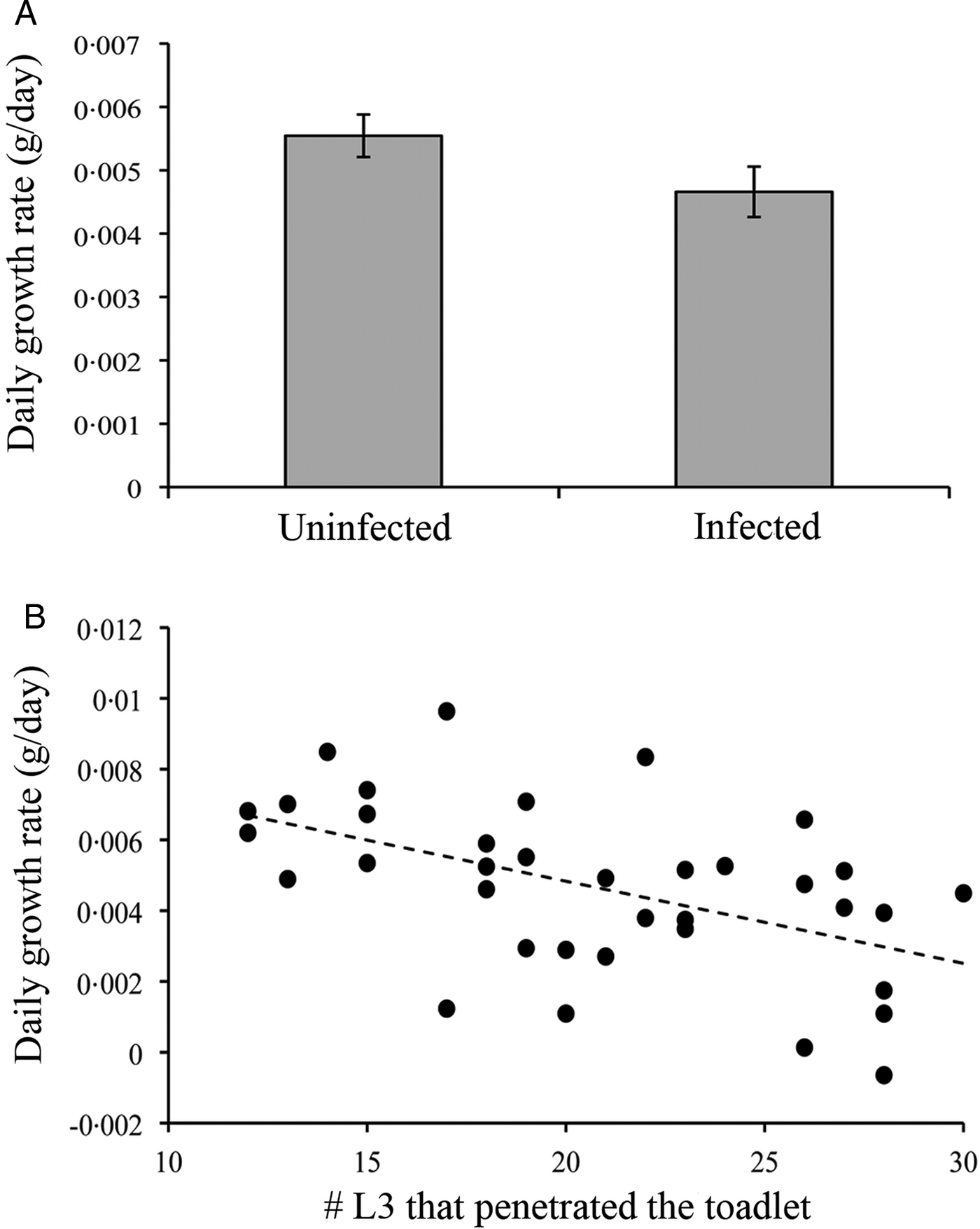
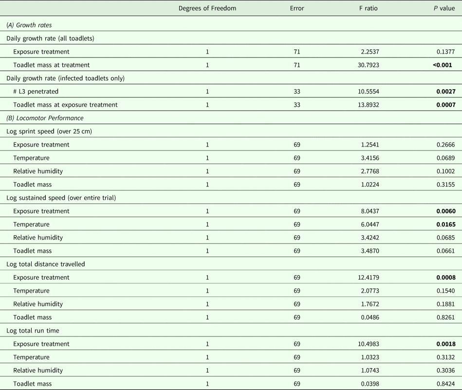
The exposure treatment did not impact toadlet daily growth rates (Fig. 1A; Table 3A). Daily growth rates were influenced by size at exposure: smaller toadlets had higher daily growth rates than larger toadlets (Table 3A). However, when considering only exposed toadlets, the number of infective larvae that penetrated the exposed toadlets was negatively correlated with daily growth rate (Fig. 1B).
(A) Daily growth rates of experimental toadlets (Rhinella horribilis) that had been exposed to no Rhabdias (uninfected) or 30 infective Rhabdias larvae (infected). Shaded columns show means and error bars show standard error. (B) Negative relationship between the number of infective Rhabdias larvae that penetrated the toadlet and subsequent daily growth rates in toadlets (R. horribilis) that had been exposed to 30 infective Rhabdias larvae (y = −0.0002x + 0.0095; R 2 = 0.26, P = 0.0027).

Results of statistical analyses investigating the impact of Rhabdias infection (treatment) on toadlet (Rhinella horribilis) locomotor performance and growth

Statistically significant P values appear in bold font.
Locomotion
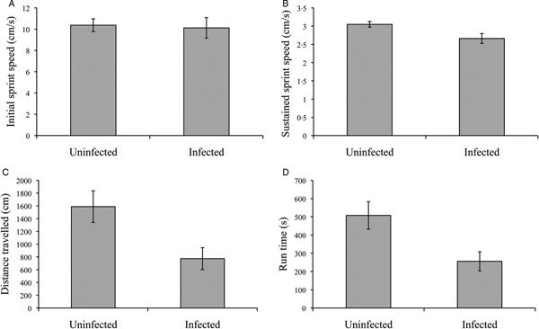
There was no effect of exposure to parasites (control vs treatment) on initial sprint speed over 25 cm (Table 3B; Fig. 2A), but sustained sprint speed over the entire trial was influenced by treatment and temperature (Table 3B, Fig. 2B). Unexposed toadlets were faster on average than exposed toadlets over the entire running trial (Fig. 2B). Sustained sprint speed decreased as temperature increased (Table 3B). Unexposed toadlets ran farther than exposed toadlets (Fig. 2C, Table 3B) and were able to run for longer before becoming exhausted (Fig. 2D, Table 3B). However, the number of L3 that penetrated the toadlet (intensity) had no influence on subsequent locomotor performance (P > 0.28 in all cases).
Locomotor performance of experimental toadlets (Rhinella horribilis) that had been exposed to no Rhabdias (uninfected) or 30 infective Rhabdias larvae (infected). (A) Initial sprint speed measured as the fastest 25 cm of the first 50 cm travelled, (B) Sustained sprint speed measured as average speed over the total distance travelled, (C) Distance travelled shows total distance travelled up until the point of exhaustion, (D) Run time shows total time ran up until the point of exhaustion. Trials were conducted 15 days post-exposure. Shaded columns show means and error bars show standard error.

Survival
Toadlets that were larger at the time of exposure were more likely to survive than those that were smaller (χ2 = 9.82, df = 1, P = 0.0017) but there was no influence of exposure treatment on toadlet survival when size was accounted for: 76% of unexposed toadlets survived vs 64.6% of exposed toadlets (χ2 = 2.38, df = 1, P = 0.1230).
Discussion
Rhabdias comparisons
Our field comparisons showed that R. horribilis are commonly infected by lung nematodes in the genus Rhabdias across Panama. Toads were infected by four genetically distinct but closely related lineages of Rhabdias across nine of 11 sampled sites in Panama. Multiple lineages of Rhabdias may be present in the same geographic locality, the same host, and even the same lung. Mean prevalence of infection per site was 63% and mean intensity of infection was 31 worms per infected host.
In general, the prevalence and intensity reported here are on the higher end of the spectrum of previous values reported for Rhabdias infections in amphibians. In particular, the number of worms that comprised infections was substantial; one adult toad collected from Gamboa hosted 941 adult Rhabdias; to our knowledge, this far exceeds published accounts of maximum infection intensity for Rhabdias in wild amphibian hosts. Pizzatto et al. (Reference Pizzatto, Kelehear and Shine2013) reported a maximum intensity of 282 R. pseudosphaerocephala infecting invasive cane toads (R. marina) in Australia, Barton (Reference Barton1998) reported a maximum of 230 in the same host-parasite system, and Ruiz-Torres et al. (Reference Ruiz-Torres, García-Prieto, Lagunas-Calvo, Violante-González and Osorio-Sarabia2017) reported a maximum of 647 R. fuelleborni in R. horribilis in Mexico. Prevalence of infection was also high compared with other studies on R. horribilis in Central America. In Costa Rica (one site, n = 77 R. horribilis examined) prevalence was only 2.6% for both R. pseudosphaerocephala and R. alabialis; mean intensity was also low: 4.5 and 1, respectively (Bursey and Brooks, Reference Bursey and Brooks2010). In Mexico R. americanus infected 11.25% (two sites, n = 34 toads examined) of R. horribilis (Espinoza-Jiménez et al., Reference Espinoza-Jiménez, García-Prieto, Osorio-Sarabia and León-Règagnon2007) and prevalence of R. fuelleborni varied from 10% (one site, n = 40 R. horribilis examined, mean intensity = 1) (Espinola-Novelo and Guillen-Hernandez, Reference Espinola-Novelo and Guillen-Hernandez2008) to 53.3% (two sites, n = 34 R. horribilis examined) (Espinoza-Jiménez et al., Reference Espinoza-Jiménez, García-Prieto, Osorio-Sarabia and León-Règagnon2007) to a maximum of 66.6% (one site, n = 66 R. horribilis examined, mean intensity = 45.9) (Ruiz-Torres et al., Reference Ruiz-Torres, García-Prieto, Lagunas-Calvo, Violante-González and Osorio-Sarabia2017). The latter study had comparable prevalence and intensity to the values that we report from Panama, though that study sampled only a single site. Interestingly, none of these prior studies on Rhabdias infections in R. horribilis specifically report co-infections with multiple species of Rhabdias in a single host
Our molecular analyses reveal that Rhabdias infections in wild adult R. horribilis in Panama are comprised of four closely related lineages or haplotypes. Despite considerable variation in key morphological features generally used for species determination in the genus Rhabdias, our molecular results suggest that the Rhabdias recovered from R. horribilis in Panama were all the same species, most likely R. pseudosphaerocephala. However, they are genetically distinct from R. pseudosphaerocephala found in Brazil (Dubey and Shine, Reference Dubey and Shine2008), with several base substitutions distinguishing the Panamanian and Brazilian haplotypes (Online Resource ESM3). Other studies using both morphological and molecular data have had similar results, where the morphological structures used for taxonomic assignment do not reflect phylogenetic patterns due to similarities and inconsistencies in the characters used for identification (Tkach et al., Reference Tkach, Halajian and Kuzmin2014; Müller et al., Reference Müller, Morais, Costa-Silva, Aguiar, Ávila and da Silva2018). Haplotype 3 was the most commonly encountered, infecting all five hosts and representing 53% of the 30 worms from which we extracted nematode DNA. Within a single host, infections were generally comprised of more than one Rhabdias haplotype (1/5 toads had a single haplotype), and as many as three.
There was no influence of host sex on the prevalence or intensity of infection. There was a significant effect of host size on infection status: larger toads were more likely to be infected with Rhabdias than were smaller conspecifics. This result mirrors the dynamics reported in R. pseudosphaerocephala in R. marina in Australia where sex did not impact the prevalence of infection but larger toads were more likely to be infected (Pizzatto et al., Reference Pizzatto, Kelehear and Shine2013). Body size can be considered a proxy for toad age such that larger toads are likely to be older. Older toads would have had more opportunities to encounter L3 in the wild and may potentially experience a reduction in immunocompetence with age, as reported in other hosts (Sorci, Reference Sorci1996; Wilson et al., Reference Wilson, Bjørnstad, Dobson, Merler, Poglayen, Randolph, Read, Skorping, Hudson, Rizzoli, Grenfell, Heesterbeek and Dobson2002; Ujvari and Madsen, Reference Ujvari and Madsen2006; Raffel et al., Reference Raffel, LeGros, Love, Rohr and Hudson2009). In contrast to the findings of Pizzatto et al. (Reference Pizzatto, Kelehear and Shine2013) there was no influence of host size on the intensity of Rhabdias infections.
Experimental infections
In our exposure experiments, on average, 21 of the 30 Rhabdias L3 succeeded in penetrating toadlets. Treatment alone did not impact daily growth rates. This result differs from a previous experimental study on introduced cane toad toadlets (R. marina) where toadlets exposed to R. pseudosphaerocephala grew less than control toadlets that were not exposed to Rhabdias (Kelehear et al., Reference Kelehear, Webb and Shine2009). However, in the current study, when considering the treatment toadlets only, the toadlets who were penetrated by the greatest number of infective Rhabdias larvae grew less than those who were penetrated by few larvae. This indicates that Rhabdias larvae impact the growth of their host in a dose-dependent manner. The same dose-dependent reduction in growth has been observed in adult cane toads (R.marina) experimentally exposed to R. pseudosphaerocephala in Australia (Kelehear et al., Reference Kelehear, Brown and Shine2011).
Exposure to these nematodes reduced toadlet locomotor performance both in terms of overall speed and endurance. Initial burst speed did not differ with treatment but sustained speed over the duration of the trial was reduced in those toadlets that had been exposed to Rhabdias. In addition, toadlets that had been exposed to Rhabdias suffered a reduction in endurance performance. On average, infected toadlets ran less than half as far as the control toadlets that had not been exposed to Rhabdias (7.7 m vs 15.9 m, respectively). Infected toadlets also tired faster than control toadlets, control toadlets ran for nearly twice as long as infected toadlets (256 s vs 508 s, respectively). Previous studies on bufonid hosts also showed impaired locomotor performance in toadlets infected with Rhabdias (Goater and Ward, Reference Goater and Ward1992; Goater et al., Reference Goater, Semlitsch and Bernasconi1993; Kelehear et al., Reference Kelehear, Webb and Shine2009).
Locomotor performance is imperative to the survival of toadlets in the wild. At this stage in their life cycle toadlets are small (<1 g) and diurnal and must stay close to water to avoid desiccation. Studies on metamorph R. marina in the dry season in tropical Australia showed that toadlets left the natal pond in the morning, travelling at least 15 m from the pond (Child et al., Reference Child, Phillips, Brown and Shine2008a), but returned to rehydrate during the middle of the day (Child et al., Reference Child, Phillips and Shine2009). A strong motivator for dispersal away from the pond edge is the presence of larger cannibalistic conspecifics (Child et al., Reference Child, Phillips and Shine2008b; Pizzatto et al., Reference Pizzatto, Child and Shine2008). The impaired locomotor performance caused by infection with Rhabdias could thus make R. horribilis more vulnerable to desiccation if they are unable to return to the pond to hydrate. Alternatively, it may prevent them from moving away from the pond edge, rendering them more likely to be cannibalized by conspecifics. Additionally, impaired locomotion would make them less capable of fleeing a predator.
There was no influence of treatment on toadlet survival in this benign laboratory setting where predation or desiccation was not a risk, indicating that the direct effects of Rhabdias infection are sublethal in R. horribilis. This mirrors the results of a study on R. bufonis infections in Bufo bufo metamorphs (Goater, Reference Goater1994) but contrasts results on the same study system where R. bufonis significantly reduced B. bufo survival (Goater and Ward, Reference Goater and Ward1992). In another host-parasite system, R. pseudosphaerocephala had significant negative effects on survival in metamorph R. marina (Kelehear et al., Reference Kelehear, Webb and Shine2009) suggesting potential variation in species-level effects on host fitness.
Conclusions
Significant progress has been made in understanding the role of pathogens on amphibian health, particularly B. dendrobatidis. However, while we often know what parasites infect tropical amphibians we know surprisingly little about their effects. Here we show that a common lung nematode has significant sub-lethal effects on toadlet performance that may translate into lifelong fitness consequences for individual toads.
Supplementary material
The supplementary material for this article can be found at https://doi.org/10.1017/S0031182019000106
Author ORCIDs
Crystal Kelehear, 0000-0003-3343-131X
Acknowledgements
We thank Roberto Ibáñez for generously providing us with crickets, advice, and field assistance. We are grateful to Jorge Ceballos for the s.e.m. work. Charles Bursey kindly instructed CK on Rhabdias identification techniques. We thank the staff of Naos Marine Laboratory, Barro Colorado Island, Bocas del Toro Research Station, and the Galeta Marine Laboratory for logistical support. We are especially indebted to Carlos Bonilla and Anibal Velarde for assistance with tadpole husbandry and infrastructure. We thank Carmen Schlöder, Ross Robertson, Diana Sharpe, Giomar Borrero, Tanyusha Grenald, and Jodi Kelehear for logistical support and help with collecting toads. We thank Rick Shine for materials.
Financial support
CK is extremely grateful for the financial support received from the Global Invasions Research Coordination Network, the A. Stanley Rand Fellowship, the George E. Burch Postdoctoral Fellowship, the Australian Society for Parasitology Network Researcher Exchange, Training, and Travel Award, and a National Geographic Society Grant for Research and Exploration [9945-16].
Conflict of interest
The authors declare they have no conflict of interest.
Ethical standards
All procedures contributing to this work comply with the ethical standards of the relevant national and institutional guides on the care and use of laboratory animals; specifically, this research was approved by STRI IACUC (2013-0815-2016 and 2013-0815-2016-A1). Our experiments involved the minimum number of animals to produce statistically reproducible results. Animals were collected under Autoridad Nacional del Ambiente permit numbers SC/A-27-13, s.e./A-12-15 and s.e./A-45-16.