




No CrossRef data available.
Article contents
Invasion of the shovelnose ray (Rhinobatos typus) by Neoheterocotyle rhinobatidis and Merizocotyle icopae (Monogenea: Monocotylidae)
Published online by Cambridge University Press: 05 December 2003
Abstract
This study examined the route of infection by free-swimming larvae of 2 monocotylid monogeneans that inhabit the gills (Neoheterocotyle rhinobatidis) and the nasal tissue (Merizocotyle icopae) of the shovelnose ray, Rhinobatos typus, from Heron Island on the Great Barrier Reef, Australia. Larvae of N. rhinobatidis and M. icopae attached directly to the gills and the nasal tissue of the ray, respectively, and did not first settle on the skin. Initial development of the post-oncomiracidium of N. rhinobatidis was rapid and hamuli formed between 6 and 24 h p.i. at a mean temperature of 26 °C. However, growth then slowed markedly and was variable; only 2 fully mature individuals were found 20 days p.i. at a mean temperature of 24·5 °C. Development of M. icopae was slow and variable throughout; hamuli did not appear until 10 days p.i. and no mature individuals were obtained even 22 days p.i. at a mean temperature of 24·5 °C. No character could be found as an indicator of parasite age for N. rhinobatidis or M. icopae due to the high variability in development in both species.
Information
- Type
- Research Article
- Information
- Copyright
- 2003 Cambridge University Press
INTRODUCTION
Monogenean (platyhelminth) parasites have a single-host life-cycle. The Monogenea are among the most host-specific of parasitic organisms (Rohde, 1978). The usually ciliated free-swimming larva is responsible for locating and attaching to its host. It is well documented that monogenean eggs may hatch in response to various cues such as light, mechanical disturbance or host secretions to enhance chances of the larva locating its specific host (see Whittington, Chisholm & Rohde, 2000). However, charting the actual route that the monogenean larva follows from first encounter with the host to the final attachment site has received far less attention. One of the more extreme invasion routes is that of the polystomatid Pseudodiplorchis americanus, which, after attaching to the skin of its amphibian host, undergoes a complex migration via the nostrils, lungs and intestine to its final site, the urinary bladder (Tinsley & Earle, 1983; Tinsley & Jackson, 1986). To date, all studies on host invasion by monopisthocotylean monogeneans have demonstrated that, regardless of the final site of attachment of the adult parasite, the first point of contact for the larva is the body surface of the host. For example, the larvae of Urocleidus adspectus (Dactylogyridae) attach to the skin of their teleost host and then migrate to their final attachment site, the gills (Cone & Burt, 1981) and the larvae of Entobdella soleae (Capsalidae) migrate from the upper to the lower surface of their teleost flatfish host (Kearn, 1984). Whittington & Ernst (2002) demonstrated that larvae of the ‘skin’ parasite Benedenia lutjani (Capsalidae) initially attach anywhere on the body surface of their teleost host, but then migrate to the branchiostegal membranes via the pelvic fins.
Host invasion routes have not been determined for monogeneans from elasmobranchs. Kearn (1987) tried to determine, but could not resolve, how the monocotylid Calicotyle kroyeri reached its definitive site, the cloaca, in rajids. It has been assumed that monopisthocotylean monogeneans from elasmobranchs will first attach to the body surface and then migrate to their final microhabitat like their counterparts on teleosts, but this assumption has not been tested experimentally. The giant shovelnose ray Rhinobatos typus and its monogenean community provides an excellent host–parasite model to investigate these questions because the rays keep well in captivity and we know much about the biology of the larval and adult monogeneans that infect them (e.g. Chisholm & Whittington, 1996, 1997, 1998, 1999, 2000). This study uses experimental infection methods similar to those developed by Whittington & Ernst (2002) to elucidate and compare the route of infection of 2 monocotylid species, the gill parasite, Neoheterocotyle rhinobatidis, and the nasal parasite, Merizocotyle icopae, on R. typus. Larval behaviour and relationships between parasite age and development are also discussed.
MATERIALS AND METHODS
Collection and maintenance of hosts
Infection experiments were carried out at the Heron Island Research Station on the Great Barrier Reef (23°27′S, 151°55′E), Queensland, Australia between November 1998 and December 2001. Juvenile shovelnose rays, R. typus, between 40 and 60 cm long were caught by seine net in Shark Bay or with hand nets between the beach rock and the beach. Fish were identified according to Last & Stevens (1994). Rays were transferred to tanks (1·8×0·8×0·25 m; 5 rays per tank maximum) and supplied with a continuous flow of fresh seawater. Two small pieces of netting were placed in each tank to trap parasite eggs and promote heavy parasite infections (see Ernst & Whittington, 1996). At least 3 infected ‘seeding’ rays were always kept in each tank to maintain a supply of adult parasites to lay eggs for experiments. All rays were fed twice daily on chopped pilchard or prawn. Some rays were kept in captivity for more than 90 days.
Establishment of parasite-free hosts
Parasites were removed from wild rays by treating them in two 40 h baths (48 h apart) in 5 mg/l praziquantel in seawater, following the protocol of Chisholm & Whittington (2002). After the second treatment, rays were rinsed with fresh seawater and transferred to a 200 l tank with a continuous flow of seawater for a minimum duration of 24 h before their exposure to live larvae for infection experiments. Hands and arms were washed and nets and siphoning hoses were soaked in hot water before and after introduction into tanks to prevent contamination by eggs between treatments and during infection experiments (see below). Dissections of 11 rays treated as described above were done to ensure that all parasites from the gills and nasal tissues were removed.
Host infection procedures
To minimize the number of fish required, heavily infected rays from the ‘seeding’ tanks were used to provide a ready supply of egg-laying parasites. The rays were pithed and adult N. rhinobatidis and M. icopae were removed from the gills and nasal tissue, respectively, and placed in separate glass Petri dishes to lay eggs. Eggs of each species were collected, counted and kept in separate glass crystallizing dishes following methods outlined by Chisholm & Whittington (2000). They were incubated at 25 °C in a 12[ratio ]12 light/dark regime until hatching occurred. Parasite-free rays were exposed to larvae during the first 2 h of the light period to ensure that larvae were freshly hatched and active. Either 1 or 2 rays were exposed to a single batch of larvae. A 50 l tank, filled to 30 l with fresh seawater, was used to expose a single ray to larvae and a 100 l tank filled to 50 l was used to expose 2 rays simultaneously. When hatching had commenced, the continual flow of seawater was stopped and the dish of larvae was secured in a Perspex holder centred in the bottom of the tank holding the ray(s). The behaviour of the ray was recorded at regular intervals during exposure and an airstone was placed in the tank after the first hour. The ray(s) was/were left in the infection tank for 4 h (except in the case of the 1 h p.i. experiments). After this time, rays were transferred to a 200 l tank of fresh seawater supplied with a continuous flow of fresh seawater. The egg dish was then resealed and the numbers of hatched and unhatched eggs were counted. Experimentally infected rays were transferred to a new 200 l tank every 3 days to prevent reinfection by larvae that may have hatched from eggs laid by mature parasites. The rays were also rinsed thoroughly between transfers to prevent the possible introduction of eggs via the body surface.
Various infection experiments were carried out for each parasite species. Rays infected with N. rhinobatidis were examined 1, 6, 24 h, 10, 15 and 20 days p.i. and rays infected with M. icopae were examined 1, 24 h, 10, 17 and 22 days p.i. Mean water temperatures during these experiments were recorded. Only a single post-larva of M. icopae was recovered in the first 1 h p.i. experiment. Therefore, we repeated this experiment using the following infection protocol to ensure that a sufficiently large number of larvae were available for infection. The ‘seeding’ rays were removed from one of the large ‘seeding’ tanks and the flow-through seawater system was turned off. A single parasite-free ray was infected by placing it directly into the ‘seeding’ tank where infections were established; the ray was put in the tank at 07.00 h, was left for 1 h and then dissected immediately. The 24 h p.i. M. icopae experiment was also done using the ‘seeding’ tank method, but airstones were introduced after the first hour of exposure. The ray was placed in the tank at 08.00 h, was left in the ‘seeding’ tank for 24 h and was dissected immediately on removal. This method could not be used for N. rhinobatidis infections because N. rhinobatidis post-larvae could not be distinguished from post-larvae of N. rhynchobatis and Troglocephalus rhinobatidis which also infect the gills of R. typus. The morphology of the hooklets on the haptor readily distinguishes M. icopae from the 3 gill species, even at the larval stage.
Recovery of parasites
At the completion of an infection experiment, the ray was pithed and each gill arch and both nasal rosettes were quickly removed and placed in separate dishes of filtered seawater. The ray was then cut into 7 sections (Fig. 1) and each section was placed in a separate container of fresh filtered seawater. All tissues (gills, nasal rosettes, external body surfaces, mouth and cloaca) and their containers were examined carefully for live parasites using a dissecting microscope and fibre-optic light source. The exact position of each monogenean found was noted before the parasite was removed, flattened under slight cover-slip pressure and transferred to a vial of 10% formalin for future processing. When the tissues had been examined fully, they were then immersed in separate dishes of 20 mg/l praziquantel in seawater for 30 min. The tissue was shaken gently and the contents of the dish were examined for any monogeneans that may have been overlooked.

Fig. 1. Body sections of Rhinobatos typus examined immediately under a dissecting microscope for newly-settled Neoheterocotyle rhinobatidis or Merizocotyle icopae larvae after 1, 6 and 24 h infection experiments.
Preserved monogeneans were stained with acetocarmine, dehydrated in an ethanol series, cleared in cedarwood oil and mounted in Canada balsam. They were examined using a Nikon compound photomicroscope with Nomarski or phase-contrast optics and their developmental characteristics including the absence/presence of haptoral sclerites and loculi and the form of the reproductive system were assessed. Measurements (total length, width, sclerite length, male copulatory organ and accessory piece length) were made using a computerized digitizing system similar to that described by Roff & Hopcroft (1986) and are presented in micrometres.
Larval behaviour and longevity
Responses of N. rhinobatidis and M. icopae larvae to light, water currents and host tissues were investigated. Ten larvae of each species were placed in separate small glass Petri dishes (45 mm diameter). In a dark room, light from a focussed fibre optic source was shone on alternate sides of the small Petri dishes and the behaviour of the larvae recorded. Water currents were created using a Pasteur pipette. Behaviour of larvae of both species when exposed to skin, gill and nasal tissue and mucus from the body surface was also noted. Swimming speeds for M. icopae larvae were calculated and their response to light recorded at 0, 6, 9, 12 and 24 h post-hatch. Swimming speed was determined by timing how long a larva took to swim across a 40 mm diameter Petri dish when light was shone on alternate sides of the dish; average swimming speed was determined from 50 trials. The longevity for N. rhinobatidis larvae was determined and swimming behaviour was recorded at regular intervals during these experiments. Larvae were considered dead if they remained unresponsive when squirted with water from a Pasteur pipette and touched by a fine needle.
RESULTS
Neoheterocotyle rhinobatidis invasion, development and microhabitat selection
Experimental details including mean incubation temperature, numbers of larvae hatched and number of specimens recovered after infection are presented in Table 1. A summary of the presence of selected morphological features is given in Table 2. Statistical analyses of worm distributions on the gills could not be made due to the low numbers of parasites recovered, but general observations could be made.
Table 1. Infection experiments with Neoheterocotyle rhinobatidis and Merizocotyle icopae detailing the number of parasites recovered (Number of eggs hatched not determined in M. icopae experiments. All N. rhinobatidis were found on gills and all M. icopae were found on nasal tissue. Note: not all parasites recovered were mounted.)

Table 2. Presence of morphological structures in Neoheterocotyle rhinobatidis examined from infection experiments (An entry for presence does not indicate that the structure is fully formed.)

Careful inspection of all R. typus skin surfaces 1, 6 and 24 h p.i. revealed that the larvae attached to the gills directly and there appears to be no skin phase. Post-oncomiracidia were found on the free edge of the gill filament (Fig. 2) within 1 h of exposure and there was no apparent preference for the gills from the left or right side of the fish or for any particular gill arch. The ciliated cells had been shed but hamuli were absent. Post-oncomiracidia had developed little by 6 h p.i. and there was no indication of hamulus formation. These specimens were also found on the free edge of the gill filaments with no preference for any particular gill arch. At 24 h p.i., hamuli, which varied greatly in morphology (Fig. 3A–C), were present in all specimens, loculi were beginning to form on the ventral surface of the haptor and some differentiation of cells in the region of the ejaculatory bulb could be seen. Worms were randomly distributed generally on the free edge of the gill filaments; the largest specimen collected at 24 h p.i. was found wedged between the secondary lamellae of a gill filament approximately ¼ of the distance between the free edge of the gill filament and the septal canal (A in Fig. 2). Only 7 worms were retrieved at 10 days p.i. (Table 1); they were wedged between the secondary gill lamellae and located generally half-way between the free edge of the gill filament and the septal canal (B in Fig. 2). Five of the seven 10-day-old specimens were found on gill arch number 4 from the left side of the fish. The haptoral loculi were fully formed and there was further differentiation of the ejaculatory bulb, but the zig-zag sclerotized septal ridge on the haptor, the dorsal haptoral accessory spines and the male copulatory organ were absent (Table 2). The majority of worms examined 15 days p.i. had a heavily sclerotized zig-zag septal ridge and dorsal accessory spines on the haptor. However, the 3 smallest 15-day-old specimens (155–198 μm body length) only had a faint sclerotized septal ridge and no dorsal haptoral accessory spines (Table 2). The form of the male copulatory organ and the accessory piece varied considerably between 15-day-old specimens. The male copulatory organ was absent in the 3 smallest specimens and other worms of varying body length (251–293 μm) had a lightly sclerotized male copulatory organ without an accessory piece or an accessory piece that was only starting to form (Table 2). Only two 15-day-old worms had what appeared to be a fully formed male copulatory organ and accessory piece. The smallest worms were found ¼ of the distance between the free edge of the gill filament and the septal canal (A in Fig. 2). As the worms matured they appeared to migrate towards the septal canal and also move proximally towards the gill arch; the 3 largest worms were found very close to the septal canal at the proximal end of the gill filament (Fig. 2). All worms recovered 20 days p.i. had a sclerotized zig-zag septal ridge, dorsal accessory spines on the haptor and a male copulatory organ, but the state of development of all of these structures also varied considerably (e.g. Figs 3D–G and 4A). Only 2 of the 14 worms examined were mature and one of these laid 2 eggs in vitro. Again the distribution of the worms on the gills was highly variable. Smaller worms were found near the free edge of the gill filament and more developed worms were closer to the septal canal at the proximal end of the gill filament; the largest worm, which laid eggs, was found in the septal canal close to the gill arch (C in Fig. 2). Data above demonstrate that the development of N. rhinobatidis is extremely variable (see Table 2) and no character can be used as an accurate measure of parasite age. For example, body length (at 24 h, 10, 15 and 20 days p.i.) and male copulatory organ length (at 15 and 20 days p.i.) vary greatly between and within the different age cohorts (Figs 3D–G and 4A).

Fig. 2. General distribution of Neoheterocotyle rhinobatidis on Rhinobatos typus gills showing gill filament (gf), secondary gill lamella (sgl) and septal canal (sc). Larvae of N. rhinobatidis first attach to the free edge of a gill filament (1) and then begin to migrate (2) towards the septal canal and the gill arch (ga) as they grow and mature. A – 24 h p.i. and small 15-day-old worms. B – 10-day and some 15- and 20-day-old worms. C – the largest 20-day-old worm with egg. Note that distributions are generalized and actual locations of specific worms varied slightly from that shown.

Fig. 3. Variation in development of Neoheterocotyle rhinobatidis recovered from gills during experimental infections. (A–C) Hamulus of different specimens 24 h p.i. (D–G) Male copulatory organ of different specimens 20 days p.i. Note presence of accessory piece (ap) which runs from the proximal (p) to distal (d) end of the largest male copulatory organ. Scale bars=20 μm. Photos taken using phase-contrast microscopy.

Fig. 4. Total length ([bull ]) and male copulatory organ length ([utrif ]) of worms examined from infection experiments. (A) Neoheterocotyle rhinobatidis. (B) Merizocotyle icopae. Those specimens/structures that could not be measured accurately are not included.
Merizocotyle icopae invasion, development and microhabitat selection
Data on infection experiments and number of worms recovered are presented in Table 1. A summary of the presence of selected morphological features is detailed in Table 3. Statistical analyses of worm distributions on the nasal tissues could not be done due to rapid mucus formation after the ray was killed, which made determining the exact location of worm attachment difficult, but general observations could be made. The terminology of the structures associated with the nasal rosette follows that reported by Zeiske, Theisen & Gruber (1987). The nasal rosette, which sits in the cartilaginous nasal capsule, is composed of 2 rows of lamellae arising perpendicularly from a central raphe. The lamellae are divided into secondary folds. There are no folds where the lamellae attach to the capsule wall. This forms a region between adjacent lamellae called the peripheral canal (see Figs 4B and 5A in Zeiske et al. 1987).
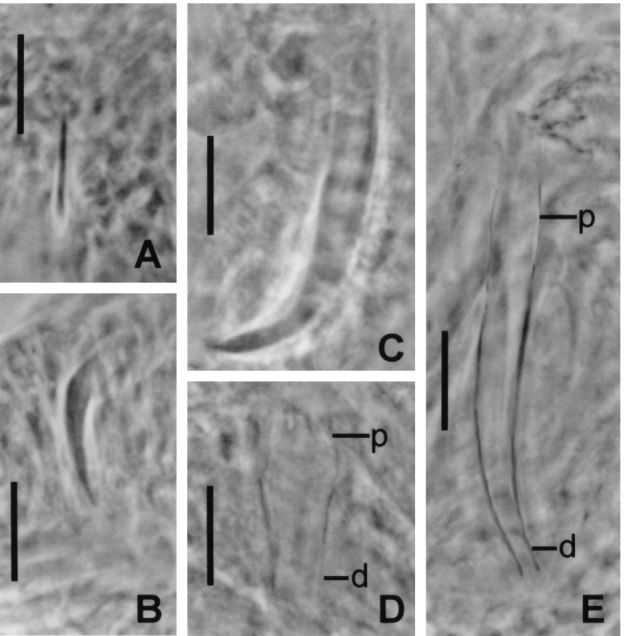
Fig. 5. Variation in development of Merizocotyle icopae recovered from nasal tissue during experimental infections. (A–C) Hamulus of different specimens 17 days p.i. (D and E) Male copulatory organ of different specimens 22 days p.i with proximal (p) and distal (d) ends indicated. Scale bars=20μm. Photos taken using phase-contrast microscopy.
Table 3. Presence of morphological structures in Merizocotyle icopae examined from infection experiments (An entry for presence does not indicate that the structure is fully formed.)

No larvae were found attached to skin surfaces 1 h or 24 h p.i.; they were randomly distributed near the free edge of the nasal lamellae with their haptor between the secondary folds. Development of most structures in M. icopae is considerably slower than in N. rhinobatidis (cf. Tables 3 and 2, respectively). Hamuli were not evident in specimens of M. icopae until at least 10 days p.i. Only 19 of 29 10-day-old worms possessed very narrow hamuli that were presumably newly formed. At 10 days p.i., loculi were observed in only 10 of 29 specimens and no reproductive structures were observed (Table 3). These worms were randomly distributed near the free edge of the nasal lamellae. All worms collected 17 days p.i. had loculi and hamuli (Table 3), but hamulus length and morphology varied considerably (Fig. 5A–C) and none was fully formed. While reproductive structures appeared to be differentiating in worms 17 days p.i., only a single specimen possessed a minute (34 μm long) male copulatory organ (Table 3). No distinct female reproductive structures were evident. The smaller, less developed worms were found near the free edge of the nasal lamellae while the larger parasites were located closer to the peripheral canal between adjacent lamellae. At 22 days p.i., all 26 specimens examined had hamuli, loculi and an ejaculatory bulb. Approximately 70% of the specimens had a male copulatory organ (Table 3) at various stages of development (Figs 4B and 5D, E) and the beginnings of the female reproductive system (i.e. ovary, oötype, vaginae) were seen in approximately 50% (Table 3). No specimens were fully mature. Only very early signs of the vitellarium (scattered single granules) were seen in 4 specimens 22 days p.i.; none laid eggs and no eggs were observed in the oötype of mounted worms (Table 3). These 4 largest, most mature specimens were found near the peripheral canal whereas the smaller worms were distributed nearer to the free edge of the nasal lamellae. Again, we found no character that provided a reliable indicator of parasite age because of the variability of development within a single cohort of parasites (Figs 4B and 5).
Larval behaviour and longevity
Larvae of N. rhinobatidis and M. icopae were photopositive throughout the 24 h period following hatching. Merizocotyle icopae larvae had an average swimming speed of 4 mm/s during this first 24 h period of activity. Freshly hatched larvae of N. rhinobatidis and M. icopae showed no response to currents generated by a pipette or to host skin, gill or nasal tissue or to host skin mucus. Longevity of N. rhinobatidis larvae was determined using 13 larvae. All larvae were alive and active 24 h after hatching. At 33 h after hatching, 11 larvae swam actively but one was dead and the other lay at the bottom of the dish. Beyond 33 h after hatching, the remaining larvae became progressively less active, swam near or settled on the bottom of the dish and began to die. Four were still swimming slowly or resting close to the bottom of the dish 55.5 h after hatching but all were dead after 60 h. No hamuli were found in any of the larvae even 60 h after hatching. None of the larvae had shed their ciliated cells.
DISCUSSION
Most monogeneans are strictly host-specific (Whittington et al. 2000) and initial location of their appropriate fish host is an extraordinary task that a monogenean larva must surmount in a short period of time. Therefore, it is reasonable to assume that when the larva finds its host fish, the first priority is to quickly attach to the body surface. Once on the host, parasites generally migrate to another ‘preferred’ site (Sukhdeo & Sukhdeo, 2002). Indeed, the few studies which have examined the route by which monopisthocotylean monogeneans infect their fish hosts confirm that infection occurs in this way (e.g. Cone & Burt, 1981; Kearn, 1984). Polystomes, internal polyopisthocotylean monogenean parasites of anurans, also first attach to the skin of their host and then undergo a series of complex migrations (Euzet & Combes, 1998). On the other hand, it has been considered that larvae of polyopisthocotyleans that live on fish gills attach to the gills directly with no skin phase. This assumption is supported by experiments on the infection route of Discocotyle sagittata from the gills of trout (see Paling, 1969; Gannicott & Tinsley, 1998) and by the study of Bovet (1967) who watched the large larvae of Diplozoon paradoxum invade and settle on the gills of their teleost host. Recently, however, Chigasaki et al. (2000) found that larvae of the gill dwelling polyopisthocotylean, Heterobothrium okamotoi, settle on both the body surface and the gills and that the body surface was the favoured first point of attachment. It seems, therefore, that the invasion route by larvae cannot be generalized across the Monogenea.
Chisholm & Whittington (2000) demonstrated that the monogeneans N. rhinobatidis from the gills and M. icopae from the nasal tissue of the elasmobranch, R. typus, share a similar hatching strategy, emerging mainly during the first 2 h of light. They postulated that the 2 species might therefore have a similar infection route and that host skin would be the first point of attachment. However, our experiments clearly demonstrate that neither species has a skin phase or, if there is attachment to the skin, it is remarkably short (see below). Larvae of N. rhinobatidis infect the gills directly and the first point of attachment for M. icopae larvae is the nasal tissue. After exposure for only 1 h, larvae of each species were already found on their respective sites. If the first point of attachment was the body surface, it is unlikely that all larvae would have migrated to the gills or nasal tissue in such a short period of time. This assumption is supported further by the fact that we found no post-larval monogeneans on the skin of the rays that we infected in the seeding tank (for the 1 h and 24 h p.i. M. icopae experiments) where large numbers of larvae of both species plus larvae of the other gill monogeneans, N. rhynchobatis and Troglocephalus rhinobatidis, had accumulated. We dissected these rays immediately upon removal from the tank during the first 2 h of light when peak hatching for all these species occurs (Chisholm & Whittington, 2000). If larvae do attach initially to the skin, we would have found them during these experiments.
This is the first time monopisthocotylean monogeneans from the gills and the nasal tissue have been shown to attach to these sites directly. We cannot determine whether the larvae actively seek out their specific site on the host or whether they are drawn into the gill and nasal areas passively with the inhalant currents. Neither the path nor the strength of water flow over the gills or the nasal tissue for R. typus or any other ray species has been documented. Ciliated larvae of N. rhinobatidis and M. icopae showed no response to host skin, gill, nasal tissue or mucus from the body surface in our behaviour experiments and therefore the cue(s) responsible for host recognition is unknown. This is not surprising because as Sukhdeo & Sukhdeo (2002) noted, despite considerable research over the past 50 years, specific signals that attract parasites to a particular site on their host have yet to be isolated. Bovet (1967) found that Diplozoon paradoxum larvae stopped swimming when entering the gill ventilating current and observed the larvae being drawn into the gill chamber of the bream passively. We did not observe such behaviour when we exposed larvae to simulated inhalent water currents created by a Pasteur pipette. If larvae are drawn in passively, they must be able to detect immediately if they are in the appropriate habitat, because on no occasion did we find larval N. rhinobatidis on nasal tissue or larval M. icopae on gill tissue.
Chisholm (1998) mapped the distribution of the dorsal sensory sensilla for a number of monocotylid larvae including N. rhinobatidis and M. icopae using silver staining techniques. We have demonstrated using scanning and transmission electron microscopy of M. icopae larvae that these sensilla are uniciliate and that most are lost approximately 24 h after infection (Cribb et al. 2003). Loss of dorsal sensilla upon attachment to the nasal rosettes of R. typus suggests that the sensilla may provide sensory feedback during swimming or may be chemo- or mechanoreceptors responsible for host location and/or for microhabitat recognition. However, because M. icopae larvae did not appear to respond to currents or to a diversity of host tissues, the cue(s) responsible for host recognition remain a mystery.
At 1 h p.i., N. rhinobatidis were found without their ciliated cells, attached to the free edge of the gill filaments. As the parasites mature (i.e. 10 and 15 days p.i.), they migrate towards the septal canal and proximally towards the gill arch. The 2 mature 20-day-old specimens were located close to the septal canal near the gill arch. Kearn (1978) and Chisholm & Whittington (1998) have documented the differences in microhabitat selection on the gills by different stages of N. rhinobatidis. Two other species, N. rhynchobatis and T. rhinobatidis, also inhabit the gills of R. typus and studies are currently underway by us to determine whether the migration of N. rhinobatidis is influenced by the presence of other species. We found a similar scenario of movements for M. icopae where larvae attached initially to the free edge of the nasal lamellae, but as the parasites matured they moved towards the peripheral canals between the nasal lamellae. The factors responsible for this migration are unclear. No other monogenean species has been reported from nasal tissue of R. typus. The distribution of M. icopae may be related to water currents over nasal tissue, but water flow has not been documented in R. typus.
We have demonstrated that once established on the host, development of N. rhinobatidis and M. icopae is significantly slower and extremely variable compared to what has been determined for other monogenean species at similar temperatures. Neobenedenia girellae (Capsalidae) from the skin of the amberjack, Seriola dumerili, took 10–11 days to reach sexual maturity at 25 °C (Bondad-Reantaso et al. 1995). The capsalid, Benedenia lutjani, reached sexual maturity between 12 and 14 days p.i. at 24 °C and between 8 and 10 days p.i. at 27 °C (Whittington & Ernst, 2002). Furthermore, the length of the anterior hamulus of B. lutjani is an excellent index of parasite age (Whittington & Ernst, 2002) as is the case for other capsalids (Ogawa, 1984 for B. hoshinai; Kearn, 1990 for E. soleae). Our work with the monogeneans of R. typus was done at the same location and at similar temperatures (24–27·5 °C) to those of Whittington & Ernst (2002) on B. lutjani. Only 2 specimens of N. rhinobatidis reached sexual maturity after 20 days p.i. and after 22 days, no M. icopae were mature. Therefore, time to maturity must be significantly longer for these monocotylid species than for the capsalid species. Variability in development between individuals of N. rhinobatidis within a single infection cohort was also remarkable. At 6 h p.i., there is no discernible change in the size or development of the post-oncomiracidia but, somewhere between 6 and 24 h p.i., the hamuli form rapidly and considerable variation in the size and morphology of hamuli is already evident. This variability is magnified later and a few 15-day-old individuals still did not have dorsal haptoral accessory spines or a male copulatory organ. At 20 days p.i., all worms had a male copulatory organ but the length varied considerably. As a result of this variability, we could find no character that could be used as an index of parasite age. Like N. rhinobatidis, development of M. icopae was also extremely variable within a single infection cohort and no predictor of parasite age could be found.
Jackson & Tinsley (2001) demonstrated that a primary infection with the polystomatid Protopolystoma xenopodis in Xenopus laevis resulted in prolonged protective immunity against re-infection. It is possible that the slow and variable parasite development observed here is a result of host ray immune responses because all rays used in our experimental infections had previous exposure to monogeneans in the wild. Little is known about immunity in elasmobranchs and there is no information about whether an immune response may retard parasite development on rays. However, Whittington & Ernst (2002) used non-naïve fish in their experimental infections of the teleost Lutjanus carponotatus with B. lutjani and, as discussed above, development within the B. lutjani cohorts was markedly uniform.
There were also some significant developmental differences between the 2 monocotylid species. While developing loculi were evident in most N. rhinobatidis 24 h p.i., loculi were still absent in about 50% of the M. icopae specimens 10 days p.i. Hamuli were present in only 19 of 29 M. icopae 10 days p.i. unlike N. rhinobatidis which all possessed hamuli 24 h p.i. By 20 days p.i., all N. rhinobatidis possessed a male copulatory organ whereas at 22 days, only 70% of M. icopae had a male copulatory organ. Slower water flow over the nasal rosettes, relative to the gills, might explain the delayed formation (in comparison to N. rhinobatidis at the same temperature) of haptoral structures such as hamuli and loculi in M. icopae because the need for powerful attachment to host nasal tissue would be reduced. But reasons for the striking delays in the development of the reproductive structures remain unclear.
We do not know how monogenean larvae locate R. typus initally, but what we do know about their biology suggests that chances of encounters with their ray host are low. First, N. rhinobatidis and M. icopae produce far fewer eggs in vitro than B. lutjani (unpublished data); whether this is the case in vivo is unknown. We determined that some N. rhinobatidis larvae can live up to 55·5 h, but they are most active during the first 24 h after hatching; the larvae must locate and attach to a shovelnose ray in this brief period. At Heron Island, juvenile R. typus come in to feed on the rising tide and congregate in Shark Bay near the waters edge. They are often buried in the sand and this would be a logical time for infection to occur since a non-moving or slow-swimming ray would be an easier target for a larva with an average swimming speed of 4 mm/s. Despite numerous investigations, we have found no monogenean eggs in the water or in sediments collected in these areas. Neoheterocotyle rhinobatidis and M. icopae eggs hatch mainly during the first 2 h of light (Chisholm & Whittington, 2000), but we do not know if this relates to host behaviour. It is also possible that rhythmical hatching is instead a response to selective pressures related to predator avoidance (e.g. Shostak & Esch, 1990).
The period of development for monogeneans from the gills and nasal tissue and, moreover, for monogeneans from elasmobranchs in general, has not been documented previously. We cannot, therefore, comment whether the variability observed here is characteristic of all monogeneans from the gills and nasal tissues of fish or specifically of monogeneans from elasmobranchs. Variable development may reflect a fundamental strategy. From an evolutionary standpoint, the adaptive value of variable development is considerable in situations where the chances of free-swimming larvae encountering their hosts are low. If parasites from a single infection event develop at differing rates, this essentially lengthens the overall duration of infection. This subsequently increases the amount of time eggs are deposited into the environment and may enhance chances for re-infection.
We thank the Director and staff of Heron Island Research Station for their kind assistance and access to facilities. Special thanks go to Kathy Townsend who, on occasion, collected rays prior to our arrival on the island and to Collette Bagnato who cared for rays during our absences. Ingo Ernst processed some of the parasite material and offered some advice on the intricacies of infection experiments. Two anonymous reviewers made useful comments on the manuscript. This study was funded by Australian Research Council grant nos A19801424 for 1998–2000 and A00104437 for 2001–2003.
References
REFERENCES
BONDAD-REANTASO, M. G.,
OGAWA, K.,
FUKUDOME, M. &
WAKABAYASHI, H.
(
1995).
Reproduction and growth of Neobenedenia girellae (Monogenea: Capsalidae), skin parasite of cultured marine fishes of Japan.
Fish Pathology 30, 227–231.CrossRefGoogle Scholar
BOVET, J.
(
1967).
Contribution à la morphologie et à la biologie de Diplozoon paradoxum v. Nordmann, 1832.
Bulletin Société Neuchâteloise des Sciences Naturelles 90, 63–159.Google Scholar
CHIGASAKI, M.,
NAKANE, M.,
OGAWA, K. &
WAKABAYSAHI, H.
(
2000).
Standardized method for experimental infection of tiger puffer Takifugu rubripes with oncomiracidia of Heterobothrium okamotoi (Monogenea: Diclidophoridae) with some data on the oncomiracidial biology.
Fish Pathology 36, 215–221.CrossRefGoogle Scholar
CHISHOLM, L. A.
(
1998).
Ciliated cells and chaetotaxy of the larvae of seven species of monocotylid monogeneans (Platyhelminthes) from Heron Island, Great Barrier Reef, Australia.
Parasitology Research 84, 828–834.CrossRefGoogle Scholar
CHISHOLM, L. A. &
WHITTINGTON, I. D.
(
1996).
Descriptions of the larvae of six species of monocotylid monogeneans from Himantura fai (Dasyatididae) and Rhinobatos typus (Rhinobatidae) from Heron Island, Great Barrier Reef, Australia.
Systematic Parasitology 35, 145–156.CrossRefGoogle Scholar
CHISHOLM, L. A. &
WHITTINGTON, I. D.
(
1997).
A revision of Neoheterocotyle (Monogenea: Monocotylidae) with descriptions of the larvae of N. rhinobatis and N. rhynchobatis from Heron Island, Great Barrier Reef, Australia.
International Journal for Parasitology 27, 1041–1060.Google Scholar
CHISHOLM, L. A. &
WHITTINGTON, I. D.
(
1998).
Morphology and development of the haptors among the Monocotylidae (Monogenea).
Hydrobiologia 383, 251–261.CrossRefGoogle Scholar
CHISHOLM, L. A. &
WHITTINGTON, I. D.
(
1999).
A revision of the Merizocotylinae Johnston and Tiegs, 1922 (Monogenea: Monocotylidae) with descriptions of new species of Empruthotrema Johnston and Tiegs, 1922 and Merizocotyle Cerfontaine, 1894.
Journal of Natural History 33, 1–33.Google Scholar
CHISHOLM, L. A. &
WHITTINGTON, I. D.
(
2000).
Egg hatching in three species of monocotylid monogenean parasites from the shovelnose ray Rhinobatos typus at Heron Island, Australia.
Parasitology 121, 303–313.CrossRefGoogle Scholar
CHISHOLM, L. A. &
WHITTINGTON, I. D.
(
2002).
Efficacy of Praziquantel bath treatments for monogenean infections of the Rhinobatos typus.
Journal of Aquatic Animal Health 14, 230–234.2.0.CO;2>CrossRefGoogle Scholar
CONE, D. K. &
BURT, M. D. B.
(
1981).
The invasion route of the gill parasite Urocleidus adspectus Mueller, 1936 (Monogenea: Ancyrocephalinae).
Canadian Journal of Zoology 59, 2166–2171.CrossRefGoogle Scholar
CRIBB, B. W.,
CHISHOLM, L. A.,
GOULD, R. &
WHITTINGTON, I. D.
(
2003).
Morphology, ultrastructure and implied function of ciliated sensory structures on the developmental stages of Merizocotyle icopae (Monogenea: Monocotylidae).
Microscopy Research and Technique 62, 267–276.CrossRefGoogle Scholar
ERNST, I. &
WHITTINGTON, I. D.
(
1996).
Hatching rhythms in the capsalid monogeneans Benedenia lutjani from the skin and B. rohdei from the gills of Lutjanus carponotatus at Heron Island, Queensland, Australia.
International Journal for Parasitology 26, 1191–1204.Google Scholar
EUZET, L. &
COMBES, C.
(
1998).
Selection of habitats among the Monogenea.
International Journal for Parasitology 28, 1645–1652.CrossRefGoogle Scholar
GANNICOTT, A. M. &
TINSLEY, R. C.
(
1998).
Larval survival characteristics and behaviour of the gill monogenean Discocotyle sagittata. Parasitology 117, 491–498.CrossRefGoogle Scholar
JACKSON, J. A. &
TINSLEY, R. C.
(
2001).
Protopolystoma xenopodis (Monogenea) primary and secondary infections in Xenopus laevis. Parasitology 123, 455–463.CrossRefGoogle Scholar
KEARN, G. C.
(
1978).
Early development and microhabitat of the monogenean Horricauda rhinobatidis, with observations on Troglocephalus rhinobatidis, from Rhinobatos batillum from Queensland, Australia.
International Journal for Parasitology 8, 305–311.CrossRefGoogle Scholar
KEARN, G. C.
(
1984).
The migration of the monogenean Entobdella soleae on the surface of its host, Solea solea. International Journal for Parasitology 14, 63–69.CrossRefGoogle Scholar
KEARN, G. C.
(
1987).
The site of development of the monogenean Calicotyle kroyeri, a parasite of rays.
Journal of the Marine Biological Association of the United Kingdom 67, 77–87.CrossRefGoogle Scholar
KEARN, G. C.
(
1990).
The rate of development and longevity of the monogenean skin parasite Entobdella soleae. Journal of Helminthology 64, 340–342.CrossRefGoogle Scholar
LAST, P. R. &
STEVENS, J. D.
(
1994).
Sharks and Rays of Australia.
CSIRO Division of Fisheries,
Australia.
OGAWA, K.
(
1984).
Development of Benedenia hoshinai (Monogenea) with some notes on its occurrence on the host.
Bulletin of the Japanese Society of Scientific Fisheries 50, 2005–2011.CrossRefGoogle Scholar
PALING, J. E.
(
1969).
The manner of infection of trout gills by the monogenean parasite Discocotyle sagittata.
Journal of Zoology 159, 293–309.CrossRefGoogle Scholar
ROFF, J. C. &
HOPCROFT, R. R.
(
1986).
High precision microcomputer based measuring system for ecological research.
Canadian Journal of Fisheries and Aquatic Sciences 43, 2044–2048.CrossRefGoogle Scholar
ROHDE, K.
(
1978).
Latitudinal differences in host-specificity of marine Monogenea and Digenea.
Marine Biology 47, 125–134.CrossRefGoogle Scholar
SHOSTAK, A. W. &
ESCH, G. W.
(
1990).
Photocycle dependent emergence by cercariae of Halipegus occidualis from Helisoma anceps, with special reference to cercarial emergence patterns as adaptations for transmission.
Journal of Parasitology 76, 790–795.CrossRefGoogle Scholar
SUKHDEO, M. V. K. &
SUKHDEO, S. C.
(
2002).
Fixed behaviours and migration in parasitic flatworms.
International Journal for Parasitology 32, 329–342.CrossRefGoogle Scholar
TINSLEY, R. C. &
EARLE, C. M.
(
1983).
Invasion of vertebrate lungs by the polystomatid monogeneans Pseudodiplorchis americanus and Neodiplorchis scaphiopodis.
Parasitology 86, 501–517.CrossRefGoogle Scholar
TINSLEY, R. C. &
JACKSON, H. C.
(
1986).
Intestinal migration in the life-cycle of Pseudodiplorchis americanus (Monogenea).
Parasitology 93, 451–469.CrossRefGoogle Scholar
WHITTINGTON, I. D.,
CHISHOLM, L. A. &
ROHDE, K.
(
2000).
The larvae of Monogenea (Platyhelminthes).
Advances in Parasitology 44, 139–232.Google Scholar
WHITTINGTON, I. D. &
ERNST, I.
(
2002).
Migration, site-specificity and development of Benedenia lutjani (Monogenea: Capsalidae) on the surface of its host, Lutjanus carponotatus (Pisces: Lutjanidae).
Parasitology 124, 423–434.CrossRefGoogle Scholar
ZEISKE, E.,
THEISEN, B. &
GRUBER, S. H.
(
1987).
Functional morphology of the olfactory organ of two carcharhinid shark species.
Canadian Journal of Zoology 65, 2406–2412.CrossRefGoogle Scholar

Fig. 1. Body sections of Rhinobatos typus examined immediately under a dissecting microscope for newly-settled Neoheterocotyle rhinobatidis or Merizocotyle icopae larvae after 1, 6 and 24 h infection experiments.

Table 1. Infection experiments with Neoheterocotyle rhinobatidis and Merizocotyle icopae detailing the number of parasites recovered

Table 2. Presence of morphological structures in Neoheterocotyle rhinobatidis examined from infection experiments

Fig. 2. General distribution of Neoheterocotyle rhinobatidis on Rhinobatos typus gills showing gill filament (gf), secondary gill lamella (sgl) and septal canal (sc). Larvae of N. rhinobatidis first attach to the free edge of a gill filament (1) and then begin to migrate (2) towards the septal canal and the gill arch (ga) as they grow and mature. A – 24 h p.i. and small 15-day-old worms. B – 10-day and some 15- and 20-day-old worms. C – the largest 20-day-old worm with egg. Note that distributions are generalized and actual locations of specific worms varied slightly from that shown.
Fig. 3. Variation in development of Neoheterocotyle rhinobatidis recovered from gills during experimental infections. (A–C) Hamulus of different specimens 24 h p.i. (D–G) Male copulatory organ of different specimens 20 days p.i. Note presence of accessory piece (ap) which runs from the proximal (p) to distal (d) end of the largest male copulatory organ. Scale bars=20 μm. Photos taken using phase-contrast microscopy.
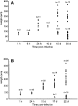
Fig. 4. Total length ([bull ]) and male copulatory organ length ([utrif ]) of worms examined from infection experiments. (A) Neoheterocotyle rhinobatidis. (B) Merizocotyle icopae. Those specimens/structures that could not be measured accurately are not included.

Fig. 5. Variation in development of Merizocotyle icopae recovered from nasal tissue during experimental infections. (A–C) Hamulus of different specimens 17 days p.i. (D and E) Male copulatory organ of different specimens 22 days p.i with proximal (p) and distal (d) ends indicated. Scale bars=20μm. Photos taken using phase-contrast microscopy.
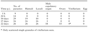
Table 3. Presence of morphological structures in Merizocotyle icopae examined from infection experiments
 You have
Access
- 6
- Cited by