Introduction
Buffalo is an important livestock resource, which occupies a critical niche in many ecologically disadvantaged agricultural systems, providing milk, meat and work power (Gasparrini, Reference Gasparrini2002), it remain as the backbone of the rural economy. Problems in buffalo reproduction include delayed puberty, silent estrous, a long post-partum period before return to estrous and low conception rates. Reduced fertility is due to lower and less reliable response to superovulation (Drost et al., Reference Drost, Wright and and Elsden1986; Karaivanov, Reference Karaivanov1986).
To overcome this inherent difficulty in buffalo reproduction, in vitro embryo production (IVEP) is gaining importance. In the last three decades there has been increasing interest shown on IVEP leading to better understanding of successful in vitro production of embryos. There has been increasing evidence on impairment of embryo development in several species during in vitro embryo culture by reactive oxygen species (ROS)-mediated cytotoxicity. Oxidative stress occurs to cells in vivo and in vitro from exposure to free radicals generated by exogenous agents such as chemicals, hyperoxia and endogenous process such as normal cellular metabolism. Under extreme oxidative conditions or if the antioxidant protective mechanisms of cells are compromised, cellular injury and cell death may occur (Chow, Reference Chow1979; Halliwell et al., Reference Halliwell, Gutteridge and Cross1992). Early mammalian embryos are susceptible to damage from ROS (Johnson and Nasr-Esfahani, Reference Johnson and Nasr-Esfahani1994) and they increase the production of oxygen-free radicals when cultured in vitro (Goto et al., Reference Goto, Noda, Mori and Nakano1993). Since little information is available on in vitro maturation (IVM) and in vitro fertilization (IVF) of buffalo oocytes (Totey et al., Reference Totey, Singh, Taneja, Pawshe and Talwar1992), understanding the embryo physiology, metabolism and culture requirements is critical to overcome the difficulties faced in in vitro production of buffalo embryos.
Retinoids are regulators of cellular growth, embryonic morphogenesis and differentiation in many types of cells and they are essential for ovarian steroid production, oocyte maturation and early embryogenesis. Deficiencies in vitamin A leads to decreased ovarian size, decreased ovarian steroid concentration and eventually lead to reproductive senescence. Several studies have indicated a positive effect of retinol supplementation when diets are adequate in vitamin A. Retinoic acid is the most important retinoid for vertebrate embryogenesis (Morris-Kay and Ward, Reference Morris-Kay and Ward1999). Administration of retinol with super ovulation increased the number of transferable embryos but did not affect ovulation rate (Shaw et al., Reference Shaw, Farin, Washburn and Britt1995). Retinol treatment in combination with super ovulation improves the developmental competence of resultant embryos (Eberhardt et al., Reference Eberhardt, Will and Godkin1999). Earlier studies have reported the presence of retinol in follicular fluid (Schweigert and Zucker, Reference Schweigert and Zucker1988; Brown et al., Reference Brown, Eberhardt, Schrick, Roberts and Godkin2003), stating that vitamin-A concentrations to be highest in large nonatretic follicles and lowest in small atretic follicles correlated with estradiol concentrations. Retinol improves embryonic development of bovine oocytes in vitro, indicated by increased blastocyst rate (Livingston et al., Reference Livingston, Eberhardt, Edwards and Godkin2004). Retinoids may act through their modulatory effects on the gene expression of gonadotropins receptors in cumulus cells (Minegishi et al., Reference Minegishi, Hirakawa, Kishi, Abe, Tano, Abe and Miyamoto2000). Biologically active retinoids mediate their effects on target cells through binding of two sets of nuclear receptors, namely retinoic acid receptors (RARs) and retinoid X receptors (RXRs). Ligand-bound RARs and RXRs function as transcription factors by binding to cis-acting DNA sequences called retinoic acid response elements (RAREs), located within the transcriptional regulation of genes (Xiao et al., Reference Xiao, Durand, Chambon and Voorhees1995). More over the role of retinoids in assisted reproductive technology has been demonstrated through their effect on cytoplasmic maturation and antioxidant property (Cetica et al., Reference Cetica, Pintos, Dalvit and Beconi2001; Ikeda et al., Reference Ikeda, Kitagawa, Imai and Yamada2005). Hence, an attempt has been made to assess the role of all-trans retinol in in vitro production of buffalo embryos through its supplementation in maturation and embryo culture medium.
Material and methods
All chemicals used in the experiment were purchased from Sigma chemicals, unless otherwise indicated.
Oocyte collection
Buffalo ovaries were obtained from a local abattoir (Chennai Corporation abattoir, Chennai, Tamilnadu) and transported to the laboratory in 0.9% saline supplemented with 50 μg/ml gentamycin, within 2 h of slaughter. The ovaries were washed repeatedly in normal saline, cumulus oocyte complex (COC) were isolated from the follicles by slicing method (Pawshe et al., Reference Pawshe, Totey and Jain1994).
The isolated oocytes were washed three times in Tyrode’s lactate–N-[2-hydroxyethyl] piperazine-N’-[2-ethanesulphonic acid] (TL–HEPES) medium and classified into three categories (good, fair and poor) according to the character of the cumulus cells. Oocytes with more than four layers of compact cumulus cells and uniform cytoplasm were classified as good quality oocytes, oocytes with less than four layers or with a partial cumulus mass were classified as fair quality oocytes and oocytes without cumulus cells with a uniform cytoplasm were classified as poor quality oocytes.
In vitro maturation
Oocytes that were completely surrounded by at least four to five layers of unexpanded cumulus cells and had a homogenous ooplasm alone were used for maturation. The collected oocytes were washed three times in TL–HEPES medium. The last two washings were in the maturation medium composed of TCM 199 supplemented with 10% fetal calf serum (FCS)a, 0.5 μg/ml FSH, 5.0 μg/ml LH and 1 μg/ml Estradiol (E2). Twenty oocytes were placed in 100 μl droplets of the maturation medium covered with sterile paraffin oil and incubated at 39°C under 5% CO2 in air and 95% relative humidity for 24 h.
In vitro fertilization
Frozen straws (0.5 ml) of buffalo semen were procured from Tamilnadu Veterinary and Animal Sciences University, Chennai. Two 0.5 ml straws thawed for 60 s at 35°C were emptied into 15 ml air-tight tube and washed with Brackett and Oliphant (BO) medium containing heparin 10 μg/ml (Brackett and Oliphant, Reference Brackett and Oliphant1975) without BSA by centrifugation at 700 × g for 5 min. The sperm pellet was mixed with 2 ml of BSA-free BO medium containing 10 mM of caffeine (sodium caffeine benzoate) and 10 μg/ml heparin in air tight tube and placed into the incubator at 38.5°C for swim-up for 1 h, after which the top 1.8 ml of the semen suspension containing motile spermatozoa were removed through micropipettes and washed twice with BSA-free BO medium. The sperm pellets were resuspended in 1 ml of BSA-free BO medium supplemented with caffeine and heparin in the same concentrations. This was further diluted with 1 ml of BO medium containing 20 mg/ml BSA supplemented with 10 μg/ml heparin. Matured buffalo oocytes were washed in BO medium. The oocytes were then distributed to 10 per 50 μl drop of fertilization medium under paraffin oil. Two microliters of swim-up spermatozoa was then added to 50 μl fertilization drops to give a final sperm concentration of 2 × 10⁶. The oocyte and spermatozoa were incubated at 39°C under 5% CO2 in air for 24 h.
In vitro embryo culture
After incubation with spermatozoa, the ova were removed from the fertilization drops. The cumulus cells were stripped off by repeated pipetting through a small-bore pipette and cultured with modified synthetic oviductal fluid (Tervit et al., Reference Tervit, Whittingham and Rowson1972), containing 3 mg/ml of BSA, 0.6 mM sodium pyruvate, 2% (v/v) BME essential amino acids and 1% (v/v) MEM non-essential amino acids, 100 μg/ml penicillin and streptomycin and cultured for 8 days at 39°C under 5% CO2, 5% O2 and 90% N2 (or) 5% CO2 in air, which is approximately 20% O2.
Cell number analysis
Embryos were evaluated for the appearance and morphological quality by determining the cell numbers. Expanded day 8 blastocysts from each treatment groups were fixed and stained as per the method described (Mermillod et al., Reference Mermillod, Vansteenbrugge, Wils, Mourmeaux, Massip and Dessy1993). These expanded blastocysts were then transferred individually onto glass microscopic slides and after drying at room temperature, fixed with 70% ethanol for 24 h. The fixed blastocysts were stained with 10 μg/ml bisbenzamide (Hoechst 33342) and 2.3% sodium citrate. The slides were observed under an epifluorescence microscope, fitted with excitation filter (330 to 380 nm) and barrier filter (420 nm). The total numbers of nuclei were counted in each blastocyst.
Comet assay for detecting DNA damage in individual embryos
DNA damage in individual embryos developed under high oxygen concentration was assessed by comet assay (Takahashi et al., Reference Takahashi, Keicho, Takahashi, Ogawa, Schultz and Okano2000). After the 3-day of culture, embryos cultured under 20% O2 with/without retinol supplement were removed and used for analysis of DNA damage by comet assay. Ten to 20 embryos were washed twice in PBS plus polyvinylpyrrolidone (4 mg/ml). Embryos in each experimental group were then transferred to a 200 μl drop of 1% low-melting temperature agarose (Genei, Bangalore, Karnataka, India) in PBS at 39°C; the agarose drop was placed on a 35-mm plastic Petri dish. Using a stereo dissecting microscope to visualize the embryos, the embryos were gently mixed with the 1% low-melting temperature agarose and then captured in a total volume of about 10 μl with a mouth-operated glass pipette. The embryos were then quickly placed on a microscope slide glass, which was initially coated with 1% high-melting temperature agarose (Genei). The slides were then placed on ice for 5 min to solidify the agarose. The embryos were then lysed by incubating the slides for 3 h at ambient temperature in lysing buffer, which was composed of 10 mM Tris, pH 10, containing 1% sodium sarcosinate, 2.5 mM NaCl, 100 mM Na2-EDTA, 1% Triton X-100, and 10 μg/ml proteinase K. The slides were then removed from the lysing solution and placed on a horizontal gel electrophoresis unit. The unit was filled with fresh electrophoresis buffer (1 mM Na2-EDTA, 300 mM NaOH) to a level 0.25 cm above the slides, and the slides were equilibrated in the electrophoresis buffer for 20 min. Electrophoresis was then conducted for 20 min at 25 V. The slides were then neutralized by immersing in 0.4 M Tris–HCl (pH 7.5) for 5 min at ambient temperature. DNA was stained by adding a 20 μl drop of acridine orange (5 μg/ml) to the slide for 2 min followed by 1 min of washing in distilled water. Observation of DNA was carried out under fluorescence microscope. DNA damage was quantified by measuring the length of the streak of DNA comet tail between the edge of the zona pellucida and the end of the visible comet tail. The length was calculated by referring to a photograph of a micrometer at the same magnification as the embryos.
Experimental design
All-trans retinol was dissolved in 100% ethanol; appropriate dilutions were made and stored at −80°C until use. The concentration of ethanol during maturation or culture was less than 0.05%. In control groups, 0.05% ethanol was added in maturation/culture medium according to the experimental design.
In experiment I, varying concentration of all-trans retinol (0, 1, 2, 5 and 10 μM) were added to the maturation medium and embryos were allowed to develop under 5% O2. In experiment II, varying concentrations of all-trans retinol (0, 1, 2, 5 and 10 μM) were added to embryo culture medium throughout the culture period and embryos were allowed to develop under 5% O2. In experiment III, various concentration of all-trans retinol was supplemented to the embryo culture medium and the embryos were allowed to develop at 20% O2. In experiment IV, all-trans retinol was added to the embryo culture medium for first 72 h of embryo development of 8-day culture under 5% O2. The embryo culture medium was changed after 72 h, to avoid toxic accumulation of ammonia with fresh culture medium and all-trans retinol.
Statistical analysis
In each experimental group, oocytes were randomly distributed. The percentages of oocytes fertilized and embryos developed to morula and blastocyst were subjected to arcsine transformation before analysis. The data for total cell count were directly subjected to analysis. All data were subjected to one-way ANOVA followed by Tukey test to determine differences among experimental groups using Statistical Package for Social Sciences (SPSS 11.0, Chicago, USA). Differences P < 0.05 were considered statistically significant.
Results
Experiment I
Data from Experiment I are shown in Table 1. The addition of 5 μM all-trans retinol resulted in a significant increase (P < 0.05) in the percentage of cleaved embryos compared to control and the embryos that subsequently developed to compact morulae increased significantly (P < 0.01) in 5 μM supplemented group when compared with control. Addition of 5 μM all-trans retinol improved (P < 0.001) blastocyst formation as compared with control and differ significantly with treatments, (P < 0.01) when compared with 1 μM supplemented group and (P < 0.05) when compared with 2 and 10 μM supplemented groups. A significant increase in total cell number was observed in 5 μM supplemented group when compared with control and 1 μM supplemented group.
Effect of presence of all-trans retinol in in vitro maturation medium on development of preimplantation buffalo embryos under low oxygen

Means in the same column with different superscripts were significantly different (P < 0.05).
Values are listed as mean ± s.e.m.
Experiment II
The effects of all-trans retinol in embryo culture medium (ECM), under low oxygen concentration are shown in Table 2. Cleavage rate did not differ significantly among treated groups when compared with control. The embryos that developed to compact morulae showed a significant reduction in the development rate of 10 μM supplemented groups (P < 0.05) when compared to 1 μM and (P < 0.01) when compared with 2 μM supplemented group. The proportion of embryos that developed to blastocyst showed a significant reduction in blastocyst rate in 10 μM supplemented group (P < 0.05) when compared with control and 1 μM supplemented group. The total cell number decreased significantly in 10 μM supplemented group (P < 0.05), when compared with control and 1 μM group.
Effect of presence of all-trans retinol in embryo culture medium on development of preimplantation buffalo embryos under low oxygen

Means in the same column with different superscripts were significantly different (P < 0.05).
Values are listed as mean ± s.e.m.
Experiment III
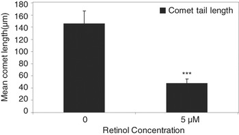
The effects of all-trans retinol in ECM under high oxygen concentration are shown in Table 3. The cleavage rate showed no significant changes, while significant change was observed in proportions of embryos that developed to morulae in 5 μM supplemented group (P < 0.01) when compared with control and (P < 0.05) when compared with 1 μM supplemented group. Supplementation of 5 μM all-trans retinol favored blastocyst formation (P < 0.001) when compared with control and treatment groups. Addition of 10 μM, tended to improve blastocyst formation (P < 0.01) when compared with control and (P < 0.05) when compared with 1 μM. The total cell numbers per blastocyst differ significantly in 5 μM supplemented group when compared with control and treatment groups. The extent of DNA damage in individual embryos on day 3 of culture under 20% O2 with or without all-trans retinol supplement is illustrated in Figure 1. Supplementation of 5 μM all-trans retinol significantly reduced the comet tail (P < 0.001) when compared with control.
Effect of all-trans retinol in embryo culture medium on development of preimplantation buffalo embryos under high oxygen concentration

Means in the same column with different superscripts were significantly different (P < 0.05).
Values are listed as mean ± s.e.m.
Effect of all-trans retinol in ECM on DNA damage in individual buffalo embryos under 20% O2. Values are expressed as mean ± standard error. ***Significant difference (P < 0.001) compared to 0 μM retinol supplemented group.

Experiment IV
Data from Experiment IV are shown in Table 4. No significant difference was observed in the rate of cleavage and development to morulae. The proportion of embryos that developed to blastocyst stage, increased significantly in 5 μM supplemented group (P < 0.001) when compared with control, 1, 2 μM and (P < 0.05) when compared with 10 μM supplemented group. Total cell number increased significantly in 5 μM supplemented group, compared to control and treatment groups.
Effect of all-trans retinol in embryo culture medium for first 72 h of culture period of embryo development of preimplantation buffalo embryos

Means in the same column with different superscripts were significantly different (P < 0.05).
Values are listed as mean ± s.e.m.
Discussion
The present study evaluated the effect of all-trans retinol in oocyte maturation and embryo culture medium on development of buffalo embryos in vitro obtained with oocytes originating from abattoir ovaries. Embryo production in buffalo is known to suffer from several limitations; still any procedures in IVEP can be practiced in buffalo but with little difficulty than other species. IVEP technology has been suggested as the best tool to accelerate genetic progress through the maternal lineage in buffalo because of the limitations now proven of multiple ovulation embryo transfer programs in this species.
The results of the study indicate that the supplementation of all-trans retinol in maturation medium at 5 μM concentration favored the blastocyst formation rate and total cell count in the blastocyst. Lower concentrations of retinol had no significant effect on blastocyst development and the higher dose had a negative effect on development. In contrast, the presence of all-trans retinol throughout the culture period under low oxygen was not beneficial to development in terms of frequency of blastocyst formation and cell count. These results are in agreement with those obtained in bovine oocytes, where retinol during IVM increases embryonic development to the blastocyst stage (Livingston et al., Reference Livingston, Eberhardt, Edwards and Godkin2004; Lima et al., Reference Lima, Oliveira, Santos, Reichenbach, Weppert, Paula-Lopes, Neto and Goncalves2006). The exact mechanism by which retinoids contribute to the developmental competence of embryo is not clearly understood. However, evidences suggest that the preparation of functional female gametes is essential for the successful IVEP. Since mammalian oocytes develop in the follicular environment surrounded by cumulus cells, these cells may exert their effect by helping oocyte to acquire developmental competence. Cumulus cells play a major role in maturation, since cumulus oocyte gap junction is one of the routes by which cumulus cells transmit metabolites (Gilula et al., Reference Gilula, Epstein and Beers1978; Coskun and Lin, Reference Coskun and Lin1994). Earlier studies suggested that both retinoic acid and retinol could act via cumulus cells, directly on the oocyte or in both ways in an autocrine or paracrine manner (Hidalgo et al., Reference Hidalgo, Diez, Duque, Facal and Gomez2003). Endogenously active retinoid receptors are present in COC, which may be competent to synthesize retinoic acid using the precursor retinol (Mohan et al., Reference Mohan, Thirumalapura and Malayer2003). These retinoid receptors being ligand-activated transcription factors are involved in regulating the transcription of several genes, which may be involved in embryo development, suggesting the positive effect of retinol to improve the developmental competence in maturation medium. Earlier studies has described the possible mechanisms by which retinoids affect the developmental capacity of oocytes and early embryos may include modulation of expression of growth factors and other developmental genes, improving mRNA quality, and direct and/or indirect affects on antioxidant defense mechanisms (Livingston et al., Reference Livingston, Eberhardt, Edwards and Godkin2004).
The present results are in disagreement with those obtained in porcine IVF, where it has been reported that treatment of oocytes with all-trans retinol resulted in better embryo cleavage compared with control, this enhancement was not reflected in blastocyst development, whereas the presence of 5 nm 9-cis retinoic acid in the IVM medium significantly increased the blastocyst formation rate, suggesting that 9-cis-retinoic acid may play an important role during IVM (Alminana et al., Reference Alminana, Gil, Cuello, Caballero, Roca, Vazquez, Gomez and Martinez2008). A possible explanation for these discrepancies could be the different IVM conditions used by these authors, where the oocytes are matured for 20–22 h with supplements and another 20–22 h without supplements.
The reduced rate of blastocyst development observed during supplementation in embryo culture medium may be due to high concentration of retinol. Earlier studies have reported that, it remains unclear whether antioxidants benefit embryo development at lower oxygen concentration, and if the protection must be extracellular or intracellular (Ali et al., Reference Ali, Bilodeau and Sirard2003). Since it appears that the balance between ROS production and scavenging is an important factor for the acquisition of fertilizing ability in vitro and antioxidants may have negative and undesirable effect if a safety threshold dosage is surpassed. Embryonic malformations also result from the presence of excessive amount of vitamin A during development.
In the present study the development of embryos to blastocyst in control group was 10–12%, which was in agreement with earlier studies by Gasparrini et al. (Reference Gasparrini, Boccia, Marchandise, Di Palo, George, Donnay and Zicarelli2006), where the blastocyst rate was 11.8%. In contrast with these results the earlier studies by Neglia et al. (Reference Neglia, Gasparrini, Caracciolo di Brienza, Di Palo, Campanile, Antonio Presicce and Zicarelli2003) yielded blastocyst rate of 19.9% in control group. The discrepancies in the blastocyst yield may be attributed to the quality of oocytes obtained from abattoir and culture conditions. In the present study, ethanol has been used to dissolve retinol at a concentration of less than 0.05% in the culture medium; such a low concentration may not cause retinol to concentrate in the oil overlay. The concentrations of retinol in culture droplets have not been quantified in the present study. From our results, it is clear that increasing concentration of retinol either enhances the developmental competence or unfavorable to development, which clearly demonstrates that with increase in retinol concentration the availability of retinol increases in the developing oocytes/embryos.
The supplementation of all-trans retinol in ECM under high oxygen concentration resulted in enhanced blastocyst formation and total cell numbers in 5 μM supplemented group. Earlier studies suggest that, oxygen at atmospheric level is known to exert harmful effects on the development of mammalian embryos, probably due to formation of free oxygen radicals (Johnson and Nasr-Esfahani, Reference Johnson and Nasr-Esfahani1994). The enhanced blastocyst formation under high oxygen concentration in presence of retinol, suggest that retinol might act as an antioxidant. In our study the comet assay revealed, that embryos cultured under 20% oxygen concentration without retinol are subjected to more DNA damage when compared to embryos with retinol supplement. It seems likely that embryos cultured under high oxygen concentration may be exposed to oxidative stress for which their defense mechanisms are insufficient to protect their delicate structures. Retinoids may act as antioxidants by scavenging free radicals and preventing lipid peroxidation (Kartha and Krishnamurthy, Reference Kartha and Krishnamurthy1977). Preimplantation embryos are very sensitive to ROS and that they convert reduced glutathione (GSH) to oxidized glutathione (GSSG) and GSS protein under oxidative stress. In addition, glutathione levels change dramatically during preimplantation development, and this may be related to a role for oxygen radicals or thiol status in differentiation. Culturing embryos in vitro decreased their glutathione content; this may be a partial cause of the retarded development observed in vitro (Gardiner and Reed, Reference Gardiner and Reed1994). Retinoids may protect the oxidative damage by maintaining adequate endogenous levels of antioxidant compounds and enzymes. Retinoic acid inhibited staurosporine-induced GSH depletion in neuronal cells, preventing oxidative damage and apoptosis (Ahlemeyer and Krieglstein, Reference Ahlemeyer and Krieglstein2000). A RARE has been identified in the promoter region of a specific isoform of glutathione-S-transferase-pi (GSTp) in glioblastoma cells (Lo and Ali-Osman, Reference Lo and Ali-Osman1997). Retinoic acid also reduced staurosporine-induced oxidative stress and apoptosis by preventing the decrease in the protein levels of superoxide dismutase-1 (SOD-1) and superoxide dismutase-2 (SOD-2), and thus supported the antioxidant defense system (Ahlemeyer et al., Reference Ahlemeyer, Bauerbach, Plath, Steuber, Heers, Tegtmeier and Krieglstein2001). Hence, the observed increase in blastocyst development by supplementation of all-trans retinol in ECM under high oxygen concentration may be due to its antioxidant property, which is evident from the comet assay.
The results of the study, in which embryos were supplemented with all-trans retinol in ECM for the first 72 h of 8-day culture under 5% O2 revealed; 5 μM all-trans retinol enhanced blastocyst formation and total cell count suggesting that, supplement timing and duration of all-trans retinol play an important role in developmental competence. Earlier studies have reported that the concentration and timing of antioxidants requirements varies with the stage of development (Hossein et al., Reference Hossein, Hashem, Jeong, Lee, Kim, Kim, Koo, Park, Lee, Park, Kang, Lee and Hwang2007). The beneficial effects of all-trans retinol on embryo development for the first 72 h, compared to its presence throughout the culture period, could have helped the embryos to overcome the developmental block.
Earlier studies have reported that, successful development of preimplantation embryos from fertilization to the blastocyst stage has been hampered by developmental arrest at a specific cell stage, i.e. two-cell stage in mice, four- to eight-cell stage in human (Braude et al., Reference Braude, Bolton and Moore1988), eight- to 16-cell stage in sheep and goat (Sakkas et al., Reference Sakkas, Batt and Cameron1989) and four- to eight-cell stage in cow (Camous et al., Reference Camous, Heyman, Meziou and Menezo1984). Although specific mechanism underlying the developmental block in various species has not been established, it is clear from the in vitro cell line studies that ROS such as superoxide anions, hydroxyl radicals and hydrogen peroxide (H2O2) are involved in macromolecular damage and apoptosis. ROS production by early embryos and their susceptibility to ROS varies with the stage of development. Previous reports suggest the possible relationship between the block and the potential rise in damaging free radicals generated from the H2O2 in two- to four-cell stage compared with other stages (Nasr-Esfahani et al., Reference Nasr-Esfahani, Aitken and Johnson1990). Thus all-trans retinol may act as an antioxidant, thereby protecting the embryos from oxidative stress. To confirm the present hypothesis further investigations are needed to elucidate the mechanism by which retinol supplementation improves embryo development in buffalo species.
On the basis of our results, it may be suggested that all-trans retinol supplementation in maturation/culture medium may have the potential to impact on in vitro production of embryos. Deciphering the mechanism by which all-trans retinol contributes to the developmental competence of preimplantation buffalo embryos would be the interest of future work.
Acknowledgements
The technical expertize provided by Dr A. Palanisamy, Department of Animal Biotechnology, Tamilnadu veterinary and Animal sciences university, Chennai, S. Yuvaraj, Department of Medical Biochemistry, Dr A. L. M., PGIBMS, University of Madras, Chennai and N. Rajesh, Huclin Research Ltd, Ticel Biopark, Chennai are greatly acknowledged.