Coarctation of the aorta is a common congenital heart disease that may be diagnosed later in life during evaluation for systemic hypertension or a murmur. Often, coarctation diagnosed later in life have associated vascular pathology.Reference Pu, Chen, Chen and Feng1 Late aneurysm formation in the proximal or distal aortic arch is a well-recognised sequela of untreated coarctation, and it is associated with an increased risk of aortic rupture and death.Reference Hormann, Pavlidis, Brunkwall and Gawenda2 Presence of aneurysm in patients with coarctation of the aorta has management challenges and poses high risk of aortic rupture.Reference Di Tommaso, Mannacio, Di Tommaso, Pinna, Fontana and Iannelli3,Reference Bai, Liu, Jin, Li, Ji and Qu4 We present a case of coarctation of the aorta diagnosed in a teenage girl with the combination of an aortic arch coarctation, a “cauliflower-like” saccular aneurysm, and stenosis at the origin of the left subclavian artery, which was successfully treated with a hybrid approach.
Case
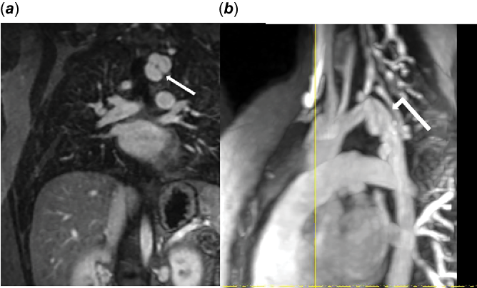
An 11-year old girl was referred to our centre from outside facility for evaluation of hypertension. Her initial blood pressure in the right arm was 118/85 mmHg and leg was 123/67. On echocardiogram, she had a tortuous aortic arch and the Doppler interrogation of the descending aorta revealed a velocity of 2.7 m/s consistent with coarctation. Intracardiac anatomy was normal with a normal left ventricular dimensions and systolic function with no evidence of left ventricular hypertrophy. There was a tricommisural aortic valve, normal mitral valve, and no obvious aneurysm on the echocardiogram. As the aortic arch was tortuous with mild increase in the descending aortic velocity, a cardiac MRI was obtained. Cardiac MRI revealed a left-sided tortuous aortic arch with hypoplasia of the transverse arch and multiple saccular aneurysms of the distal aortic arch and proximal descending thoracic aortic segment (the aneurysms measured between 5 and 11 mm in diameter) (Fig 1). The aortic aneurysms began approximately 1 cm distal to the left common carotid artery origin and extended over a length of 3.7 cm. Within this area, turbulent flow was seen and there was a narrow appearance of the main lumen of the aorta measuring between 11 and 12 mm. At the level of the diaphragm, the aorta measured 13 mm in diameter. The left subclavian artery arose from the distal portion of this abnormal segment. There were no major collaterals arising from the aorta. Left ventricular function was normal. The initial plan was to follow her conservatively.
Over the course of 3 years, there was a significant increase in her systolic BP to 140 mmHg in right arm and there was also a blood pressure gradient from her right arm to her left arm and the left leg of > 20 mmHg and narrowing at the origin of the left subclavian artery. There was no change in her cardiac function or left ventricular mass. A repeat cardiac MRI showed progressive increase in the size of aortic arch aneurysms. The largest dimension of the aneurysm increased steadily from 19 to 27 mm diameter. A significant stenosis was noted just after the take-off of the left common carotid artery before the subclavian artery. Thus, it was decided to take her to the cardiac catheterisation lab for a hybrid intervention.
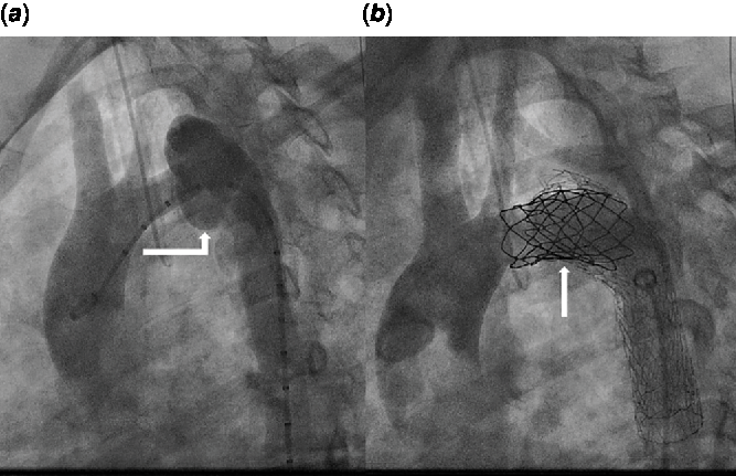
The procedure was performed under general anesthesia and a lumbar drain was put in place for spinal protection. She initially underwent the left carotid artery to subclavian artery bypass graft by the vascular surgeon. Dissection of the subclavian artery was performed medially until 1 cm proximal to the vertebral artery. The carotid sheath was opened with care and the carotid artery was dissected for a length of about 3–4 cm and encircled with vessel loops. A 6-mm Propaten graft was selected and anastomosis was performed between the graft and the left subclavian artery. The graft was then tunneled beneath the internal jugular vein and end-to-side anastomosis was performed between the left common carotid artery and the graft. The flow was established to the subclavian artery via the graft. Cardiac catheterisation was performed using a left femoral artery cut-down and placing an 18Fr sheath under general anesthesia. A 4Fr glide catheter with a Lunderquist wire was advanced to the aortic root. The glidecath was exchanged over the wire for a Gore TAG vascular graft, which was deployed in the aortic arch just distal to the take-off of the left common carotid artery. Balloon dilation of the 21-mm Gore TAG graft was performed and noted that there was foreshortening of the proximal segment so that the superior portion of the distal graft protruded into the aneurysm. This was confirmed by angiography. Due to the short landing zone (13.1 mm) and discrepant diameters between the vessel (11 mm) and the TAG graft (21 mm), we felt that a more suitable landing zone would be required. We placed a balloon expandable 39 mm covered Cheatham-Platinum stent, on a NuMed Z-Med II balloon 20 mm × 4 cm over the proximal coarctation segment, just distal to the left common carotid artery, to open up of the coarctation segment. We then placed a second Gore TAG graft in the previously placed CP covered stent and successfully treated both the coarctation and aneurysmic segments (Fig 2). At the end of the case, there was no ascending to descending aortic gradient and follow-up angiogram noted complete elimination of the narrowed segment of the aorta and no leak. At 3-year follow-up, her BP was 120/69 mmHg and there was no gradient between right and left arm nor the lower extremities. The echocardiogram showed normal flow across the stent with no LV dimensions and systolic function.
Cardiac MRI images showing the multiple aneurysms in the (a) coronal view (depicted by the white arrow) and (b) lateral view (depicted by the white elbowed arrow).

Cardiac catheterization lateral view images of the coarctation with aneurysms (a) pre-stent (white arrow) and (b) post-stent placement (white elbowed arrow).

Discussion
The aneurysm formation is a rare complication of coarctation of the aorta and has an undefined risk of rupture, particularly if they are adjacent to a residual haemodynamic obstruction.Reference Rothman, Ciccolo, Galindo and Evans5 Surgical correction of such aneurysms poses significant challenges of bleeding and risks of rupture.Reference Di Tommaso, Mannacio, Di Tommaso, Pinna, Fontana and Iannelli3 This may be further complicated by the aneurysm being located adjacent to the left subclavian artery.Reference Rothman, Ciccolo, Galindo and Evans5 Our case highlights the role of hybrid procedures for managing this situation and may be considered an alternative to open surgery.
The prevalence of aneurysms in the proximal or distal aorta is about 5% in the post-surgical era.Reference Preventza, Livesay, Cooley, Krajcer, Cheong and Coselli6 The distribution of the aneurysms are as follows: 32% proximal to the coarctation, 51% distal, and 17% involve the left subclavian artery rather than the aorta.Reference Di Tommaso, Mannacio, Di Tommaso, Pinna, Fontana and Iannelli3,Reference Preventza, Livesay, Cooley, Krajcer, Cheong and Coselli6 Inflammation of the aortic wall, congenital weakness of the aortic arterial wall, or asymmetric and enhanced shear stress may play a significant role in the development of aneurysm.Reference Pu, Chen, Chen and Feng1 A 16-month-old female was described with a saccular aortic aneurysm distal to her aortic coarctation without any history of intervention or vascular inflammatory disease.Reference Ozyuksel, Canturk, Dindar and Akcevin7 The pathology report demonstrated remarkable basophilic degeneration and vacuole formation at the transition zone between the coarctation and aneurysmal segments suggesting possible inflammation is the leading cause of the localised weakness of the aortic wall and subsequent aneurysm formation.Reference Ozyuksel, Canturk, Dindar and Akcevin7
Endovascular approach has been previously described in a 44-year-old man with Jehovah’s Witness who had unrepaired coarctation of the aorta and presented with a pre-stenotic dissecting thoracic aortic aneurysm.Reference Di Tommaso, Mannacio, Di Tommaso, Pinna, Fontana and Iannelli3 An endovascular aortic repair in which two stent grafts were placed in the descending aorta was successfully performed. Another case reported a 46-year-old man with coarctation of aorta concurrent with aortic arch aneurysm invading the left subclavian artery in whom a novel fenestration device was utilised to create fenestration of left common carotid artery.Reference Bai, Liu, Jin, Li, Ji and Qu4 There are few other cases of this approach in the literature; however, they were all in adults with significant co-morbidities who were considered high-risk surgical patients.Reference Khavandi, Bentham and Marlais8,Reference Bell, Taylor, Aukett, Young, Anderson and Reidy9 In our case, the use of covered balloon or self-expanding stents would have jailed off the subclavian artery. Thus, a vascular graft was necessary to provide blood blow into the subclavian artery prior to stent implantation. Due to the curvature of the aortic arch, we felt that use of a self-expanding endograft would be the preferred approach as opposed to using multiple stiffer balloon expanding CP-covered stents. Though, the smallest diameter endograft that we had available was the 21-mm Gore TAG endograft. Due to the mismatch between the “normal” transverse aortic arch of 11 mm and the short landing zone between the origin of the left common carotid and aneurysm (13.3 mm), the TAG graft jumped distal into the proximal aneurysm. The decision was made to place a covered CP stent just distal to the take-off of the left common carotid artery and to dilate the segment up to 18 mm, thus allowing for a better landing zone for the TAG graft. Once a better landing zone had been established, a second 21-mm Gore TAG graft was able to be properly positioned within the aortic arch segment, thus entirely covering the involved aortic segment. Our case highlights that hybrid approach is a safe approach in children to avoid open-heart surgery.
There is another case with two-stage hybrid management for an adult patient with a large single aortic arch aneurysm that was located at the greater curvature close to left subclavian artery, and was complicated by aortic arch coarctation.Reference Pu, Chen, Chen and Feng1 This is similar to our case where the aneurysm was located at the greater curvature as well. Endovascular approach, such as percutaneous balloon dilatation and stent implantation, is not suitable in cases with extensive calcification and is associated with higher procedural risk of aortic rupture.Reference Pu, Chen, Chen and Feng1 Our patient was young and did not have any calcifications and thus, the hybrid approach was utilised. The stenotic left subclavian artery was intimately associated with the aortic arch aneurysms and thus, the combined surgical/endovascular approach was utilised for this patient. Though we felt we would be far superior to the origin of the anterior spinal artery, we placed a lumbar drain for spinal protection.
The management of patients with complex coarctation of aorta with aneurysms requires multidisciplinary care including vascular and interventional cardiologists. This is demonstrated well in our patient with successful outcome with collaborative approach. There are many variables influencing the treatment strategy and include the degree of coarctation; location of the aneurysm relative to the coarctation; the shape, size, and degree of calcification of the aneurysm; suitability of landing zones for percutaneous devices deployment; patient age; and presence of collateral flow if let subclavian artery exclusion is required.
Conclusion
The use of a hybrid approach integrating endovascular repair with percutaneous intervention for the treatment of a complex aortic coarctation appears feasible and safe with successful results in the short-term follow-up. This hybrid approach is advantageous over isolated conventional surgical repair as it avoids the morbidity associated with it.
Acknowledgements
None.
Financial Support
This research received no specific grant from any funding agency, commercial or not-for-profit sectors.
Conflicts of Interest
None.