Implications
This study shows that the rumen bacterial community adapts rapidly to the consumption of oxalic acid (OA), a potentially toxic secondary compound occurring in a wide range of plants. Oxalate-rich plants may become important forage resources for ruminants when other feedstuffs are in short supply, especially in saline lands or in arid and semi-arid regions. In such occasions, the adaptation of the ruminal microbiota to OA would most likely prevent animal poisoning and impairment of diet utilization. Understanding how rapidly the rumen microbial ecosystem responds to this compound suggests focusing on management during the first days of oxalate-rich plants introduction in the diet of ruminants.
Introduction
Oxalic acid (OA) is an organic dicarboxylic acid occurring in a wide range of plants consumed by ruminants, especially in saline lands or in arid and semi-arid regions (Ben Salem et al., Reference Ben Salem, Norman, Nefzaoui, Mayberry, Pearce and Revell2010; El Shaer, Reference El Shaer2010). Although most oxalate-rich feed resources present low levels of this secondary compound (around 10 to 20 g/kg dry matter (DM); Libert and Franceschi, Reference Libert and Franceschi1987; Rahman and Kawamura, Reference Rahman and Kawamura2011), some of them, such as Atriplex spp., Setaria spp. or Halogeton spp., can accumulate oxalates to potentially toxic concentrations (up to 300 g/kg DM; Cheeke, Reference Cheeke1995; Ben Salem et al., Reference Ben Salem, Norman, Nefzaoui, Mayberry, Pearce and Revell2010; Rahman and Kawamura, Reference Rahman and Kawamura2011). In such cases, the abrupt consumption of a sufficient quantity of these plants will most probably cause poisoning in livestock, the main signs including hypocalcaemia and renal toxicity (James, Reference James1972; Cheeke, Reference Cheeke1995).
However, gradual exposure to increasing levels of OA leads to an adaptation of the ruminal microbiota and a consequent greater breakdown of this compound, which protects the host animal from toxicity (Allison et al., Reference Allison, Littledike and James1977; Duncan et al., Reference Duncan, Frutos and Young1997). It has been assumed that OA is readily degraded in the rumen by the bacteria Oxalobacter formigenes (Daniel et al., Reference Daniel, Cook, Hartman and Allison1989), a specialist oxalotroph that uses oxalates as its sole or major carbon and energy source (Allison et al., Reference Allison, Dawson, Mayberry and Foss1985). Thus, although other less specialized oxalate-degrading bacteria have been described in the gastrointestinal tract of non-ruminants, especially humans (Sahin, Reference Sahin2003; Abratt and Reid, Reference Abratt and Reid2010), very less work of a similar nature has been published concerning ruminants. Furthermore, available studies in the latter rely exclusively on traditional microbiology culture-based techniques (Dawson et al., Reference Dawson, Allison and Hartman1980; Daniel et al., Reference Daniel, Cook, Hartman and Allison1989), and, to our knowledge, the impact of OA on the structure of the rumen microbial community and its changes over time, as well as the potential consequences on rumen function, remain largely unknown.
Therefore, this study was conducted in sheep to examine the effect of OA on the rumen bacterial community, using cultivation-independent molecular techniques and paying particular attention to O. formigenes, and on ruminal fermentation and degradation of feedstuffs.
Material and methods
Animals, diets and experimental design
All experimental procedures were conducted in accordance with Spanish Royal Decree 1201/2005 for the protection of animals used for experimental purposes.
Five individually penned Assaf ewes (mean BW, 80 ± 13.9 kg), fitted with a ruminal cannula (40-mm internal diameter), were used in this study. The animals were fed low-quality grass hay (929 g organic matter, 74 g CP, 592 g NDF and 337 g ADF per kg DM) at 1.2 times their estimated maintenance energy requirements (Agricultural and Food Research Council (AFRC, 1993)) for 18 days before commencing the assay and throughout the 14-day experimental period. The grass hay was offered in two equal meals at 0900 and 1800 h, and clean water was always available.
OA (75688; Sigma-Aldrich, Steinheim, Germany), at 0.6 mmol/kg BW per day, was dissolved in 100 ml of water and administered twice daily through the rumen cannula before each meal. The dose was gradually increased (from 20% to 100%) over the first 5 days. In vivo and in situ assays were performed on days 0 (immediately before starting the administration of OA, control), 4 (Oxa4), 7 (Oxa7) and 14 (Oxa14) of the dosing period. Even though treatment may be confounded with experimental period, this approach was used, rather than a switchback or change-over design, to avoid possible residual carry-over effects of OA on ruminal microbiota. The same experimental design has been used previously to examine the rates of OA degradation in the rumen of sheep and goats (Duncan et al., Reference Duncan, Frutos and Young1997).
Experimental procedures
In vivo studies
Samples of ruminal digesta (25 ml) were collected via the cannula at 0, 3, 6 and 9 h after the morning feeding and immediately frozen at −80°C. Digesta was then pooled to provide composite samples for each animal and sampling day, freeze-dried and stored again at −80°C until microbiology analysis. With the same frequency, rumen fluid was strained through two layers of muslin to collect samples for pH measurement and for ammonia (4 ml, acidified with 4 ml 0.2 M HCl) and volatile fatty acid (VFA; 0.8 ml, deproteinized with 0.5 ml of 20 g/l metaphosphoric and 4 g/l crotonic acids in 0.5 M HCl) determinations.
In situ studies
In situ rumen degradation of two structurally different feeds, alfalfa hay (132 g CP, 503 g NDF and 401 g ADF per kg DM) and barley straw (49 g CP, 739 g NDF and 430 g ADF per kg DM), were estimated using nylon bags (150 × 100 mm, pore size of 50 μm, Ankom Technology Corp., Macedon, NY, USA). Two bags containing 4 g of either alfalfa hay or barley straw, ground to pass 2-mm screen, were incubated in the rumen of each ewe for 12 and 24 h. After removal from the rumen, they were washed with cold tap water and frozen (−30°C) for 24 h to help remove microbial attachment to feed particles. Once defrosted, bags were washed again with cold water in a commercial washing machine, dried and weighed to determine DM losses.
Feed intake was monitored daily by weighing and drying the refusals.
Chemical analysis
The experimental diet and incubated feeds were analysed for DM (ISO 6496:1999), ash (ISO 5984:2002) and CP (ISO 5983-2:2009). NDF and ADF were determined by the methods described by Mertens (Reference Mertens2002) and the Association of Official Analytical Chemists (Official Method 973.18), respectively, using the Ankom technology (Ankom Technology Corp.). NDF was assayed with sodium sulphite and α-amylase and expressed with residual ash (the latter also for ADF). Ammonia concentrations were determined by a colorimetric method (Weatherburn, Reference Weatherburn1967) and VFA by gas chromatography, with crotonic acid as internal standard (Ottenstein and Bartley, Reference Ottenstein and Bartley1971).
Microbial community analysis
After thorough mixing and DNA extraction (Belenguer et al., Reference Belenguer, Hervás, Yañez-Ruiz, Toral, Ezquerro and Frutos2010), duplicates were combined and used as templates for quantitative real-time PCR (qPCR) amplification and terminal restriction fragment length polymorphism (T-RFLP) analysis. Sample DNA concentration was determined by spectrophotometry (NanoDrop ND-1000 Spectrophotometer, Nanodrop Technologies, Wilmington, DE, USA).
For T-RFLP analysis of 16S rRNA genes, a universal bacteria-specific primer pair set (Hongoh et al., Reference Hongoh, Yuzawa, Okhuma and Kudo2003) and three restriction enzymes (HhaI, MspI and HaeIII) were used (Belenguer et al., Reference Belenguer, Hervás, Yañez-Ruiz, Toral, Ezquerro and Frutos2010). The lengths of the fluorescently labelled terminal restriction fragments (T-RF) were determined with the size standard ET-900-R (GE Healthcare Life Sciences, Buckinghamshire, UK) using the GeneMarker Analysis software (SoftGenetics, State College, PA, USA).
In silico restriction for the major rumen bacteria with the primers and enzymes used in the analysis, obtained from the Ribosomal Database Project II website (URL: http://rdp.cme.msu.edu/index.jsp; Cole et al., Reference Cole, Wang, Cardenas, Fish, Chai, Farris, Kulam-Syed-Mohideen, McGarrell, Marsh, Garrity and Tiedje2009), were used to infer the bacterial composition of ruminal digesta.
The qPCR analysis was carried out using the Applied Biosystems StepOne Plus Real-Time PCR system (Applied Biosystems, Foster City, CA, USA) to investigate the abundance of O. formigenes. The primer sets used in this study included 16S rRNA-gene targeted oligonucleotides for total bacteria (McSweeney and Denman, Reference McSweeney and Denman2007) and others that were designed on the basis of the sequence of the gene encoding for the oxalyl-CoA decarboxylase (oxc) for O. formigenes (Jiang et al., Reference Jiang, Knight, Easter, Neiberg, Holmes and Assimos2011). The PCR reactions were performed in triplicate with SYBR Green PCR Mastermix (Applied Biosystems, Foster City, CA, USA), using a 20 μl reaction mixture with 0.6 μl of each primer (10 μM) for total bacteria and 2.5 μl for O. formigenes, as described previously (Belenguer et al., Reference Belenguer, Hervás, Yañez-Ruiz, Toral, Ezquerro and Frutos2010). Dilutions of samples and purified genomic DNA extracted from a pure culture of the O. formigenes strain DSM 4420 (DSMZ Bacteria Collection, Braunschweig, Germany) were used to construct species-specific calibration curves and to check the PCR amplification efficiency for the relative quantification of specific DNA in rumen DNA preparations, as reported previously (Belenguer et al., Reference Belenguer, Hervás, Yañez-Ruiz, Toral, Ezquerro and Frutos2010). The qPCR efficiency ranged between 85% and 100%, and negative controls without DNA template were run with every assay to assess the overall specificity.
Calculations and statistical analysis
Data from T-RFLP (size, bp, and peak area for each T-RF) were analysed as outlined by Abdo et al. (Reference Abdo, Schüette, Bent, Williams, Forney and Joyce2006), and used to determine the relative abundance of each fragment over the total peak area, as well as the diversity indices (number of T-RF or richness, and the Shannon–Wiener and Shannon evenness indices; Hill et al., Reference Hill, Walsh, Harris and Moffett2003). Hierarchical clustering analysis with the Ward's method on the basis of Jaccard or Bray–Curtis distances was performed using R-project software (www.r-project.org, version 2.13.1) to build a dendrogram. Analysis of similarity (ANOSIM) was also carried out with the same software, which produces a test statistic (R-value), indicating the extent to which the groups differ (R > 0.75 groups are well separated, 0.75 > R > 0.50 groups are overlapping but clearly different, and R < 0.25 groups are barely separable; Frey et al., Reference Frey, Pell, Berthiaume, Lapierre, Lee, HaJ, Mendell and Angert2009). The level of significance was based on the Bray–Curtis similarity matrix.
In addition to these statistical analyses of some microbiological results, T-RF relative abundances and all the remaining data (i.e. those from in vivo and in situ assays) were analysed by one-way analysis of variance (ANOVA), using the MIXED procedure of the SAS software package (version 9.2, SAS Institute Inc., Cary, NC, USA). As some qPCR results did not satisfy the assumptions of data normality, data were transformed to log10 before ANOVA. The statistical model included the fixed effect of OA administration (Control, Oxa4, Oxa7 and Oxa14) and the random effect of animal. Furthermore, in vivo data taken over time (hours post feeding) were analysed by repeated measures, using the MIXED procedure. The statistical model included the fixed effects of OA administration, hours post feeding and their interaction. Means were separated using the ‘pdiff’ option of the ‘lsmeans’ statement of the MIXED procedure. Differences were declared significant at P < 0.05 and considered a trend towards significance at P < 0.10. Least square means are reported.
Results
Rumen fermentation and degradation parameters
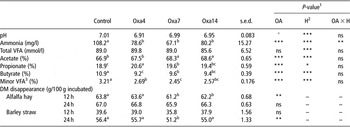
Variations due to hours post feeding were observed in rumen pH, and ammonia and VFA concentrations (P < 0.01); however, for clarity, only mean values are shown in Table 1. The administration of OA reduced ammonia mean concentration (P < 0.001), although this effect was not significant at all collection times (interaction, P < 0.01), and it did not affect (P > 0.05) the mean values of pH and total VFA, acetate and propionate concentrations. However, the molar proportions of acetic and propionic acids increased (P < 0.001), and both the concentrations and molar proportions of butyric acid and the sum of minor VFA (valeric, isobutyric, isovaleric and caproic acids) were reduced in response to OA (P < 0.001).
Mean rumen fermentation characteristics (pH, ammonia concentration and VFA concentration and molar proportions), and DM disappearance from alfalfa hay and barley straw incubated for 12 and 24 h in the rumen of sheep on days 0 (Control), 4 (Oxa4), 7 (Oxa7) and 14 (Oxa14) of OA dosing

VFA = volatile fatty acid; DM = dry matter; OA = oxalic acid.
1Probability of significant effects because of OA administration (Control, Oxa4, Oxa7 and Oxa14), hours post feeding (H; 0, 3, 6 and 9) and their interaction (OA × H): ns = not significant (P > 0.10); +P < 0.10; *P < 0.05; **P < 0.01; ***P < 0.001.
2Although, as expected, variations because of hours post feeding (H) were observed in rumen pH, and ammonia and VFA concentrations (P < 0.01); for clarity, only mean values are shown.
3Calculated as the sum of valeric, isobutyric, isovaleric and caproic acids.
a,b,cMean values with different superscripts in each row differ significantly (P < 0.05).
Table 1 also shows the effect of OA on ruminal degradation. DM disappearance of alfalfa hay was slightly reduced (−3%) after 12 h of incubation on Oxa7 and Oxa14 (P < 0.01), but this effect was not observed in the 24-h incubations. On the other hand, DM disappearance of barley straw was only transiently decreased (P < 0.01) on Oxa7 and after 24 h of incubation, with no significant differences by the end of the experiment (Oxa14).
The administration of OA had no effect on feed intake (P > 0.10).
Bacterial community analysis by T-RFLP
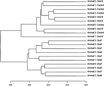
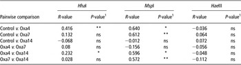
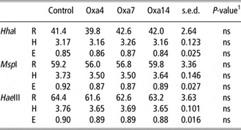
Rumen bacterial T-RFLP analysis of 16S rRNA genes generated on average 41.5 ± 0.89, 58.0 ± 1.15 and 63.0 ± 1.20 fragments with the enzymes HhaI, MspI and HaeIII, respectively. Hierarchical clustering indicated that OA altered only transiently the structure of the rumen bacterial community (Figure 1), as the analysis resulted in two major clusters, and samples obtained on days 0 and 14 of OA dosing were grouped together and separated from those collected on days 4 and 7. Although the diversity indices (richness, Shannon–Weiner and Shannon Evenness) were not modified by the treatment after either HhaI, MspI or HaeIII digestions (P > 0.10; Table 2), the ANOSIM (pairwise comparisons) indicates significant differences in the bacterial community between sampling days (Table 3). In this regard, the enzyme MspI showed the most consistent results, with low and non-significant R-values for the comparison of initial (control) and final (Oxa14) structures. Similar results were observed between bacterial profiles after 4 (Oxa4) and 7 (Oxa7) days on OA, whereas comparisons between control or Oxa14 and either Oxa4 or Oxa7 resulted in significant R-values higher than 0.5 (P < 0.05). Only the comparison control v. Oxa4 was significantly different (R > 0.25 and P < 0.05) with HhaI, and R-values were always low and not significant with HaeIII.
Cluster analysis of the terminal restriction fragment length polymorphism (T-RFLP) profiles, based on the Ward's method and the Jaccard distances, of total bacteria present in the ruminal digesta of sheep on days 0 (Control), 4 (Oxa4), 7 (Oxa7) and 14 (Oxa14) of oxalic acid dosing.

Analysis of similarity (pairwise comparisons) of the total bacteria T-RFLP profile of ruminal digesta of sheep on days 0 (Control), 4 (Oxa4), 7 (Oxa7) and 14 (Oxa14) of OA dosing

T-RFLP = terminal restriction fragment length polymorphism; OA = oxalic acid.
1Level of significance of R-value: ns = not significant (P > 0.10); *P < 0.05; **P < 0.01.
Diversity indices (richness, R; Shannon–Wiener, H; and Shannon evenness, E) calculated from the total bacteria T-RFLP profiles of ruminal digesta of sheep on days 0 (Control), 4 (Oxa4), 7 (Oxa7) and 14 (Oxa14) of OA dosing

T-RFLP = terminal restriction fragment length polymorphism; OA = oxalic acid.
1Probability of significant effect due to oxalic acid administration (Control, Oxa4, Oxa7 and Oxa14): ns = not significant (P > 0.10).
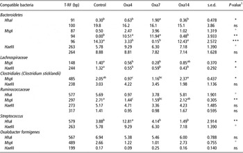
Several different T-RF exhibited variations in their relative frequency out of the total peak area because of the presence of OA (Table 4). These changes were only transient in many cases, with the initial values being recovered by the end of the experiment (e.g. 91 and 579 bp with HhaI; and 96 and 297 bp with MspI). The T-RF compatible with O. formigenes obtained with HhaI (567 bp) and HaeIII (199 bp) showed no variations with OA (P > 0.10), although that obtained with MspI (489 bp) tended to decrease its relative abundance on Oxa4 and Oxa7 (P < 0.10).
Relative frequencies over the total peak area (%) of some T-RF identified by T-RF length polymorphism profiles in DNA samples of ruminal digesta of sheep on days 0 (Control), 4 (Oxa4), 7 (Oxa7) and 14 (Oxa14) of OA dosing

T-RF = terminal restriction fragment; OA = oxalic acid.
1Probability of significant effect due to oxalic acid administration (Control, Oxa4, Oxa7 and Oxa14): ns = not significant (P > 0.10); +P < 0.10; *P < 0.05; **P < 0.01; ***P < 0.001.
a,b,cMean values with different superscripts in each row differ significantly (P < 0.05).
O. formigenes quantification by qPCR
The DNA abundance of the oxc gene of O. formigenes was very small relative to that of total bacterial 16S rRNA gene (mean 0.005 ± 0.0007%); however, it was significantly increased over time with the administration of OA (P < 0.001; Table 5). Similar results were obtained when the abundance of the specific oxc gene DNA of these oxalate-degrading bacteria was expressed as pg/ng total DNA.
Abundance of the oxc gene DNA of Oxalobacter formigenes quantified by real-time PCR (expressed as percentage of the total bacteria 16S rDNA, or as specific DNA concentration) in the ruminal digesta of sheep on days 0 (Control), 4 (Oxa4), 7 (Oxa7) and 14 (Oxa14) of oxalic acid dosing

1Probability of significant effect due to oxalic acid administration (Control, Oxa4, Oxa7 and Oxa14): **P < 0.01; ***P < 0.001.
a,b,cMean values with different superscripts in each row differ significantly (P < 0.05).
Discussion
Previous research has shown that adaptation of the rumen environment to the consumption of increasing levels of OA prevents poisoning of the host animal (Duncan et al., Reference Duncan, Frutos and Young1997). However, little is known on the action of this secondary compound on the rumen bacterial community and its potential consequences on ruminal function. In this experiment, the level of OA was chosen on the basis of preceding studies to simulate moderate ingestion rates by ruminants under field conditions (Libert and Franceschi, Reference Libert and Franceschi1987; Duncan et al., Reference Duncan, Frutos and Young1997 and Reference Duncan, Frutos and Young2000).
Changes in the ruminal microbiota occurred rapidly with OA dosing (Allison et al., Reference Allison, Littledike and James1977; Duncan et al., Reference Duncan, Frutos and Young1997; Frutos et al., Reference Frutos, Duncan, Kyriazakis and Gordon1998); however, interestingly, they were reversed on day 14, which suggests an adaptation of the rumen bacterial community along with the increase in the abundance of O. formigenes and therefore with the ability to degrade OA. This contrasts with the rumen degradation of other plant secondary compounds, such as tannins, involving a complex consortium of microorganisms (Belenguer et al., Reference Belenguer, Hervás, Yañez-Ruiz, Toral, Ezquerro and Frutos2010) and the consequent need of longer periods for adaptation of the whole rumen microbial community.
Changes in concentrations and molar proportions of specific VFA by OA administration suggest divergences in fermentation pathways probably because of a stimulation of specific microbial groups to the detriment of others (Russell and Wallace, Reference Russell and Wallace1997). This is supported by some T-RFLP results. For instance, T-RF compatible with uncultured bacteria of the family Lachnospiraceae (148 bp with MspI and 244 bp with HaeIII), which includes important ruminal butyrate producers, such as Butyrivibrio spp. (Mrazek et al., Reference Mrazek, Tepsi, Avgustin and Kopecny2006), decreased its relative abundance with OA, indicating a potential relationship with reductions in butyrate concentration.
The decrease caused by OA in ammonia concentration, together with the lower levels of minor VFA that originate mostly from deamination of specific amino acids, would be consistent with an inhibition of rumen proteolysis. This effect was initially related to variations of a T-RF (485 bp with MspI) compatible with Clostridium sticklandii, which belongs to rumen hyper-ammonia-producing bacteria (Krause and Russell, Reference Krause and Russell1996). However, changes in this fragment were inconsistent and prevented the establishment of a clear relationship.
OA reduced the ruminal DM disappearance of the two reference feedstuffs, but initial values were only recovered for barley straw. As both feedstuffs differ substantially in their fibrous fraction, these results would suggest a faster adaptation of hemicellulolytic bacteria to OA, probably because their presence was favoured by the low-quality forage offered to the animals (Tajima et al., Reference Tajima, Aminov, Nagamine, Matsui, Nakamura and Benno2001). The lack of differences in alfalfa DM disappearance after 24 h of incubation was probably because of this feed reaching its maximum degradation before that time.
Shifts in ruminal fermentation could be speculated to be partially explained by the effect of OA administration on calcium and other cation levels in the rumen and the consequent impact on rumen function and bacterial community structure (Sahin, Reference Sahin2003).
Variations in ruminal degradation may also be related to changes in the relative frequencies of a number of T-RF showing significant differences on days 4 and 7 of OA administration, compared with the control, but recovering the initial values on day 14. Example of this are two fragments (94 and 96 bp with MspI) that are compatible with Bacteroidetes, a large rumen microbial group (Edwards et al., Reference Edwards, McEwan, Travis and Wallace2004), or another T-RF (297 bp with MspI) compatible with species of the family Ruminococcacea, which includes some of the main cellulolytic bacteria (Krause et al., Reference Krause, Denman, Mackie, Morrison, Rae, Attwood and McSweeney2003). It is probably worth mentioning here that, despite methodological biases and some remarkable differences in the relative abundances of particular T-RF (e.g. 94 and 96 bp with MspI), the T-RFLP technique appears to be able to reliably detect the relative changes induced to individual components of complex microbial communities (Hartmann and Widmer, Reference Hartmann and Widmer2008).
Although different oxalotroph bacteria have been isolated from the digestive tract of humans (e.g. Eubacterium lentum or Enterococcus faecalis; Sahin, Reference Sahin2003; Abratt and Reid, Reference Abratt and Reid2010), rumen degradation of OA has been considered to rely only on O. formigenes (Dawson et al., Reference Dawson, Allison and Hartman1980; Daniel et al., Reference Daniel, Cook, Hartman and Allison1989). In this study, however, none of the three T-RFs that may correspond to O. formigenes (567 with HhaI, 489 with MspI, and 199 with HaeIII) was significantly increased by OA administration. Nevertheless, it is possible that other bacteria of the phylum Firmicutes, a broad and diverse rumen microbial group (Edwards et al., Reference Edwards, McEwan, Travis and Wallace2004), may result in similarly sized fragments and mask potential changes in O. formigenes. Moreover, T-RFLP may miss populations present in small proportions, unlike real-time PCR, which is more sensitive and accurate. In fact, results from the latter technique showed a low abundance of O. formigenes and a rapid increase with OA administration.
Real-time PCR analysis was based on the measurement of the abundance of the gene encoding oxalyl-CoA decarboxylase (oxc gene) in O. formigenes (Jiang et al., Reference Jiang, Knight, Easter, Neiberg, Holmes and Assimos2011). As these bacteria use that enzyme for the breakdown of OA (Allison et al., Reference Allison, Dawson, Mayberry and Foss1985), changes in the relative abundance of the oxc gene would be a direct reflection of variations in their relative abundance in the rumen. As shown in Table 5, oxc gene represented <0.007% of total bacterial 16S rDNA, which was expected given the likely differences in the number of copies of both genes in the bacterial cell (Khammar et al., Reference Khammar, Martin, Ferro, Job, Aragno and Verrecchia2009), the dependency of O. formigenes on OA as source of energy, and the low intake of this compound in animals fed conventional diets. This agrees with earlier studies reporting very low proportions of oxalate degraders, likely O. formigenes, in the rumen (on average, 0.38% of total bacteria; Daniel et al., Reference Daniel, Cook, Hartman and Allison1989). Furthermore, on the basis of previous measurements (Jiang et al., Reference Jiang, Knight, Easter, Neiberg, Holmes and Assimos2011) reporting that 1 ng DNA of oxc equals 4.82 × 10⁵ cells of O. formigenes, the mean number of cells in the rumen of sheep was estimated to range between 0.2 and 4.1 × 10⁸ cells/g after 0 or 14 days of OA administration, respectively. According to the rates of OA degradation in sheep reported by Duncan et al. (Reference Duncan, Frutos and Young1997), probably a little proportion of OA escaped ruminal breakdown. Notwithstanding, if all the OA were metabolized by O. formigenes, the rates of degradation per cell would be up to 0.06 pmol/day, which is well below the maximum capacity estimated by Daniel et al. (Reference Daniel, Cook, Hartman and Allison1989) in these bacteria (0.94 pmol/day). This supports that rumen O. formigenes would be capable of detoxifying the oxalates consumed by sheep and may represent an example of a single organism taking an extremely specialized metabolic role in the presence of many other species (Wallace, Reference Wallace2008).
Conclusion
The administration of OA altered the rumen environment, including the bacterial community. However, the slight changes observed in certain ruminal fermentation and degradation parameters did not seem to compromise the rumen function. The modifications of the bacterial community structure were interestingly reversed in <14 days, suggesting a speedy adaptation of the ruminal microbiota to the consumption of this compound. This rapid adaptation was likely linked to the rapid increase in the abundance of O. formigenes, which is assumed to be responsible for oxalate breakdown.
Acknowledgements
M. Ben Bati gratefully acknowledges receipt of a grant from the Mediterranean Agronomic Institute of Zaragoza (IAMZ, Spain).